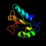
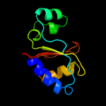
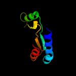
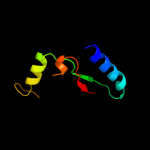
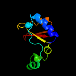
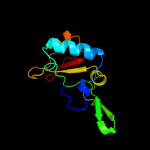
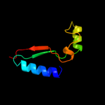
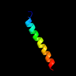
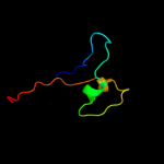
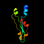
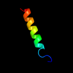
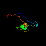
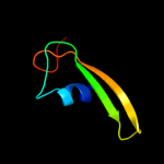
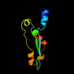
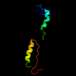
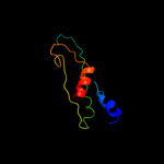
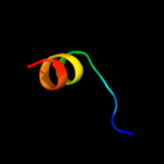
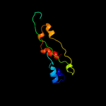
1 d1lbua2
100.0
26
Fold: Hedgehog/DD-peptidaseSuperfamily: Hedgehog/DD-peptidaseFamily: Muramoyl-pentapeptide carboxypeptidase2 c1lbuA_
99.9
25
PDB header: hydrolaseChain: A: PDB Molecule: muramoyl-pentapeptide carboxypeptidase;PDBTitle: hydrolase metallo (zn) dd-peptidase
3 c2vo9C_
98.2
21
PDB header: hydrolaseChain: C: PDB Molecule: l-alanyl-d-glutamate peptidase;PDBTitle: crystal structure of the enzymatically active domain of the2 listeria monocytogenes bacteriophage 500 endolysin ply500
4 d2vo9a1
98.1
25
Fold: Hedgehog/DD-peptidaseSuperfamily: Hedgehog/DD-peptidaseFamily: VanY-like5 d2ibge1
97.2
16
Fold: Hedgehog/DD-peptidaseSuperfamily: Hedgehog/DD-peptidaseFamily: Hedgehog (development protein), N-terminal signaling domain6 c3m1nB_
97.2
22
PDB header: signaling proteinChain: B: PDB Molecule: sonic hedgehog protein;PDBTitle: crystal structure of human sonic hedgehog n-terminal domain
7 d3d1ma1
97.1
20
Fold: Hedgehog/DD-peptidaseSuperfamily: Hedgehog/DD-peptidaseFamily: Hedgehog (development protein), N-terminal signaling domain8 d1r44a_
96.0
27
Fold: Hedgehog/DD-peptidaseSuperfamily: Hedgehog/DD-peptidaseFamily: VanX-like9 c2e76D_
59.7
17
PDB header: photosynthesisChain: D: PDB Molecule: cytochrome b6-f complex iron-sulfur subunit;PDBTitle: crystal structure of the cytochrome b6f complex with tridecyl-2 stigmatellin (tds) from m.laminosus
10 c2f2fC_
53.4
23
PDB header: toxinChain: C: PDB Molecule: cytolethal distending toxin c;PDBTitle: crystal structure of cytolethal distending toxin (cdt) from2 actinobacillus actinomycetemcomitans
11 d2f2fc1
53.4
23
Fold: beta-TrefoilSuperfamily: Ricin B-like lectinsFamily: Ricin B-like12 d1gtma2
43.3
29
Fold: Aminoacid dehydrogenase-like, N-terminal domainSuperfamily: Aminoacid dehydrogenase-like, N-terminal domainFamily: Aminoacid dehydrogenases13 c1p84E_
39.2
13
PDB header: oxidoreductaseChain: E: PDB Molecule: ubiquinol-cytochrome c reductase iron-sulfurPDBTitle: hdbt inhibited yeast cytochrome bc1 complex
14 d1sr4c_
36.1
21
Fold: beta-TrefoilSuperfamily: Ricin B-like lectinsFamily: Ricin B-like15 c3g7kD_
30.3
15
PDB header: isomeraseChain: D: PDB Molecule: 3-methylitaconate isomerase;PDBTitle: crystal structure of methylitaconate-delta-isomerase
16 d1bvua2
29.7
28
Fold: Aminoacid dehydrogenase-like, N-terminal domainSuperfamily: Aminoacid dehydrogenase-like, N-terminal domainFamily: Aminoacid dehydrogenases17 c2h31A_
29.1
9
PDB header: ligase, lyaseChain: A: PDB Molecule: multifunctional protein ade2;PDBTitle: crystal structure of human paics, a bifunctional carboxylase and2 synthetase in purine biosynthesis
18 c3k8zD_
28.0
20
PDB header: oxidoreductaseChain: D: PDB Molecule: nad-specific glutamate dehydrogenase;PDBTitle: crystal structure of gudb1 a decryptified secondary glutamate2 dehydrogenase from b. subtilis
19 d1am7a_
26.8
21
Fold: Lysozyme-likeSuperfamily: Lysozyme-likeFamily: Lambda lysozyme20 d1euza2
23.8
25
Fold: Aminoacid dehydrogenase-like, N-terminal domainSuperfamily: Aminoacid dehydrogenase-like, N-terminal domainFamily: Aminoacid dehydrogenases21 d1m0wa1
not modelled
22.5
19
Fold: PreATP-grasp domainSuperfamily: PreATP-grasp domainFamily: Eukaryotic glutathione synthetase, substrate-binding domain22 c2fynO_
not modelled
21.5
21
PDB header: oxidoreductaseChain: O: PDB Molecule: ubiquinol-cytochrome c reductase iron-sulfurPDBTitle: crystal structure analysis of the double mutant rhodobacter2 sphaeroides bc1 complex
23 d2hgsa1
not modelled
20.6
26
Fold: PreATP-grasp domainSuperfamily: PreATP-grasp domainFamily: Eukaryotic glutathione synthetase, substrate-binding domain24 c2fyuE_
not modelled
19.5
7
PDB header: oxidoreductaseChain: E: PDB Molecule: ubiquinol-cytochrome c reductase iron-sulfur subunit,PDBTitle: crystal structure of bovine heart mitochondrial bc1 with jg1442 inhibitor
25 c3kalB_
not modelled
18.4
22
PDB header: ligaseChain: B: PDB Molecule: homoglutathione synthetase;PDBTitle: structure of homoglutathione synthetase from glycine max in2 closed conformation with homoglutathione, adp, a sulfate3 ion, and three magnesium ions bound
26 c3a7kD_
not modelled
14.3
11
PDB header: membrane proteinChain: D: PDB Molecule: halorhodopsin;PDBTitle: crystal structure of halorhodopsin from natronomonas2 pharaonis
27 c3aoeC_
not modelled
14.3
27
PDB header: oxidoreductaseChain: C: PDB Molecule: glutamate dehydrogenase;PDBTitle: crystal structure of hetero-hexameric glutamate dehydrogenase from2 thermus thermophilus (leu bound form)
28 c2pw0A_
not modelled
12.7
9
PDB header: unknown functionChain: A: PDB Molecule: prpf methylaconitate isomerase;PDBTitle: crystal structure of trans-aconitate bound to methylaconitate2 isomerase prpf from shewanella oneidensis
29 c2pq4B_
not modelled
12.4
29
PDB header: chaperone/oxidoreductaseChain: B: PDB Molecule: periplasmic nitrate reductase precursor;PDBTitle: nmr solution structure of napd in complex with napa1-352 signal peptide
30 c2c7hA_
not modelled
12.4
33
PDB header: ubiquitin-like proteinChain: A: PDB Molecule: retinoblastoma-binding protein 6, isoform 3;PDBTitle: solution nmr structure of the dwnn domain from human rbbp6
31 c3n6jA_
not modelled
11.2
10
PDB header: isomeraseChain: A: PDB Molecule: mandelate racemase/muconate lactonizing protein;PDBTitle: crystal structure of mandelate racemase/muconate lactonizing protein2 from actinobacillus succinogenes 130z
32 c3mfnD_
not modelled
11.2
20
PDB header: structural genomics, unknown functionChain: D: PDB Molecule: uncharacterized protein;PDBTitle: dfer_2879 protein of unknown function from dyadobacter fermentans
33 d2h9fa1
not modelled
10.0
18
Fold: Diaminopimelate epimerase-likeSuperfamily: Diaminopimelate epimerase-likeFamily: PA0793-like34 d1b26a2
not modelled
10.0
34
Fold: Aminoacid dehydrogenase-like, N-terminal domainSuperfamily: Aminoacid dehydrogenase-like, N-terminal domainFamily: Aminoacid dehydrogenases35 c2hgsA_
not modelled
9.9
26
PDB header: amine/carboxylate ligaseChain: A: PDB Molecule: protein (glutathione synthetase);PDBTitle: human glutathione synthetase
36 d2hrkb1
not modelled
9.9
47
Fold: GST C-terminal domain-likeSuperfamily: GST C-terminal domain-likeFamily: Arc1p N-terminal domain-like37 c3bbnP_
not modelled
9.7
20
PDB header: ribosomeChain: P: PDB Molecule: ribosomal protein s16;PDBTitle: homology model for the spinach chloroplast 30s subunit2 fitted to 9.4a cryo-em map of the 70s chlororibosome.
38 d1u02a_
not modelled
9.5
21
Fold: HAD-likeSuperfamily: HAD-likeFamily: Trehalose-phosphatase39 d2r25b1
not modelled
9.4
14
Fold: Flavodoxin-likeSuperfamily: CheY-likeFamily: CheY-related40 c1bvuF_
not modelled
9.3
29
PDB header: oxidoreductaseChain: F: PDB Molecule: protein (glutamate dehydrogenase);PDBTitle: glutamate dehydrogenase from thermococcus litoralis
41 c1nr1A_
not modelled
9.2
17
PDB header: oxidoreductaseChain: A: PDB Molecule: glutamate dehydrogenase 1;PDBTitle: crystal structure of the r463a mutant of human glutamate2 dehydrogenase
42 d1okga3
not modelled
8.6
44
Fold: FKBP-likeSuperfamily: FKBP-likeFamily: 3-mercaptopyruvate sulfurtransferase, C-terminal domain43 d1qw1a1
not modelled
8.6
18
Fold: SH3-like barrelSuperfamily: C-terminal domain of transcriptional repressorsFamily: FeoA-like44 d1v9la2
not modelled
8.4
25
Fold: Aminoacid dehydrogenase-like, N-terminal domainSuperfamily: Aminoacid dehydrogenase-like, N-terminal domainFamily: Aminoacid dehydrogenases45 c1xf7A_
not modelled
8.4
57
PDB header: transcriptionChain: A: PDB Molecule: wilms' tumor protein;PDBTitle: high resolution nmr structure of the wilms' tumor2 suppressor protein (wt1) finger 3
46 d1xf7a_
not modelled
8.4
57
Fold: beta-beta-alpha zinc fingersSuperfamily: beta-beta-alpha zinc fingersFamily: Classic zinc finger, C2H247 c1m0tB_
not modelled
7.7
19
PDB header: ligaseChain: B: PDB Molecule: glutathione synthetase;PDBTitle: yeast glutathione synthase
48 c3s2xB_
not modelled
7.7
32
PDB header: transferaseChain: B: PDB Molecule: acetyl-coa synthase subunit alpha;PDBTitle: structure of acetyl-coenzyme a synthase alpha subunit c-terminal2 domain
49 c3hdvB_
not modelled
7.0
12
PDB header: transcription regulatorChain: B: PDB Molecule: response regulator;PDBTitle: crystal structure of response regulator receiver protein from2 pseudomonas putida
50 d1bgva2
not modelled
7.0
23
Fold: Aminoacid dehydrogenase-like, N-terminal domainSuperfamily: Aminoacid dehydrogenase-like, N-terminal domainFamily: Aminoacid dehydrogenases51 c2wyoC_
not modelled
7.0
19
PDB header: ligaseChain: C: PDB Molecule: glutathione synthetase;PDBTitle: trypanosoma brucei glutathione synthetase
52 d1sp2a_
not modelled
6.6
57
Fold: beta-beta-alpha zinc fingersSuperfamily: beta-beta-alpha zinc fingersFamily: Classic zinc finger, C2H253 d1dz3a_
not modelled
6.6
16
Fold: Flavodoxin-likeSuperfamily: CheY-likeFamily: CheY-related54 d1qcza_
not modelled
6.5
19
Fold: Flavodoxin-likeSuperfamily: N5-CAIR mutase (phosphoribosylaminoimidazole carboxylase, PurE)Family: N5-CAIR mutase (phosphoribosylaminoimidazole carboxylase, PurE)55 c2jx5A_
not modelled
6.5
35
PDB header: ribosomal proteinChain: A: PDB Molecule: glub(s27a);PDBTitle: solution structure of the ubiquitin domain n-terminal to2 the s27a ribosomal subunit of giardia lamblia
56 c1arfA_
not modelled
6.1
43
PDB header: transcription regulationChain: A: PDB Molecule: yeast transcription factor adr1;PDBTitle: structures of dna-binding mutant zinc finger domains:2 implications for dna binding
57 d2adra1
not modelled
5.8
43
Fold: beta-beta-alpha zinc fingersSuperfamily: beta-beta-alpha zinc fingersFamily: Classic zinc finger, C2H258 c3dclC_
not modelled
5.8
40
PDB header: structural genomics, unknown functionChain: C: PDB Molecule: tm1086;PDBTitle: crystal structure of tm1086
59 d1x6ha2
not modelled
5.7
57
Fold: beta-beta-alpha zinc fingersSuperfamily: beta-beta-alpha zinc fingersFamily: Classic zinc finger, C2H260 c1areA_
not modelled
5.7
43
PDB header: transcription regulationChain: A: PDB Molecule: yeast transcription factor adr1;PDBTitle: structures of dna-binding mutant zinc finger domains:2 implications for dna binding
61 d1ynja1
not modelled
5.5
7
Fold: DCoH-likeSuperfamily: RBP11-like subunits of RNA polymeraseFamily: RNA polymerase alpha subunit dimerisation domain62 c3rggD_
not modelled
5.3
13
PDB header: lyaseChain: D: PDB Molecule: phosphoribosylaminoimidazole carboxylase, pure protein;PDBTitle: crystal structure of treponema denticola pure bound to air
63 c3thaB_
not modelled
5.3
13
PDB header: lyaseChain: B: PDB Molecule: tryptophan synthase alpha chain;PDBTitle: tryptophan synthase subunit alpha from campylobacter jejuni.
64 c3lk2B_
not modelled
5.3
44
PDB header: protein bindingChain: B: PDB Molecule: f-actin-capping protein subunit beta isoforms 1 and 2;PDBTitle: crystal structure of capz bound to the uncapping motif from carmil
65 c2g9mB_
not modelled
5.1
11
PDB header: electron transportChain: B: PDB Molecule: phycoerythrin;PDBTitle: crystal structure of the pigment protein phycoerythrin from2 cyanobacterium at 2.6a resolution


