Phyre Home Retrieve Phyre Job Id
Emaill.a.kelley@imperial.ac.uk
DescriptionP41069
DateThu Jan 5 12:01:26 GMT 2012
Unique Job IDcb2e943934353d74

Summary 

Top model
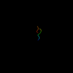
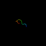
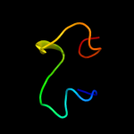
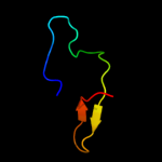
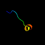
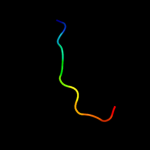
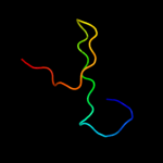
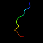
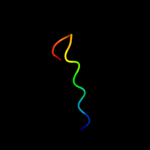
Image coloured by rainbow N → C terminus
Model (left) based on template d1jb0a_
Top template information
Fold:Photosystem I subunits PsaA/PsaB
Superfamily:Photosystem I subunits PsaA/PsaB
Family:Photosystem I subunits PsaA/PsaB
Confidence and coverage
Confidence: 23.6% Coverage: 7%
12 residues ( 7% of your sequence) have been modelled with 23.6% confidence by the single highest scoring template.
You may wish to submit your sequence to Phyrealarm. This will automatically scan your sequence every week for new potential templates as they appear in the Phyre2 library.
Please note: You must be registered and logged in to use Phyrealarm.
Warning: 61% of your sequence is predicted disordered. Disordered regions cannot be meaningfully predicted.
3D viewing
Interactive 3D view in Jmol

Sequence analysis 

Secondary structure and disorder prediction 

   1........10.........20.........30.........40.........50.........60
Sequence  MKQTSFFIPLLGTLLLYGCAGTSTEFECNATTSDTCMTMEQANEKAKKLERSSEAKPVAA
Secondary structure 


SS confidence 


Disorder  ???


?


??????
????


??????????????
Disorder confidence 


   .........70.........80.........90.........100.........110.........120
Sequence  SLPRLAEGNFRTMPVQTVTATTPSGSRPAVTAHPEQKLLAPRPLFTAAREVKTVVPVSSV
Secondary structure 


SS confidence 


Disorder  ??

????????????????????????????????????????????????????????
Disorder confidence 


   .........130.........140.........150.........160.........170.
Sequence  TPVTPPRPLRTGEQTAALWIAPYIDNQDVYHQPSSVFFVIKPSAWGKPRIN
Secondary structure 


SS confidence 


Disorder  ???????????


?


???????
Disorder confidence 


Confidence Key
High(9)                    Low (0)
?Disordered
Alpha helix
Beta strand

Domain analysis 

Hover over an aligned region to see model and summary info

Please note, only up to the top 20 hits are modelled to reduce computer load

RankAligned region

PDB 1jb0 chain A

3D model

Region: 25 - 36
Aligned: 12
Modelled: 12
Confidence: 23.6%
Identity: 33%
Fold: Photosystem I subunits PsaA/PsaB
Superfamily: Photosystem I subunits PsaA/PsaB
Family: Photosystem I subunits PsaA/PsaB

Phyre2

PDB 1jb0 chain B

3D model

Region: 25 - 36
Aligned: 12
Modelled: 12
Confidence: 21.2%
Identity: 33%
Fold: Photosystem I subunits PsaA/PsaB
Superfamily: Photosystem I subunits PsaA/PsaB
Family: Photosystem I subunits PsaA/PsaB

Phyre2

PDB 2l61 chain A

3D model

Region: 17 - 38
Aligned: 17
Modelled: 22
Confidence: 19.6%
Identity: 35%
PDB header:metal binding protein
Chain: A: PDB Molecule:ec protein i/ii;
PDBTitle: protein and metal cluster structure of the wheat metallothionein2 domain g-ec-1. the second part of the puzzle.

Phyre2

PDB 3d0f chain A

3D model

Region: 139 - 170
Aligned: 32
Modelled: 32
Confidence: 16.7%
Identity: 6%
PDB header:transferase
Chain: A: PDB Molecule:penicillin-binding 1 transmembrane protein mrca;
PDBTitle: structure of the big_1156.2 domain of putative penicillin-binding2 protein mrca from nitrosomonas europaea atcc 19718

Phyre2

PDB 2vsa chain A

3D model

Region: 134 - 148
Aligned: 15
Modelled: 15
Confidence: 14.3%
Identity: 27%
PDB header:toxin
Chain: A: PDB Molecule:mosquitocidal toxin;
PDBTitle: structure and mode of action of a mosquitocidal holotoxin

Phyre2

PDB 3e9v chain A domain 1

3D model

Region: 133 - 144
Aligned: 12
Modelled: 12
Confidence: 12.8%
Identity: 33%
Fold: BTG domain-like
Superfamily: BTG domain-like
Family: BTG domain-like

Phyre2

PDB 2z15 chain A domain 1

3D model

Region: 133 - 144
Aligned: 12
Modelled: 12
Confidence: 11.8%
Identity: 33%
Fold: BTG domain-like
Superfamily: BTG domain-like
Family: BTG domain-like

Phyre2

PDB 2ktr chain A

3D model

Region: 143 - 151
Aligned: 9
Modelled: 9
Confidence: 9.2%
Identity: 11%
PDB header:signaling protein, transport protein
Chain: A: PDB Molecule:sequestosome-1;
PDBTitle: nmr structure of p62 pb1 dimer determined based on pcs

Phyre2

PDB 1q90 chain C

3D model

Region: 137 - 147
Aligned: 11
Modelled: 11
Confidence: 9.0%
Identity: 0%
Fold: ISP domain
Superfamily: ISP domain
Family: Rieske iron-sulfur protein (ISP)

Phyre2

PDB 2jvy chain A

3D model

Region: 23 - 30
Aligned: 8
Modelled: 8
Confidence: 8.7%
Identity: 38%
PDB header:metal binding protein
Chain: A: PDB Molecule:nf-kappa-b essential modulator;
PDBTitle: solution structure of the eda-id-related c417f mutant of2 human nemo zinc finger

Phyre2

PDB 2aap chain A

3D model

Region: 18 - 30
Aligned: 13
Modelled: 13
Confidence: 8.4%
Identity: 38%
PDB header:toxin
Chain: A: PDB Molecule:jingzhaotoxin-vii;
PDBTitle: solution structure of jingzhaotoxin-vii

Phyre2

PDB 1e1h chain C

3D model

Region: 137 - 157
Aligned: 21
Modelled: 21
Confidence: 8.2%
Identity: 19%
PDB header:hydrolase
Chain: C: PDB Molecule:botulinum neurotoxin type a light chain;
PDBTitle: crystal structure of recombinant botulinum neurotoxin type2 a light chain, self-inhibiting zn endopeptidase.

Phyre2

PDB 1wmh chain A

3D model

Region: 130 - 142
Aligned: 13
Modelled: 13
Confidence: 8.0%
Identity: 8%
Fold: beta-Grasp (ubiquitin-like)
Superfamily: CAD & PB1 domains
Family: PB1 domain

Phyre2

PDB 3k3q chain B

3D model

Region: 137 - 157
Aligned: 21
Modelled: 21
Confidence: 7.8%
Identity: 19%
PDB header:immune system
Chain: B: PDB Molecule:botulinum neurotoxin type a;
PDBTitle: crystal structure of a llama antibody complexed with the c.2 botulinum neurotoxin serotype a catalytic domain

Phyre2

PDB 1ymz chain A

3D model

Region: 144 - 170
Aligned: 24
Modelled: 27
Confidence: 7.6%
Identity: 17%
PDB header:unknown function
Chain: A: PDB Molecule:cc45;
PDBTitle: cc45, an artificial ww domain designed using statistical2 coupling analysis

Phyre2

PDB 2iyj chain A

3D model

Region: 143 - 157
Aligned: 15
Modelled: 15
Confidence: 7.6%
Identity: 13%
PDB header:isomerase
Chain: A: PDB Molecule:thiol disulfide interchange protein dsbc;
PDBTitle: crystal structure of the n-terminal dimer domain of e.coli2 dsbc

Phyre2

PDB 3ic3 chain C

3D model

Region: 134 - 141
Aligned: 8
Modelled: 8
Confidence: 7.2%
Identity: 0%
PDB header:oxidoreductase
Chain: C: PDB Molecule:putative pyruvate dehydrogenase;
PDBTitle: structure of a putative pyruvate dehydrogenase from the photosynthetic2 bacterium rhodopseudomonas palustrus cga009

Phyre2

PDB 1e0m chain A

3D model

Region: 144 - 170
Aligned: 24
Modelled: 27
Confidence: 7.1%
Identity: 21%
PDB header:de novo protein
Chain: A: PDB Molecule:wwprototype;
PDBTitle: prototype ww domain

Phyre2

PDB 1rfs chain A

3D model

Region: 137 - 147
Aligned: 11
Modelled: 11
Confidence: 7.0%
Identity: 9%
Fold: ISP domain
Superfamily: ISP domain
Family: Rieske iron-sulfur protein (ISP)

Phyre2

PDB 1cew chain I

3D model

Region: 136 - 146
Aligned: 11
Modelled: 11
Confidence: 7.0%
Identity: 9%
Fold: Cystatin-like
Superfamily: Cystatin/monellin
Family: Cystatins

Phyre2
1

d1jb0a_
2

d1jb0b_
3

c2l61A_
4

c3d0fA_
5

c2vsaA_
6

d3e9va1
7

d2z15a1
8

c2ktrA_
9

d1q90c_
10

c2jvyA_
11

c2aapA_
12

c1e1hC_
13

d1wmha_
14

c3k3qB_
15

c1ymzA_
16

c2iyjA_
17

c3ic3C_
18

c1e0mA_
19

d1rfsa_
20

d1cewi_
21


22


23


24


25


26


27


28


29


30


31


32


33


Detailed template information 

#
Template Alignment Coverage3D Model Confidence
% i.d. Template Information
1d1jb0a_


23.6 33 Fold:Photosystem I subunits PsaA/PsaB
Superfamily:Photosystem I subunits PsaA/PsaB
Family:Photosystem I subunits PsaA/PsaB
2d1jb0b_


21.2 33 Fold:Photosystem I subunits PsaA/PsaB
Superfamily:Photosystem I subunits PsaA/PsaB
Family:Photosystem I subunits PsaA/PsaB
3c2l61A_


19.6 35 PDB header:metal binding protein
Chain: A: PDB Molecule:ec protein i/ii;
PDBTitle: protein and metal cluster structure of the wheat metallothionein2 domain g-ec-1. the second part of the puzzle.
4c3d0fA_


16.7 6 PDB header:transferase
Chain: A: PDB Molecule:penicillin-binding 1 transmembrane protein mrca;
PDBTitle: structure of the big_1156.2 domain of putative penicillin-binding2 protein mrca from nitrosomonas europaea atcc 19718
5c2vsaA_


14.3 27 PDB header:toxin
Chain: A: PDB Molecule:mosquitocidal toxin;
PDBTitle: structure and mode of action of a mosquitocidal holotoxin
6d3e9va1


12.8 33 Fold:BTG domain-like
Superfamily:BTG domain-like
Family:BTG domain-like
7d2z15a1


11.8 33 Fold:BTG domain-like
Superfamily:BTG domain-like
Family:BTG domain-like
8c2ktrA_


9.2 11 PDB header:signaling protein, transport protein
Chain: A: PDB Molecule:sequestosome-1;
PDBTitle: nmr structure of p62 pb1 dimer determined based on pcs
9d1q90c_


9.0 0 Fold:ISP domain
Superfamily:ISP domain
Family:Rieske iron-sulfur protein (ISP)
10c2jvyA_


8.7 38 PDB header:metal binding protein
Chain: A: PDB Molecule:nf-kappa-b essential modulator;
PDBTitle: solution structure of the eda-id-related c417f mutant of2 human nemo zinc finger
11c2aapA_


8.4 38 PDB header:toxin
Chain: A: PDB Molecule:jingzhaotoxin-vii;
PDBTitle: solution structure of jingzhaotoxin-vii
12c1e1hC_


8.2 19 PDB header:hydrolase
Chain: C: PDB Molecule:botulinum neurotoxin type a light chain;
PDBTitle: crystal structure of recombinant botulinum neurotoxin type2 a light chain, self-inhibiting zn endopeptidase.
13d1wmha_


8.0 8 Fold:beta-Grasp (ubiquitin-like)
Superfamily:CAD & PB1 domains
Family:PB1 domain
14c3k3qB_


7.8 19 PDB header:immune system
Chain: B: PDB Molecule:botulinum neurotoxin type a;
PDBTitle: crystal structure of a llama antibody complexed with the c.2 botulinum neurotoxin serotype a catalytic domain
15c1ymzA_


7.6 17 PDB header:unknown function
Chain: A: PDB Molecule:cc45;
PDBTitle: cc45, an artificial ww domain designed using statistical2 coupling analysis
16c2iyjA_


7.6 13 PDB header:isomerase
Chain: A: PDB Molecule:thiol disulfide interchange protein dsbc;
PDBTitle: crystal structure of the n-terminal dimer domain of e.coli2 dsbc
17c3ic3C_


7.2 0 PDB header:oxidoreductase
Chain: C: PDB Molecule:putative pyruvate dehydrogenase;
PDBTitle: structure of a putative pyruvate dehydrogenase from the photosynthetic2 bacterium rhodopseudomonas palustrus cga009
18c1e0mA_


7.1 21 PDB header:de novo protein
Chain: A: PDB Molecule:wwprototype;
PDBTitle: prototype ww domain
19d1rfsa_


7.0 9 Fold:ISP domain
Superfamily:ISP domain
Family:Rieske iron-sulfur protein (ISP)
20d1cewi_


7.0 9 Fold:Cystatin-like
Superfamily:Cystatin/monellin
Family:Cystatins
21c2j4mA_


not modelled 7.0 24 PDB header:protein binding
Chain: A: PDB Molecule:endoglucanase 45a;
PDBTitle: double dockerin from piromyces equi cel45a
22c2wwaJ_


not modelled 6.8 33 PDB header:ribosome
Chain: J: PDB Molecule:60s ribosomal protein l19;
PDBTitle: cryo-em structure of idle yeast ssh1 complex bound to the2 yeast 80s ribosome
23d1p90a_


not modelled 6.6 26 Fold:Ribonuclease H-like motif
Superfamily:Nitrogenase accessory factor-like
Family:Nitrogenase accessory factor
24d1hc1a3


not modelled 6.6 14 Fold:Immunoglobulin-like beta-sandwich
Superfamily:E set domains
Family:Arthropod hemocyanin, C-terminal domain
25d2jmfa1


not modelled 6.4 13 Fold:WW domain-like
Superfamily:WW domain
Family:WW domain
26d1plqa2


not modelled 6.3 15 Fold:DNA clamp
Superfamily:DNA clamp
Family:DNA polymerase processivity factor
27d2rm0w1


not modelled 6.0 21 Fold:WW domain-like
Superfamily:WW domain
Family:WW domain
28c2kykA_


not modelled 5.8 20 PDB header:ligase
Chain: A: PDB Molecule:e3 ubiquitin-protein ligase itchy homolog;
PDBTitle: the sandwich region between two lmp2a py motif regulates the2 interaction between aip4ww2domain and py motif
29d1mzga_


not modelled 5.7 16 Fold:SufE/NifU
Superfamily:SufE/NifU
Family:SufE-like
30d1t3ba2


not modelled 5.6 13 Fold:Cystatin-like
Superfamily:DsbC/DsbG N-terminal domain-like
Family:DsbC/DsbG N-terminal domain-like
31c2djyA_


not modelled 5.2 13 PDB header:ligase/signaling protein
Chain: A: PDB Molecule:smad ubiquitination regulatory factor 2;
PDBTitle: solution structure of smurf2 ww3 domain-smad7 py peptide2 complex
32d1pqsa_


not modelled 5.1 20 Fold:beta-Grasp (ubiquitin-like)
Superfamily:CAD & PB1 domains
Family:PB1 domain
33d1r46a2


not modelled 5.0 14 Fold:TIM beta/alpha-barrel
Superfamily:(Trans)glycosidases
Family:Amylase, catalytic domain

Binding site prediction 

Due to computational demand, binding site predictions are not run for batch jobs

If you want to predict binding sites, please manually submit your model of choice to 3DLigandSite


Phyre is for academic use only

Please cite: Protein structure prediction on the web: a case study using the Phyre server
Kelley LA and Sternberg MJE. Nature Protocols 4, 363 - 371 (2009) [pdf] [Import into BibTeX]
 
If you use the binding site predictions from 3DLigandSite, please also cite:
3DLigandSite: predicting ligand-binding sites using similar structures.
Wass MN, Kelley LA and Sternberg MJ Nucleic Acids Research 38, W469-73 (2010) [PubMed]
 
© Structural Bioinformatics Group
Imperial College London
Lawrence Kelley, Benjamin Jefferys
Disclaimer
Terms and Conditions
Component software
Template detection: HHpred 1.51
Secondary structure prediction: Psi-pred 2.5
Disorder prediction: Disopred 2.4
Transmembrane prediction: Memsat_SVM
Multi-template modelling and ab initio: Poing 1.0