Phyre Home Retrieve Phyre Job Id
Emaill.a.kelley@imperial.ac.uk
DescriptionP76228
DateWed Jan 25 15:21:06 GMT 2012
Unique Job IDc8b00471cc62e7e5

Summary 

Top model
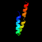
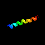
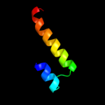
Image coloured by rainbow N → C terminus
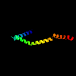
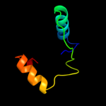
Model (left) based on template c2w8aC_
Top template information
PDB header:membrane protein
Chain: C: PDB Molecule:glycine betaine transporter betp;
PDBTitle: crystal structure of the sodium-coupled glycine betaine2 symporter betp from corynebacterium glutamicum with bound3 substrate
Confidence and coverage
Confidence: 64.8% Coverage: 23%
80 residues ( 23% of your sequence) have been modelled with 64.8% confidence by the single highest scoring template.
You may wish to submit your sequence to Phyrealarm. This will automatically scan your sequence every week for new potential templates as they appear in the Phyre2 library.
Please note: You must be registered and logged in to use Phyrealarm.
3D viewing
Interactive 3D view in Jmol

Sequence analysis 

Secondary structure and disorder prediction 

   1........10.........20.........30.........40.........50.........60
Sequence  MKKVLLQNHPGSEKYSFNGWEIFNSNFERMIKENKAMLLCKWGFYLTCVVAVMFVFAAIT
Secondary structure 


SS confidence 


Disorder  ???

??
?


????
Disorder confidence 


   .........70.........80.........90.........100.........110.........120
Sequence  SNGLNERGLITAGCSFLYLLIMMGLIVRAGFKAKKEQLHYYQAKGIEPLSIEKLQALQLI
Secondary structure 


SS confidence 


Disorder  ??
???


??????


Disorder confidence 


   .........130.........140.........150.........160.........170.........180
Sequence  APYRFYHKQWSETLEFWPRKPEPGKDTFQYHVLPFDSIDIISKRRESLEDQWGIEDSESY
Secondary structure 


SS confidence 


Disorder 


?


?

???


?????


Disorder confidence 


   .........190.........200.........210.........220.........230.........240
Sequence  CALMEHFLSGDHGANTFKANMEEAPEQVIALLNKFAVFPSDYISDCANHSSGKSSAKLIW
Secondary structure 


SS confidence 


Disorder 


?


?

?????


????


Disorder confidence 


   .........250.........260.........270.........280.........290.........300
Sequence  AAELSWMISISSTAFQNGTIEEELAWHYIMLASRKAHELFESEEDYQKNSQMGFLYWHIC
Secondary structure 


SS confidence 


Disorder 


Disorder confidence 


   .........310.........320.........330.........340......
Sequence  CYRRKLTDAELEACYRYDKQFWEHYSKKCRWPIRNVPWGASSVKYS
Secondary structure 


SS confidence 


Disorder  ??????


???????


?


???????
Disorder confidence 


Confidence Key
High(9)                    Low (0)
?Disordered
Alpha helix
Beta strand

Domain analysis 

Hover over an aligned region to see model and summary info

Please note, only up to the top 20 hits are modelled to reduce computer load

RankAligned region

PDB 2w8a chain C

3D model

Region: 36 - 115
Aligned: 80
Modelled: 80
Confidence: 64.8%
Identity: 11%
PDB header:membrane protein
Chain: C: PDB Molecule:glycine betaine transporter betp;
PDBTitle: crystal structure of the sodium-coupled glycine betaine2 symporter betp from corynebacterium glutamicum with bound3 substrate

Phyre2

PDB 1jkv chain A

3D model

Region: 159 - 206
Aligned: 48
Modelled: 48
Confidence: 22.1%
Identity: 15%
Fold: Ferritin-like
Superfamily: Ferritin-like
Family: Manganese catalase (T-catalase)

Phyre2

PDB 1go5 chain A

3D model

Region: 205 - 263
Aligned: 49
Modelled: 59
Confidence: 19.7%
Identity: 8%
Fold: RuvA C-terminal domain-like
Superfamily: UBA-like
Family: TAP-C domain-like

Phyre2

PDB 2qwu chain B

3D model

Region: 131 - 156
Aligned: 26
Modelled: 25
Confidence: 17.8%
Identity: 35%
PDB header:cell invasion
Chain: B: PDB Molecule:intracellular growth locus, subunit c;
PDBTitle: crystal structure of f. tularensis pathogenicity island2 protein c

Phyre2

PDB 2cwl chain A domain 1

3D model

Region: 159 - 203
Aligned: 45
Modelled: 45
Confidence: 16.4%
Identity: 18%
Fold: Ferritin-like
Superfamily: Ferritin-like
Family: Manganese catalase (T-catalase)

Phyre2

PDB 1oai chain A

3D model

Region: 206 - 263
Aligned: 48
Modelled: 58
Confidence: 16.2%
Identity: 8%
Fold: RuvA C-terminal domain-like
Superfamily: UBA-like
Family: TAP-C domain-like

Phyre2

PDB 1jog chain A

3D model

Region: 249 - 281
Aligned: 33
Modelled: 33
Confidence: 15.3%
Identity: 15%
Fold: Four-helical up-and-down bundle
Superfamily: Nucleotidyltransferase substrate binding subunit/domain
Family: Family 1 bi-partite nucleotidyltransferase subunit

Phyre2

PDB 1wty chain A

3D model

Region: 249 - 281
Aligned: 33
Modelled: 33
Confidence: 14.7%
Identity: 18%
Fold: Four-helical up-and-down bundle
Superfamily: Nucleotidyltransferase substrate binding subunit/domain
Family: Family 1 bi-partite nucleotidyltransferase subunit

Phyre2

PDB 3gkx chain B

3D model

Region: 172 - 223
Aligned: 52
Modelled: 52
Confidence: 14.3%
Identity: 2%
PDB header:structural genomics, unknown function
Chain: B: PDB Molecule:putative arsc family related protein;
PDBTitle: crystal structure of putative arsc family related protein from2 bacteroides fragilis

Phyre2

PDB 3cuq chain A

3D model

Region: 94 - 134
Aligned: 27
Modelled: 27
Confidence: 14.2%
Identity: 30%
PDB header:protein transport
Chain: A: PDB Molecule:vacuolar-sorting protein snf8;
PDBTitle: integrated structural and functional model of the human escrt-ii2 complex

Phyre2

PDB 2zme chain A

3D model

Region: 94 - 137
Aligned: 30
Modelled: 30
Confidence: 13.5%
Identity: 30%
PDB header:protein transport
Chain: A: PDB Molecule:vacuolar-sorting protein snf8;
PDBTitle: integrated structural and functional model of the human escrt-ii2 complex

Phyre2

PDB 2gyk chain B domain 1

3D model

Region: 271 - 292
Aligned: 22
Modelled: 22
Confidence: 13.2%
Identity: 23%
Fold: His-Me finger endonucleases
Superfamily: His-Me finger endonucleases
Family: HNH-motif

Phyre2

PDB 1e8o chain A

3D model

Region: 280 - 289
Aligned: 10
Modelled: 10
Confidence: 12.9%
Identity: 10%
Fold: Signal recognition particle alu RNA binding heterodimer, SRP9/14
Superfamily: Signal recognition particle alu RNA binding heterodimer, SRP9/14
Family: Signal recognition particle alu RNA binding heterodimer, SRP9/14

Phyre2

PDB 2rdd chain B

3D model

Region: 74 - 99
Aligned: 26
Modelled: 26
Confidence: 12.4%
Identity: 8%
PDB header:membrane protein/transport protein
Chain: B: PDB Molecule:upf0092 membrane protein yajc;
PDBTitle: x-ray crystal structure of acrb in complex with a novel2 transmembrane helix.

Phyre2

PDB 2job chain A

3D model

Region: 251 - 270
Aligned: 20
Modelled: 20
Confidence: 12.1%
Identity: 30%
PDB header:lipid binding protein
Chain: A: PDB Molecule:antilipopolysaccharide factor;
PDBTitle: solution structure of an antilipopolysaccharide factor from2 shrimp and its possible lipid a binding site

Phyre2

PDB 1914 chain A

3D model

Region: 277 - 289
Aligned: 13
Modelled: 13
Confidence: 12.0%
Identity: 23%
PDB header:alu domain
Chain: A: PDB Molecule:signal recognition particle 9/14 fusion protein;
PDBTitle: signal recognition particle alu rna binding heterodimer, srp9/14

Phyre2

PDB 1zdb chain A

3D model

Region: 94 - 117
Aligned: 24
Modelled: 24
Confidence: 11.9%
Identity: 25%
PDB header:igg binding domain
Chain: A: PDB Molecule:mini protein a domain, z38;
PDBTitle: phage-selected mini protein a domain, z38, nmr, minimized2 mean structure

Phyre2

PDB 1wwp chain A

3D model

Region: 249 - 281
Aligned: 33
Modelled: 33
Confidence: 11.7%
Identity: 12%
PDB header:structural genomics, unknown function
Chain: A: PDB Molecule:hypothetical protein ttha0636;
PDBTitle: crystal structure of ttk003001694 from thermus thermophilus2 hb8

Phyre2

PDB 2rh2 chain A domain 1

3D model

Region: 9 - 19
Aligned: 11
Modelled: 11
Confidence: 10.6%
Identity: 45%
Fold: SH3-like barrel
Superfamily: Electron transport accessory proteins
Family: R67 dihydrofolate reductase

Phyre2

PDB 1qqr chain A

3D model

Region: 2 - 13
Aligned: 12
Modelled: 12
Confidence: 9.0%
Identity: 42%
Fold: beta-Grasp (ubiquitin-like)
Superfamily: Staphylokinase/streptokinase
Family: Staphylokinase/streptokinase

Phyre2
1

c2w8aC_
2

d1jkva_
3

d1go5a_
4

c2qwuB_
5

d2cwla1
6

d1oaia_
7

d1joga_
8

d1wtya_
9

c3gkxB_
10

c3cuqA_
11

c2zmeA_
12

d2gykb1
13

d1e8oa_
14

c2rddB_
15

c2jobA_
16

c1914A_
17

c1zdbA_
18

c1wwpA_
19

d2rh2a1
20

d1qqra_
21


22


23


24


25


26


27


28


29


30


31


32


33


34


35


36


37


38


39


40


41


42


43


44


45


46


47


48


49


50


Detailed template information 

#
Template Alignment Coverage3D Model Confidence
% i.d. Template Information
1c2w8aC_


64.8 11 PDB header:membrane protein
Chain: C: PDB Molecule:glycine betaine transporter betp;
PDBTitle: crystal structure of the sodium-coupled glycine betaine2 symporter betp from corynebacterium glutamicum with bound3 substrate
2d1jkva_


22.1 15 Fold:Ferritin-like
Superfamily:Ferritin-like
Family:Manganese catalase (T-catalase)
3d1go5a_


19.7 8 Fold:RuvA C-terminal domain-like
Superfamily:UBA-like
Family:TAP-C domain-like
4c2qwuB_


17.8 35 PDB header:cell invasion
Chain: B: PDB Molecule:intracellular growth locus, subunit c;
PDBTitle: crystal structure of f. tularensis pathogenicity island2 protein c
5d2cwla1


16.4 18 Fold:Ferritin-like
Superfamily:Ferritin-like
Family:Manganese catalase (T-catalase)
6d1oaia_


16.2 8 Fold:RuvA C-terminal domain-like
Superfamily:UBA-like
Family:TAP-C domain-like
7d1joga_


15.3 15 Fold:Four-helical up-and-down bundle
Superfamily:Nucleotidyltransferase substrate binding subunit/domain
Family:Family 1 bi-partite nucleotidyltransferase subunit
8d1wtya_


14.7 18 Fold:Four-helical up-and-down bundle
Superfamily:Nucleotidyltransferase substrate binding subunit/domain
Family:Family 1 bi-partite nucleotidyltransferase subunit
9c3gkxB_


14.3 2 PDB header:structural genomics, unknown function
Chain: B: PDB Molecule:putative arsc family related protein;
PDBTitle: crystal structure of putative arsc family related protein from2 bacteroides fragilis
10c3cuqA_


14.2 30 PDB header:protein transport
Chain: A: PDB Molecule:vacuolar-sorting protein snf8;
PDBTitle: integrated structural and functional model of the human escrt-ii2 complex
11c2zmeA_


13.5 30 PDB header:protein transport
Chain: A: PDB Molecule:vacuolar-sorting protein snf8;
PDBTitle: integrated structural and functional model of the human escrt-ii2 complex
12d2gykb1


13.2 23 Fold:His-Me finger endonucleases
Superfamily:His-Me finger endonucleases
Family:HNH-motif
13d1e8oa_


12.9 10 Fold:Signal recognition particle alu RNA binding heterodimer, SRP9/14
Superfamily:Signal recognition particle alu RNA binding heterodimer, SRP9/14
Family:Signal recognition particle alu RNA binding heterodimer, SRP9/14
14c2rddB_


12.4 8 PDB header:membrane protein/transport protein
Chain: B: PDB Molecule:upf0092 membrane protein yajc;
PDBTitle: x-ray crystal structure of acrb in complex with a novel2 transmembrane helix.
15c2jobA_


12.1 30 PDB header:lipid binding protein
Chain: A: PDB Molecule:antilipopolysaccharide factor;
PDBTitle: solution structure of an antilipopolysaccharide factor from2 shrimp and its possible lipid a binding site
16c1914A_


12.0 23 PDB header:alu domain
Chain: A: PDB Molecule:signal recognition particle 9/14 fusion protein;
PDBTitle: signal recognition particle alu rna binding heterodimer, srp9/14
17c1zdbA_


11.9 25 PDB header:igg binding domain
Chain: A: PDB Molecule:mini protein a domain, z38;
PDBTitle: phage-selected mini protein a domain, z38, nmr, minimized2 mean structure
18c1wwpA_


11.7 12 PDB header:structural genomics, unknown function
Chain: A: PDB Molecule:hypothetical protein ttha0636;
PDBTitle: crystal structure of ttk003001694 from thermus thermophilus2 hb8
19d2rh2a1


10.6 45 Fold:SH3-like barrel
Superfamily:Electron transport accessory proteins
Family:R67 dihydrofolate reductase
20d1qqra_


9.0 42 Fold:beta-Grasp (ubiquitin-like)
Superfamily:Staphylokinase/streptokinase
Family:Staphylokinase/streptokinase
21c3fz4A_


not modelled 8.6 2 PDB header:oxidoreductase
Chain: A: PDB Molecule:putative arsenate reductase;
PDBTitle: the crystal structure of a possible arsenate reductase from2 streptococcus mutans ua159
22d1dd4d_


not modelled 8.4 27 Fold:Ribosomal protein L7/12, oligomerisation (N-terminal) domain
Superfamily:Ribosomal protein L7/12, oligomerisation (N-terminal) domain
Family:Ribosomal protein L7/12, oligomerisation (N-terminal) domain
23d1f6va_


not modelled 8.2 20 Fold:C-terminal domain of B transposition protein
Superfamily:C-terminal domain of B transposition protein
Family:C-terminal domain of B transposition protein
24c2vqcA_


not modelled 7.9 17 PDB header:dna-binding protein
Chain: A: PDB Molecule:hypothetical 13.2 kda protein;
PDBTitle: structure of a dna binding winged-helix protein, f-112,2 from sulfolobus spindle-shaped virus 1.
25d2vqca1


not modelled 7.9 17 Fold:DNA/RNA-binding 3-helical bundle
Superfamily:"Winged helix" DNA-binding domain
Family:F112-like
26c3kcvG_


not modelled 7.9 13 PDB header:transport protein
Chain: G: PDB Molecule:probable formate transporter 1;
PDBTitle: structure of formate channel
27d1eexa_


not modelled 7.6 20 Fold:TIM beta/alpha-barrel
Superfamily:Cobalamin (vitamin B12)-dependent enzymes
Family:Diol dehydratase, alpha subunit
28d1tz7a1


not modelled 7.6 19 Fold:TIM beta/alpha-barrel
Superfamily:(Trans)glycosidases
Family:Amylase, catalytic domain
29d2p12a1


not modelled 7.5 17 Fold:FomD barrel-like
Superfamily:FomD-like
Family:FomD-like
30d2jioa2


not modelled 7.4 13 Fold:Formate dehydrogenase/DMSO reductase, domains 1-3
Superfamily:Formate dehydrogenase/DMSO reductase, domains 1-3
Family:Formate dehydrogenase/DMSO reductase, domains 1-3
31c3l78A_


not modelled 7.3 6 PDB header:transcription
Chain: A: PDB Molecule:regulatory protein spx;
PDBTitle: the crystal structure of smu.1142c from streptococcus mutans ua159
32d1eswa_


not modelled 7.2 11 Fold:TIM beta/alpha-barrel
Superfamily:(Trans)glycosidases
Family:Amylase, catalytic domain
33d1qusa_


not modelled 7.1 15 Fold:Lysozyme-like
Superfamily:Lysozyme-like
Family:Bacterial muramidase, catalytic domain
34c3sokB_


not modelled 6.9 35 PDB header:cell adhesion
Chain: B: PDB Molecule:fimbrial protein;
PDBTitle: dichelobacter nodosus pilin fima
35d2o8ia1


not modelled 6.9 21 Fold:UraD-like
Superfamily:UraD-Like
Family:UraD-like
36d1914a1


not modelled 6.7 20 Fold:Signal recognition particle alu RNA binding heterodimer, SRP9/14
Superfamily:Signal recognition particle alu RNA binding heterodimer, SRP9/14
Family:Signal recognition particle alu RNA binding heterodimer, SRP9/14
37d1oqwa_


not modelled 6.2 41 Fold:Pili subunits
Superfamily:Pili subunits
Family:Pilin
38c2v5iA_


not modelled 6.2 16 PDB header:viral protein
Chain: A: PDB Molecule:salmonella typhimurium db7155 bacteriophage det7
PDBTitle: structure of the receptor-binding protein of bacteriophage2 det7: a podoviral tailspike in a myovirus
39c3klzE_


not modelled 6.2 12 PDB header:membrane protein
Chain: E: PDB Molecule:putative formate transporter 1;
PDBTitle: pentameric formate channel with formate bound
40c1svfB_


not modelled 6.0 35 PDB header:viral protein
Chain: B: PDB Molecule:protein (fusion glycoprotein);
PDBTitle: paramyxovirus sv5 fusion protein core
41d1x1na1


not modelled 6.0 15 Fold:TIM beta/alpha-barrel
Superfamily:(Trans)glycosidases
Family:Amylase, catalytic domain
42d2fug34


not modelled 5.7 40 Fold:Ferredoxin-like
Superfamily:4Fe-4S ferredoxins
Family:Ferredoxin domains from multidomain proteins
43c2p7vA_


not modelled 5.6 33 PDB header:transcription
Chain: A: PDB Molecule:regulator of sigma d;
PDBTitle: crystal structure of the escherichia coli regulator of sigma 70, rsd,2 in complex with sigma 70 domain 4
44d2pila_


not modelled 5.6 29 Fold:Pili subunits
Superfamily:Pili subunits
Family:Pilin
45d1pyta_


not modelled 5.4 16 Fold:Ferredoxin-like
Superfamily:Protease propeptides/inhibitors
Family:Pancreatic carboxypeptidase, activation domain
46c1bhbA_


not modelled 5.3 24 PDB header:photoreceptor
Chain: A: PDB Molecule:bacteriorhodopsin;
PDBTitle: three-dimensional structure of (1-71) bacterioopsin2 solubilized in methanol-chloroform and sds micelles3 determined by 15n-1h heteronuclear nmr spectroscopy
47c2k85A_


not modelled 5.2 21 PDB header:protein binding
Chain: A: PDB Molecule:glucocorticoid receptor dna-binding factor 1;
PDBTitle: p190-a rhogap ff1 domain
48c3m8eA_


not modelled 5.1 19 PDB header:dna binding protein
Chain: A: PDB Molecule:putative dna-binding protein;
PDBTitle: protein structure of type iii plasmid segregation tubr
49d2q37a1


not modelled 5.1 21 Fold:UraD-like
Superfamily:UraD-Like
Family:UraD-like
50c2q37A_


not modelled 5.1 21 PDB header:plant protein, lyase
Chain: A: PDB Molecule:ohcu decarboxylase;
PDBTitle: crystal structure of ohcu decarboxylase in the presence of2 (s)-allantoin

Binding site prediction 

Due to computational demand, binding site predictions are not run for batch jobs

If you want to predict binding sites, please manually submit your model of choice to 3DLigandSite

Transmembrane helix prediction 

Transmembrane helices have been predicted in your sequence to adopt the topology shown below


Phyre is for academic use only

Please cite: Protein structure prediction on the web: a case study using the Phyre server
Kelley LA and Sternberg MJE. Nature Protocols 4, 363 - 371 (2009) [pdf] [Import into BibTeX]
 
If you use the binding site predictions from 3DLigandSite, please also cite:
3DLigandSite: predicting ligand-binding sites using similar structures.
Wass MN, Kelley LA and Sternberg MJ Nucleic Acids Research 38, W469-73 (2010) [PubMed]
 
© Structural Bioinformatics Group
Imperial College London
Lawrence Kelley, Benjamin Jefferys
Disclaimer
Terms and Conditions
Component software
Template detection: HHpred 1.51
Secondary structure prediction: Psi-pred 2.5
Disorder prediction: Disopred 2.4
Transmembrane prediction: Memsat_SVM
Multi-template modelling and ab initio: Poing 1.0