1 c2b664_
100.0
32
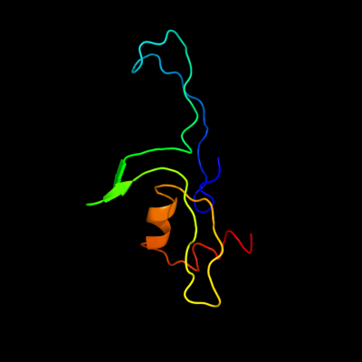
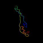
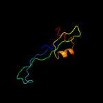
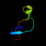
PDB header: ribosomeChain: 4: PDB Molecule: 50s ribosomal protein l31;PDBTitle: 50s ribosomal subunit from a crystal structure of release factor rf1,2 trnas and mrna bound to the ribosome. this file contains the 50s3 subunit from a crystal structure of release factor rf1, trnas and4 mrna bound to the ribosome and is described in remark 400
2 d1vs6z1
100.0
46
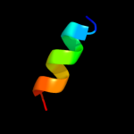
Fold: L28p-likeSuperfamily: L28p-likeFamily: Ribosomal protein L31p3 c3bbo1_
100.0
28
PDB header: ribosomeChain: 1: PDB Molecule: ribosomal protein l31;PDBTitle: homology model for the spinach chloroplast 50s subunit2 fitted to 9.4a cryo-em map of the 70s chlororibosome
4 d2j0141
99.9
39
Fold: L28p-likeSuperfamily: L28p-likeFamily: Ribosomal protein L31p5 c2j034_
99.9
39
PDB header: ribosomeChain: 4: PDB Molecule: 50s ribosomal protein l31;PDBTitle: structure of the thermus thermophilus 70s ribosome2 complexed with mrna, trna and paromomycin (part 4 of 4).3 this file contains the 50s subunit from molecule ii.
6 c3f1f4_
97.1
27
PDB header: ribosomeChain: 4: PDB Molecule: 50s ribosomal protein l31;PDBTitle: crystal structure of a translation termination complex2 formed with release factor rf2. this file contains the 50s3 subunit of one 70s ribosome. the entire crystal structure4 contains two 70s ribosomes as described in remark 400.
7 c2wh44_
97.1
27
PDB header: ribosomeChain: 4: PDB Molecule: 50s ribosomal protein l31;PDBTitle: insights into translational termination from the structure2 of rf2 bound to the ribosome
8 c2ketA_
29.0
31
PDB header: antibioticChain: A: PDB Molecule: cathelicidin-6;PDBTitle: solution structure of bmap-27
9 d1x6ha1
27.6
16
Fold: beta-beta-alpha zinc fingersSuperfamily: beta-beta-alpha zinc fingersFamily: Classic zinc finger, C2H210 c3cucB_
21.9
54
PDB header: signaling proteinChain: B: PDB Molecule: protein of unknown function with a fic domain;PDBTitle: crystal structure of a fic domain containing signaling protein2 (bt_2513) from bacteroides thetaiotaomicron vpi-5482 at 2.71 a3 resolution
11 d1tqza1
20.7
25
Fold: PH domain-like barrelSuperfamily: PH domain-likeFamily: Necap1 N-terminal domain-like12 c3g66A_
17.8
11
PDB header: transferaseChain: A: PDB Molecule: sortase c;PDBTitle: the crystal structure of streptococcus pneumoniae sortase c2 provides novel insights into catalysis as well as pilin3 substrate specificity
13 c3cngC_
16.6
14
PDB header: hydrolaseChain: C: PDB Molecule: nudix hydrolase;PDBTitle: crystal structure of nudix hydrolase from nitrosomonas europaea
14 d1eb7a2
16.5
25
Fold: Cytochrome cSuperfamily: Cytochrome cFamily: Di-heme cytochrome c peroxidase15 d2fiya1
16.5
0
Fold: FdhE-likeSuperfamily: FdhE-likeFamily: FdhE-like16 c3o0pA_
15.3
11
PDB header: transferase , hydrolaseChain: A: PDB Molecule: sortase family protein;PDBTitle: pilus-related sortase c of group b streptococcus
17 c4a1eT_
15.2
26
PDB header: ribosomeChain: T: PDB Molecule: rpl24;PDBTitle: t.thermophila 60s ribosomal subunit in complex with2 initiation factor 6. this file contains 5s rrna, 5.8s rrna3 and proteins of molecule 1
18 d1nmla2
15.0
17
Fold: Cytochrome cSuperfamily: Cytochrome cFamily: Di-heme cytochrome c peroxidase19 c2yiuE_
14.7
43
PDB header: oxidoreductaseChain: E: PDB Molecule: cytochrome c1, heme protein;PDBTitle: x-ray structure of the dimeric cytochrome bc1 complex from2 the soil bacterium paracoccus denitrificans at 2.73 angstrom resolution
20 c3cwbQ_
13.6
43
PDB header: oxidoreductaseChain: Q: PDB Molecule: mitochondrial cytochrome c1, heme protein;PDBTitle: chicken cytochrome bc1 complex inhibited by an iodinated analogue of2 the polyketide crocacin-d
21 c2w1jB_
not modelled
13.5
11
PDB header: transferaseChain: B: PDB Molecule: putative sortase;PDBTitle: crystal structure of sortase c-1 (srtc-1) from2 streptococcus pneumoniae
22 c3n3vA_
not modelled
12.7
46
PDB header: transferaseChain: A: PDB Molecule: adenosine monophosphate-protein transferase ibpa;PDBTitle: crystal structure of ibpafic2-h3717a in complex with adenylylated2 cdc42
23 c2fynH_
not modelled
12.5
29
PDB header: oxidoreductaseChain: H: PDB Molecule: cytochrome c1;PDBTitle: crystal structure analysis of the double mutant rhodobacter2 sphaeroides bc1 complex
24 c1nmlA_
not modelled
12.5
17
PDB header: oxidoreductaseChain: A: PDB Molecule: di-haem cytochrome c peroxidase;PDBTitle: di-haemic cytochrome c peroxidase from pseudomonas nautica 617, form2 in (ph 4.0)
25 d3cx5d1
not modelled
12.5
29
Fold: Cytochrome cSuperfamily: Cytochrome cFamily: Cytochrome bc1 domain26 c2kw8A_
not modelled
11.8
16
PDB header: protein bindingChain: A: PDB Molecule: lpxtg-site transpeptidase family protein;PDBTitle: solution structure of bacillus anthracis sortase a (srta)2 transpeptidase
27 c1p84D_
not modelled
11.6
29
PDB header: oxidoreductaseChain: D: PDB Molecule: cytochrome c1, heme protein;PDBTitle: hdbt inhibited yeast cytochrome bc1 complex
28 d1ppjd1
not modelled
11.5
43
Fold: Cytochrome cSuperfamily: Cytochrome cFamily: Cytochrome bc1 domain29 c1zrtD_
not modelled
11.2
43
PDB header: oxidoreductase/metal transportChain: D: PDB Molecule: cytochrome c1;PDBTitle: rhodobacter capsulatus cytochrome bc1 complex with2 stigmatellin bound
30 c3hq7A_
not modelled
10.9
17
PDB header: oxidoreductaseChain: A: PDB Molecule: cytochrome c551 peroxidase;PDBTitle: ccpa from g. sulfurreducens, g94k/k97q/r100i variant
31 c1dvbA_
not modelled
10.7
4
PDB header: electron transportChain: A: PDB Molecule: rubrerythrin;PDBTitle: rubrerythrin
32 c3izcZ_
not modelled
10.7
19
PDB header: ribosomeChain: Z: PDB Molecule: 60s ribosomal protein rpl24 (l24e);PDBTitle: localization of the large subunit ribosomal proteins into a 6.1 a2 cryo-em map of saccharomyces cerevisiae translating 80s ribosome
33 c2b9vB_
not modelled
10.6
24
PDB header: hydrolaseChain: B: PDB Molecule: alpha-amino acid ester hydrolase;PDBTitle: acetobacter turbidans alpha-amino acid ester hydrolase
34 d1oa8a_
not modelled
10.1
20
Fold: AXH domainSuperfamily: AXH domainFamily: AXH domain35 d1e7la1
not modelled
10.1
60
Fold: LEM/SAP HeH motifSuperfamily: Recombination endonuclease VII, C-terminal and dimerization domainsFamily: Recombination endonuclease VII, C-terminal and dimerization domains36 c2keqA_
not modelled
9.7
36
PDB header: splicingChain: A: PDB Molecule: dna polymerase iii alpha subunit, nucleic acidPDBTitle: solution structure of dnae intein from nostoc punctiforme
37 c3iz5Z_
not modelled
9.4
22
PDB header: ribosomeChain: Z: PDB Molecule: 60s ribosomal protein l24 (l24e);PDBTitle: localization of the large subunit ribosomal proteins into a 5.5 a2 cryo-em map of triticum aestivum translating 80s ribosome
38 d1mi8a_
not modelled
9.3
25
Fold: Hedgehog/intein (Hint) domainSuperfamily: Hedgehog/intein (Hint) domainFamily: Intein (protein splicing domain)39 c1zd7B_
not modelled
8.7
25
PDB header: transferaseChain: B: PDB Molecule: dna polymerase iii alpha subunit;PDBTitle: 1.7 angstrom crystal structure of post-splicing form of a dnae intein2 from synechocystis sp. pcc 6803
40 c3mdnD_
not modelled
8.6
23
PDB header: transferaseChain: D: PDB Molecule: glutamine aminotransferase class-ii domain protein;PDBTitle: structure of glutamine aminotransferase class-ii domain protein2 (spo2029) from silicibacter pomeroyi
41 c3rbjB_
not modelled
8.5
11
PDB header: hydrolaseChain: B: PDB Molecule: sortase family protein;PDBTitle: crystal structure of the lid-mutant of streptococcus agalactiae2 sortase c1
42 d1iufa2
not modelled
8.3
25
Fold: DNA/RNA-binding 3-helical bundleSuperfamily: Homeodomain-likeFamily: Centromere-binding43 c3eqxB_
not modelled
8.3
46
PDB header: dna binding proteinChain: B: PDB Molecule: fic domain containing transcriptional regulator;PDBTitle: crystal structure of a fic family protein (so_4266) from shewanella2 oneidensis at 1.6 a resolution
44 c1xezA_
not modelled
7.8
32
PDB header: toxinChain: A: PDB Molecule: hemolysin;PDBTitle: crystal structure of the vibrio cholerae cytolysin (hlya)2 pro-toxin with octylglucoside bound
45 c2w1kB_
not modelled
7.7
22
PDB header: transferaseChain: B: PDB Molecule: putative sortase;PDBTitle: crystal structure of sortase c-3 (srtc-3) from2 streptococcus pneumoniae
46 d1wada_
not modelled
7.7
50
Fold: Multiheme cytochromesSuperfamily: Multiheme cytochromesFamily: Cytochrome c3-like47 d1at0a_
not modelled
7.7
21
Fold: Hedgehog/intein (Hint) domainSuperfamily: Hedgehog/intein (Hint) domainFamily: Hedgehog C-terminal (Hog) autoprocessing domain48 c1iqcB_
not modelled
7.3
25
PDB header: oxidoreductaseChain: B: PDB Molecule: di-heme peroxidase;PDBTitle: crystal structure of di-heme peroxidase from nitrosomonas europaea
49 c1mpxB_
not modelled
7.2
26
PDB header: hydrolaseChain: B: PDB Molecule: alpha-amino acid ester hydrolase;PDBTitle: alpha-amino acid ester hydrolase labeled with selenomethionine
50 d1am2a_
not modelled
7.1
20
Fold: Hedgehog/intein (Hint) domainSuperfamily: Hedgehog/intein (Hint) domainFamily: Intein (protein splicing domain)51 c3dd7A_
not modelled
6.8
20
PDB header: ribosome inhibitorChain: A: PDB Molecule: death on curing protein;PDBTitle: structure of doch66y in complex with the c-terminal domain of phd
52 d1j5ya2
not modelled
6.6
19
Fold: HPr-likeSuperfamily: Putative transcriptional regulator TM1602, C-terminal domainFamily: Putative transcriptional regulator TM1602, C-terminal domain53 c1l7qA_
not modelled
6.5
29
PDB header: hydrolaseChain: A: PDB Molecule: cocaine esterase;PDBTitle: ser117ala mutant of bacterial cocaine esterase coce
54 c2lcjA_
not modelled
6.5
20
PDB header: hydrolaseChain: A: PDB Molecule: pab polc intein;PDBTitle: solution nmr structure of pab polii intein
55 c2xwgA_
not modelled
6.4
17
PDB header: hydrolaseChain: A: PDB Molecule: sortase;PDBTitle: crystal structure of sortase c-1 from actinomyces oris (formerly2 actinomyces naeslundii)
56 c3ougA_
not modelled
6.4
22
PDB header: lyaseChain: A: PDB Molecule: aspartate 1-decarboxylase;PDBTitle: crystal structure of cleaved l-aspartate-alpha-decarboxylase from2 francisella tularensis
57 c1uheA_
not modelled
6.3
22
PDB header: lyaseChain: A: PDB Molecule: aspartate 1-decarboxylase alpha chain;PDBTitle: crystal structure of aspartate decarboxylase, isoaspargine complex
58 c2jspA_
not modelled
6.2
10
PDB header: gene regulationChain: A: PDB Molecule: transcriptional regulatory protein ros;PDBTitle: the prokaryotic cys2his2 zinc finger adopts a novel fold as2 revealed by the nmr structure of a. tumefaciens ros dna3 binding domain
59 c3re9A_
not modelled
6.1
11
PDB header: transferaseChain: A: PDB Molecule: sortase-like protein;PDBTitle: crystal structure of sortasec1 from streptococcus suis
60 c2imzA_
not modelled
6.0
17
PDB header: hydrolaseChain: A: PDB Molecule: endonuclease pi-mtui;PDBTitle: crystal structure of mtu reca intein splicing domain
61 c3floD_
not modelled
5.9
15
PDB header: transferaseChain: D: PDB Molecule: dna polymerase alpha catalytic subunit a;PDBTitle: crystal structure of the carboxyl-terminal domain of yeast2 dna polymerase alpha in complex with its b subunit
62 c1iufA_
not modelled
5.8
25
PDB header: dna binding proteinChain: A: PDB Molecule: centromere abp1 protein;PDBTitle: low resolution solution structure of the two dna-binding2 domains in schizosaccharomyces pombe abp1 protein
63 c2in0A_
not modelled
5.8
20
PDB header: hydrolaseChain: A: PDB Molecule: endonuclease pi-mtui;PDBTitle: crystal structure of mtu reca intein splicing domain
64 c2c45F_
not modelled
5.7
13
PDB header: lyaseChain: F: PDB Molecule: aspartate 1-decarboxylase precursor;PDBTitle: native precursor of pyruvoyl dependent aspartate2 decarboxylase
65 d1i77a_
not modelled
5.6
67
Fold: Multiheme cytochromesSuperfamily: Multiheme cytochromesFamily: Cytochrome c3-like66 c3hq2A_
not modelled
5.6
42
PDB header: hydrolaseChain: A: PDB Molecule: bacillus subtilis m32 carboxypeptidase;PDBTitle: bsucp crystal structure
67 c2jmzA_
not modelled
5.5
30
PDB header: unknown functionChain: A: PDB Molecule: hypothetical protein mj0781;PDBTitle: solution structure of a klba intein precursor from2 methanococcus jannaschii
68 c1v0eB_
not modelled
5.4
41
PDB header: hydrolaseChain: B: PDB Molecule: endo-alpha-sialidase;PDBTitle: endosialidase of bacteriophage k1f
69 c3gycB_
not modelled
5.2
10
PDB header: hydrolaseChain: B: PDB Molecule: putative glycoside hydrolase;PDBTitle: crystal structure of putative glycoside hydrolase (yp_001304622.1)2 from parabacteroides distasonis atcc 8503 at 1.85 a resolution
70 c4znfA_
not modelled
5.2
21
PDB header: zinc finger dna binding domainChain: A: PDB Molecule: zinc finger;PDBTitle: high-resolution three-dimensional structure of a single2 zinc finger from a human enhancer binding protein in3 solution
71 c3znfA_
not modelled
5.2
21
PDB header: zinc finger dna binding domainChain: A: PDB Molecule: zinc finger;PDBTitle: high-resolution three-dimensional structure of a single2 zinc finger from a human enhancer binding protein in3 solution
72 d4znfa_
not modelled
5.2
21
Fold: beta-beta-alpha zinc fingersSuperfamily: beta-beta-alpha zinc fingersFamily: Classic zinc finger, C2H2