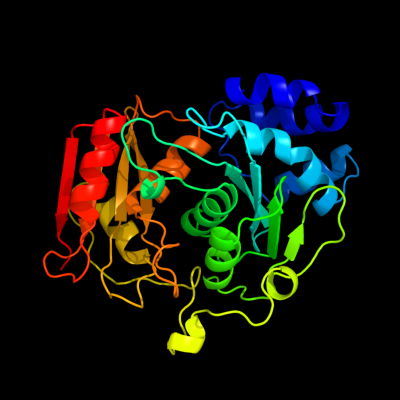
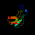
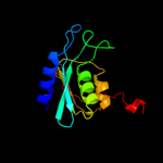
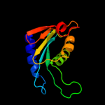
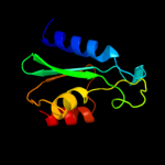
| 1 | c1alnA_
|
|
 |
100.0 |
100 |
PDB header:hydrolase
Chain: A: PDB Molecule:cytidine deaminase;
PDBTitle: crystal structure of cytidine deaminase complexed with 3-deazacytidine
|
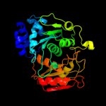
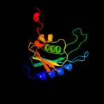
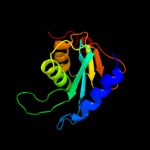
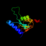
| 2 | d1alna1
|
|
 |
100.0 |
100 |
Fold:Cytidine deaminase-like
Superfamily:Cytidine deaminase-like
Family:Cytidine deaminase |
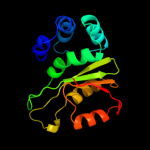
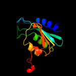
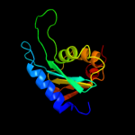
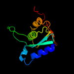
| 3 | d1alna2
|
|
 |
100.0 |
100 |
Fold:Cytidine deaminase-like
Superfamily:Cytidine deaminase-like
Family:Cytidine deaminase |
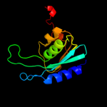
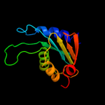
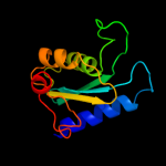
| 4 | d1r5ta_
|
|
 |
100.0 |
29 |
Fold:Cytidine deaminase-like
Superfamily:Cytidine deaminase-like
Family:Cytidine deaminase |
| 5 | d1uwza_
|
|
 |
100.0 |
28 |
Fold:Cytidine deaminase-like
Superfamily:Cytidine deaminase-like
Family:Cytidine deaminase |
| 6 | d1mq0a_
|
|
 |
100.0 |
31 |
Fold:Cytidine deaminase-like
Superfamily:Cytidine deaminase-like
Family:Cytidine deaminase |
| 7 | d2fr5a1
|
|
 |
100.0 |
31 |
Fold:Cytidine deaminase-like
Superfamily:Cytidine deaminase-like
Family:Cytidine deaminase |
| 8 | c3ijfX_
|
|
 |
100.0 |
34 |
PDB header:hydrolase
Chain: X: PDB Molecule:cytidine deaminase;
PDBTitle: crystal structure of cytidine deaminase from mycobacterium2 tuberculosis
|
| 9 | c3dmoD_
|
|
 |
100.0 |
32 |
PDB header:hydrolase
Chain: D: PDB Molecule:cytidine deaminase;
PDBTitle: 1.6 a crystal structure of cytidine deaminase from2 burkholderia pseudomallei
|
| 10 | d2d30a1
|
|
 |
100.0 |
29 |
Fold:Cytidine deaminase-like
Superfamily:Cytidine deaminase-like
Family:Cytidine deaminase |
| 11 | c3r2nC_
|
|
 |
100.0 |
29 |
PDB header:hydrolase
Chain: C: PDB Molecule:cytidine deaminase;
PDBTitle: crystal structure of cytidine deaminase from mycobacterium leprae
|
| 12 | c3b8fB_
|
|
 |
100.0 |
19 |
PDB header:hydrolase
Chain: B: PDB Molecule:putative blasticidin s deaminase;
PDBTitle: crystal structure of the cytidine deaminase from bacillus anthracis
|
| 13 | c3oj6C_
|
|
 |
100.0 |
22 |
PDB header:hydrolase
Chain: C: PDB Molecule:blasticidin-s deaminase;
PDBTitle: crystal structure of blasticidin s deaminase from coccidioides immitis
|
| 14 | d2z3ga1
|
|
 |
100.0 |
21 |
Fold:Cytidine deaminase-like
Superfamily:Cytidine deaminase-like
Family:Cytidine deaminase |
| 15 | c2hvwC_
|
|
 |
98.4 |
22 |
PDB header:hydrolase
Chain: C: PDB Molecule:deoxycytidylate deaminase;
PDBTitle: crystal structure of dcmp deaminase from streptococcus2 mutans
|
| 16 | c2w4lC_
|
|
 |
98.1 |
27 |
PDB header:hydrolase
Chain: C: PDB Molecule:deoxycytidylate deaminase;
PDBTitle: human dcmp deaminase
|
| 17 | d1z3aa1
|
|
 |
97.8 |
19 |
Fold:Cytidine deaminase-like
Superfamily:Cytidine deaminase-like
Family:Deoxycytidylate deaminase-like |
| 18 | d1p6oa_
|
|
 |
97.7 |
16 |
Fold:Cytidine deaminase-like
Superfamily:Cytidine deaminase-like
Family:Deoxycytidylate deaminase-like |
| 19 | c3ocqA_
|
|
 |
97.6 |
21 |
PDB header:hydrolase
Chain: A: PDB Molecule:putative cytosine/adenosine deaminase;
PDBTitle: crystal structure of trna-specific adenosine deaminase from salmonella2 enterica
|
| 20 | d1wkqa_
|
|
 |
97.6 |
11 |
Fold:Cytidine deaminase-like
Superfamily:Cytidine deaminase-like
Family:Deoxycytidylate deaminase-like |
| 21 | d1wwra1 |
|
not modelled |
97.5 |
20 |
Fold:Cytidine deaminase-like
Superfamily:Cytidine deaminase-like
Family:Deoxycytidylate deaminase-like |
| 22 | d2g84a1 |
|
not modelled |
97.5 |
20 |
Fold:Cytidine deaminase-like
Superfamily:Cytidine deaminase-like
Family:Deoxycytidylate deaminase-like |
| 23 | d2b3ja1 |
|
not modelled |
97.5 |
19 |
Fold:Cytidine deaminase-like
Superfamily:Cytidine deaminase-like
Family:Deoxycytidylate deaminase-like |
| 24 | d1vq2a_ |
|
not modelled |
97.4 |
21 |
Fold:Cytidine deaminase-like
Superfamily:Cytidine deaminase-like
Family:Deoxycytidylate deaminase-like |
| 25 | d2a8na1 |
|
not modelled |
97.2 |
19 |
Fold:Cytidine deaminase-like
Superfamily:Cytidine deaminase-like
Family:Deoxycytidylate deaminase-like |
| 26 | c2hxvA_ |
|
not modelled |
97.1 |
28 |
PDB header:biosynthetic protein
Chain: A: PDB Molecule:diaminohydroxyphosphoribosylaminopyrimidine deaminase/ 5-
PDBTitle: crystal structure of a diaminohydroxyphosphoribosylaminopyrimidine2 deaminase/ 5-amino-6-(5-phosphoribosylamino)uracil reductase (tm1828)3 from thermotoga maritima at 1.80 a resolution
|
| 27 | c2nx8A_ |
|
not modelled |
96.9 |
14 |
PDB header:hydrolase
Chain: A: PDB Molecule:trna-specific adenosine deaminase;
PDBTitle: the crystal structure of the trna-specific adenosine deaminase from2 streptococcus pyogenes
|
| 28 | c2o7pA_ |
|
not modelled |
96.5 |
23 |
PDB header:hydrolase, oxidoreductase
Chain: A: PDB Molecule:riboflavin biosynthesis protein ribd;
PDBTitle: the crystal structure of ribd from escherichia coli in complex with2 the oxidised nadp+ cofactor in the active site of the reductase3 domain
|
| 29 | c2d5nB_ |
|
not modelled |
95.8 |
30 |
PDB header:hydrolase, oxidoreductase
Chain: B: PDB Molecule:riboflavin biosynthesis protein ribd;
PDBTitle: crystal structure of a bifunctional deaminase and reductase2 involved in riboflavin biosynthesis
|
| 30 | d2hxva2 |
|
not modelled |
95.7 |
15 |
Fold:Cytidine deaminase-like
Superfamily:Cytidine deaminase-like
Family:Deoxycytidylate deaminase-like |
| 31 | c3dh1D_ |
|
not modelled |
95.5 |
13 |
PDB header:hydrolase
Chain: D: PDB Molecule:trna-specific adenosine deaminase 2;
PDBTitle: crystal structure of human trna-specific adenosine-34 deaminase2 subunit adat2
|
| 32 | d2b3za2 |
|
not modelled |
95.1 |
29 |
Fold:Cytidine deaminase-like
Superfamily:Cytidine deaminase-like
Family:Deoxycytidylate deaminase-like |
| 33 | d3d37a2 |
|
not modelled |
57.7 |
16 |
Fold:Phage tail proteins
Superfamily:Phage tail proteins
Family:Baseplate protein-like |
| 34 | c2d9sA_ |
|
not modelled |
45.2 |
26 |
PDB header:ligase
Chain: A: PDB Molecule:cbl e3 ubiquitin protein ligase;
PDBTitle: solution structure of rsgi ruh-049, a uba domain from mouse2 cdna
|
| 35 | c3gv1A_ |
|
not modelled |
44.3 |
17 |
PDB header:structural genomics, unknown function
Chain: A: PDB Molecule:disulfide interchange protein;
PDBTitle: crystal structure of disulfide interchange protein from neisseria2 gonorrhoeae
|
| 36 | c2do6A_ |
|
not modelled |
43.1 |
18 |
PDB header:ligase
Chain: A: PDB Molecule:e3 ubiquitin-protein ligase cbl-b;
PDBTitle: solution structure of rsgi ruh-065, a uba domain from human2 cdna
|
| 37 | c2eqjA_ |
|
not modelled |
40.7 |
29 |
PDB header:transcription
Chain: A: PDB Molecule:metal-response element-binding transcription
PDBTitle: solution structure of the tudor domain of metal-response2 element-binding transcription factor 2
|
| 38 | c2jnhA_ |
|
not modelled |
37.8 |
17 |
PDB header:ligase
Chain: A: PDB Molecule:e3 ubiquitin-protein ligase cbl-b;
PDBTitle: solution structure of the uba domain from cbl-b
|
| 39 | d2fug21 |
|
not modelled |
28.8 |
13 |
Fold:Thioredoxin fold
Superfamily:Thioredoxin-like
Family:NQO2-like |
| 40 | c3dmlA_ |
|
not modelled |
28.8 |
17 |
PDB header:oxidoreductase
Chain: A: PDB Molecule:putative uncharacterized protein;
PDBTitle: crystal structure of the periplasmic thioredoxin soxs from2 paracoccus pantotrophus (reduced form)
|
| 41 | c3g9bA_ |
|
not modelled |
27.4 |
16 |
PDB header:transferase
Chain: A: PDB Molecule:dolichyl-diphosphooligosaccharide-protein
PDBTitle: crystal structure of reduced ost6l
|
| 42 | c2hlsB_ |
|
not modelled |
26.4 |
14 |
PDB header:oxidoreductase
Chain: B: PDB Molecule:protein disulfide oxidoreductase;
PDBTitle: the crystal structure of a protein disulfide oxidoreductase from2 aeropyrum pernix k1
|
| 43 | c2r2jA_ |
|
not modelled |
26.3 |
9 |
PDB header:chaperone
Chain: A: PDB Molecule:thioredoxin domain-containing protein 4;
PDBTitle: crystal structure of human erp44
|
| 44 | c1jn9B_ |
|
not modelled |
25.9 |
19 |
PDB header:hydrolase
Chain: B: PDB Molecule:putative l-asparaginase;
PDBTitle: structure of putative asparaginase encoded by escherichia coli ybik2 gene
|
| 45 | c1t3mD_ |
|
not modelled |
25.9 |
19 |
PDB header:hydrolase
Chain: D: PDB Molecule:putative l-asparaginase;
PDBTitle: structure of the isoaspartyl peptidase with l-asparaginase2 activity from e. coli
|
| 46 | c1t3mB_ |
|
not modelled |
25.9 |
19 |
PDB header:hydrolase
Chain: B: PDB Molecule:putative l-asparaginase;
PDBTitle: structure of the isoaspartyl peptidase with l-asparaginase2 activity from e. coli
|
| 47 | c3p8dB_ |
|
not modelled |
25.4 |
16 |
PDB header:protein binding
Chain: B: PDB Molecule:medulloblastoma antigen mu-mb-50.72;
PDBTitle: crystal structure of the second tudor domain of human phf20 (homodimer2 form)
|
| 48 | d1fo5a_ |
|
not modelled |
24.5 |
13 |
Fold:Thioredoxin fold
Superfamily:Thioredoxin-like
Family:Thioltransferase |
| 49 | c2ju5A_ |
|
not modelled |
24.0 |
7 |
PDB header:oxidoreductase
Chain: A: PDB Molecule:thioredoxin disulfide isomerase;
PDBTitle: dsbh oxidoreductase
|
| 50 | c2zalB_ |
|
not modelled |
23.5 |
19 |
PDB header:hydrolase
Chain: B: PDB Molecule:l-asparaginase;
PDBTitle: crystal structure of e. coli isoaspartyl aminopeptidase/l-asparaginase2 in complex with l-aspartate
|
| 51 | c1a8yA_ |
|
not modelled |
22.2 |
11 |
PDB header:calcium-binding protein
Chain: A: PDB Molecule:calsequestrin;
PDBTitle: crystal structure of calsequestrin from rabbit skeletal muscle2 sarcoplasmic reticulum at 2.4 a resolution
|
| 52 | d1z67a1 |
|
not modelled |
21.9 |
28 |
Fold:YidB-like
Superfamily:YidB-like
Family:YidB-like |
| 53 | c3kp8A_ |
|
not modelled |
21.6 |
27 |
PDB header:oxidoreductase
Chain: A: PDB Molecule:vkorc1/thioredoxin domain protein;
PDBTitle: the thioredoxin-like domain of a vkor homolog from2 synechococcus sp.
|
| 54 | d1zmaa1 |
|
not modelled |
21.0 |
29 |
Fold:Thioredoxin fold
Superfamily:Thioredoxin-like
Family:Thioltransferase |
| 55 | c1x5eA_ |
|
not modelled |
20.7 |
22 |
PDB header:electron transport
Chain: A: PDB Molecule:thioredoxin domain containing protein 1;
PDBTitle: the solution structure of the thioredoxin-like domain of2 human thioredoxin-related transmembrane protein
|
| 56 | c3gwlB_ |
|
not modelled |
20.3 |
25 |
PDB header:oxidoreductase
Chain: B: PDB Molecule:fad-linked sulfhydryl oxidase;
PDBTitle: crystal structure of asfv pb119l, a viral sulfhydryl oxidase
|
| 57 | c3dxbE_ |
|
not modelled |
19.7 |
17 |
PDB header:splicing, transcription
Chain: E: PDB Molecule:thioredoxin n-terminally fused to puf60(uhm);
PDBTitle: structure of the uhm domain of puf60 fused to thioredoxin
|
| 58 | c2e5qA_ |
|
not modelled |
19.5 |
26 |
PDB header:transcription
Chain: A: PDB Molecule:phd finger protein 19;
PDBTitle: solution structure of the tudor domain of phd finger2 protein 19, isoform b [homo sapiens]
|
| 59 | d1jr8a_ |
|
not modelled |
19.3 |
42 |
Fold:Four-helical up-and-down bundle
Superfamily:FAD-dependent thiol oxidase
Family:FAD-dependent thiol oxidase |
| 60 | c2zalD_ |
|
not modelled |
19.2 |
19 |
PDB header:hydrolase
Chain: D: PDB Molecule:l-asparaginase;
PDBTitle: crystal structure of e. coli isoaspartyl aminopeptidase/l-asparaginase2 in complex with l-aspartate
|
| 61 | c3f8uA_ |
|
not modelled |
18.1 |
23 |
PDB header:immune system/isomerase
Chain: A: PDB Molecule:protein disulfide-isomerase a3erp57;
PDBTitle: tapasin/erp57 heterodimer
|
| 62 | c3gwnA_ |
|
not modelled |
18.0 |
33 |
PDB header:oxidoreductase
Chain: A: PDB Molecule:probable fad-linked sulfhydryl oxidase r596;
PDBTitle: crystal structure of the fad binding domain from mimivirus sulfhydryl2 oxidase r596
|
| 63 | d1sena_ |
|
not modelled |
17.7 |
18 |
Fold:Thioredoxin fold
Superfamily:Thioredoxin-like
Family:Thioltransferase |
| 64 | c1senA_ |
|
not modelled |
17.7 |
18 |
PDB header:structural genomics, unknown function
Chain: A: PDB Molecule:thioredoxin-like protein p19;
PDBTitle: endoplasmic reticulum protein rp19 o95881
|
| 65 | c2ppvA_ |
|
not modelled |
17.2 |
17 |
PDB header:transferase
Chain: A: PDB Molecule:uncharacterized protein;
PDBTitle: crystal structure of a protein belonging to the upf0052 (se_0549) from2 staphylococcus epidermidis atcc 12228 at 2.00 a resolution
|
| 66 | c1t3bA_ |
|
not modelled |
17.1 |
12 |
PDB header:isomerase
Chain: A: PDB Molecule:thiol:disulfide interchange protein dsbc;
PDBTitle: x-ray structure of dsbc from haemophilus influenzae
|
| 67 | c2nytB_ |
|
not modelled |
17.0 |
23 |
PDB header:hydrolase
Chain: B: PDB Molecule:probable c->u-editing enzyme apobec-2;
PDBTitle: the apobec2 crystal structure and functional implications2 for aid
|
| 68 | d1y0ua_ |
|
not modelled |
16.8 |
19 |
Fold:DNA/RNA-binding 3-helical bundle
Superfamily:"Winged helix" DNA-binding domain
Family:ArsR-like transcriptional regulators |
| 69 | d1hyua4 |
|
not modelled |
16.4 |
14 |
Fold:Thioredoxin fold
Superfamily:Thioredoxin-like
Family:PDI-like |
| 70 | c3l9vE_ |
|
not modelled |
15.5 |
43 |
PDB header:oxidoreductase
Chain: E: PDB Molecule:putative thiol-disulfide isomerase or thioredoxin;
PDBTitle: crystal structure of salmonella enterica serovar typhimurium srga
|
| 71 | c2zakB_ |
|
not modelled |
15.5 |
19 |
PDB header:hydrolase
Chain: B: PDB Molecule:l-asparaginase precursor;
PDBTitle: orthorhombic crystal structure of precursor e. coli isoaspartyl2 peptidase/l-asparaginase (ecaiii) with active-site t179a mutation
|
| 72 | d1v58a1 |
|
not modelled |
15.2 |
15 |
Fold:Thioredoxin fold
Superfamily:Thioredoxin-like
Family:DsbC/DsbG C-terminal domain-like |
| 73 | d1j9ba_ |
|
not modelled |
14.8 |
31 |
Fold:Thioredoxin fold
Superfamily:Thioredoxin-like
Family:ArsC-like |
| 74 | c2r41A_ |
|
not modelled |
14.4 |
17 |
PDB header:structural genomics, unknown function
Chain: A: PDB Molecule:uncharacterized protein;
PDBTitle: crystal structure of the protein of unknown function from enterococcus2 faecalis
|
| 75 | c1zy7A_ |
|
not modelled |
14.2 |
31 |
PDB header:hydrolase
Chain: A: PDB Molecule:rna-specific adenosine deaminase b1, isoform
PDBTitle: crystal structure of the catalytic domain of an adenosine2 deaminase that acts on rna (hadar2) bound to inositol3 hexakisphosphate (ihp)
|
| 76 | c1jzdA_ |
|
not modelled |
13.9 |
8 |
PDB header:oxidoreductase
Chain: A: PDB Molecule:thiol:disulfide interchange protein dsbc;
PDBTitle: dsbc-dsbdalpha complex
|
| 77 | c3f46A_ |
|
not modelled |
13.7 |
17 |
PDB header:oxidoreductase
Chain: A: PDB Molecule:5,10-methenyltetrahydromethanopterin hydrogenase;
PDBTitle: the crystal structure of c176a mutated [fe]-hydrogenase (hmd)2 holoenzyme from methanocaldococcus jannaschii
|
| 78 | c1k2xB_ |
|
not modelled |
13.7 |
17 |
PDB header:hydrolase
Chain: B: PDB Molecule:putative l-asparaginase;
PDBTitle: crystal structure of putative asparaginase encoded by escherichia coli2 ybik gene
|
| 79 | c1jn9D_ |
|
not modelled |
13.7 |
17 |
PDB header:hydrolase
Chain: D: PDB Molecule:putative l-asparaginase;
PDBTitle: structure of putative asparaginase encoded by escherichia coli ybik2 gene
|
| 80 | c1k2xD_ |
|
not modelled |
13.7 |
17 |
PDB header:hydrolase
Chain: D: PDB Molecule:putative l-asparaginase;
PDBTitle: crystal structure of putative asparaginase encoded by escherichia coli2 ybik gene
|
| 81 | c3feuA_ |
|
not modelled |
13.6 |
71 |
PDB header:oxidoreductase
Chain: A: PDB Molecule:putative lipoprotein;
PDBTitle: crystal structure of dsba-like thioredoxin domain vf_a0457 from vibrio2 fischeri
|
| 82 | c2albA_ |
|
not modelled |
13.2 |
27 |
PDB header:isomerase
Chain: A: PDB Molecule:protein disulfide-isomerase a3;
PDBTitle: nmr structure of the n-terminal domain a of the2 glycoprotein chaperone erp57
|
| 83 | c2hj3A_ |
|
not modelled |
13.1 |
33 |
PDB header:oxidoreductase
Chain: A: PDB Molecule:sulfhydryl oxidase erv1p;
PDBTitle: structure of the arabidopsis thaliana erv1 thiol oxidase
|
| 84 | d1qjta_ |
|
not modelled |
12.8 |
11 |
Fold:EF Hand-like
Superfamily:EF-hand
Family:Eps15 homology domain (EH domain) |
| 85 | c3idvA_ |
|
not modelled |
12.7 |
32 |
PDB header:isomerase
Chain: A: PDB Molecule:protein disulfide-isomerase a4;
PDBTitle: crystal structure of the a0a fragment of erp72
|
| 86 | d1rioa_ |
|
not modelled |
12.6 |
8 |
Fold:lambda repressor-like DNA-binding domains
Superfamily:lambda repressor-like DNA-binding domains
Family:Phage repressors |
| 87 | c1sjiA_ |
|
not modelled |
12.5 |
0 |
PDB header:metal binding protein
Chain: A: PDB Molecule:calsequestrin, cardiac muscle isoform;
PDBTitle: comparing skeletal and cardiac calsequestrin structures and2 their calcium binding: a proposed mechanism for coupled3 calcium binding and protein polymerization
|
| 88 | c1p4vA_ |
|
not modelled |
11.8 |
20 |
PDB header:hydrolase
Chain: A: PDB Molecule:n(4)-(beta-n-acetylglucosaminyl)-l-asparaginase
PDBTitle: crystal structure of the glycosylasparaginase precursor2 d151n mutant with glycine
|
| 89 | c3cawB_ |
|
not modelled |
11.7 |
32 |
PDB header:oxidoreductase
Chain: B: PDB Molecule:o-succinylbenzoate synthase;
PDBTitle: crystal structure of o-succinylbenzoate synthase from2 bdellovibrio bacteriovorus liganded with mg
|
| 90 | c2gezF_ |
|
not modelled |
11.6 |
24 |
PDB header:hydrolase
Chain: F: PDB Molecule:l-asparaginase beta subunit;
PDBTitle: crystal structure of potassium-independent plant asparaginase
|
| 91 | c2b5eA_ |
|
not modelled |
11.4 |
25 |
PDB header:isomerase
Chain: A: PDB Molecule:protein disulfide-isomerase;
PDBTitle: crystal structure of yeast protein disulfide isomerase
|
| 92 | c2kboA_ |
|
not modelled |
11.3 |
23 |
PDB header:hydrolase
Chain: A: PDB Molecule:dna dc->du-editing enzyme apobec-3g;
PDBTitle: structure, interaction, and real-time monitoring of the2 enzymatic reaction of wild type apobec3g
|
| 93 | c2wz9A_ |
|
not modelled |
11.2 |
13 |
PDB header:protein binding
Chain: A: PDB Molecule:glutaredoxin-3;
PDBTitle: crystal structure of the thioredoxin domain of human txnl2
|
| 94 | c1zypB_ |
|
not modelled |
11.0 |
13 |
PDB header:oxidoreductase
Chain: B: PDB Molecule:alkyl hydroperoxide reductase subunit f;
PDBTitle: synchrotron reduced form of the n-terminal domain of2 salmonella typhimurium ahpf
|
| 95 | d2hzba1 |
|
not modelled |
10.9 |
20 |
Fold:CofD-like
Superfamily:CofD-like
Family:CofD-like |
| 96 | d1f9ma_ |
|
not modelled |
10.7 |
9 |
Fold:Thioredoxin fold
Superfamily:Thioredoxin-like
Family:Thioltransferase |
| 97 | d1oqca_ |
|
not modelled |
10.5 |
25 |
Fold:Four-helical up-and-down bundle
Superfamily:FAD-dependent thiol oxidase
Family:FAD-dependent thiol oxidase |
| 98 | c1j08A_ |
|
not modelled |
10.4 |
17 |
PDB header:isomerase
Chain: A: PDB Molecule:glutaredoxin-like protein;
PDBTitle: crystal structure of glutaredoxin-like protein from2 pyrococcus horikoshii
|
| 99 | d1j08a1 |
|
not modelled |
10.2 |
17 |
Fold:Thioredoxin fold
Superfamily:Thioredoxin-like
Family:PDI-like |