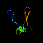
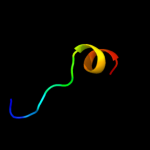
1 c1n0fF_
100.0
25
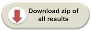
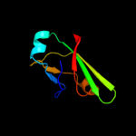
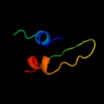
PDB header: biosynthetic proteinChain: F: PDB Molecule: protein mraz;PDBTitle: crystal structure of a cell division and cell wall2 biosynthesis protein upf0040 from mycoplasma pneumoniae:3 indication of a novel fold with a possible new conserved4 sequence motif
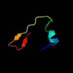
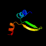
2 d1n0ea_
100.0
25
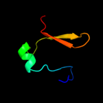
Fold: Double-split beta-barrelSuperfamily: AbrB/MazE/MraZ-likeFamily: Hypothetical protein MraZ3 c2glwA_
95.8
18
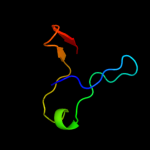
PDB header: transcriptionChain: A: PDB Molecule: 92aa long hypothetical protein;PDBTitle: the solution structure of phs018 from pyrococcus horikoshii
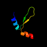
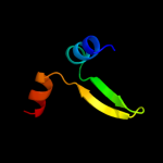
4 c2w1tB_
94.4
21
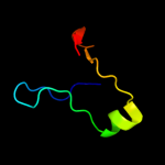
PDB header: transcriptionChain: B: PDB Molecule: stage v sporulation protein t;PDBTitle: crystal structure of b. subtilis spovt
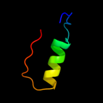
5 d1yfba1
88.9
21
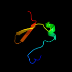
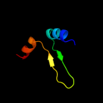
Fold: Double-split beta-barrelSuperfamily: AbrB/MazE/MraZ-likeFamily: AbrB N-terminal domain-like6 c2ro5B_
88.0
21
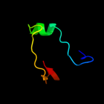
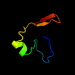
PDB header: transcriptionChain: B: PDB Molecule: stage v sporulation protein t;PDBTitle: rdc-refined solution structure of the n-terminal dna2 recognition domain of the bacillus subtilis transition-3 state regulator spovt
7 d2fy9a1
87.6
21
Fold: Double-split beta-barrelSuperfamily: AbrB/MazE/MraZ-likeFamily: AbrB N-terminal domain-like8 c2l66B_
50.8
12
PDB header: transcription regulatorChain: B: PDB Molecule: transcriptional regulator, abrb family;PDBTitle: the dna-recognition fold of sso7c4 suggests a new member of spovt-abrb2 superfamily from archaea.
9 c3hs2H_
41.5
15
PDB header: antitoxinChain: H: PDB Molecule: prevent host death protein;PDBTitle: crystal structure of phd truncated to residue 57 in an orthorhombic2 space group
10 c3hryA_
41.2
15
PDB header: antitoxinChain: A: PDB Molecule: prevent host death protein;PDBTitle: crystal structure of phd in a trigonal space group and partially2 disordered
11 c3nnqA_
33.9
21
PDB header: viral proteinChain: A: PDB Molecule: n-terminal domain of moloney murine leukemia virusPDBTitle: crystal structure of the n-terminal domain of moloney murine leukemia2 virus integrase, northeast structural genomics consortium target or3
12 d2odka1
32.2
18
Fold: YefM-likeSuperfamily: YefM-likeFamily: YefM-like13 c2odkD_
30.3
18
PDB header: structural genomics, unknown functionChain: D: PDB Molecule: hypothetical protein;PDBTitle: putative prevent-host-death protein from nitrosomonas europaea
14 c3qq5A_
27.5
21
PDB header: oxidoreductaseChain: A: PDB Molecule: small gtp-binding protein;PDBTitle: crystal structure of the [fefe]-hydrogenase maturation protein hydf
15 c2k2eA_
22.9
32
PDB header: structural genomics, unknown functionChain: A: PDB Molecule: uncharacterized protein bp2786;PDBTitle: solution nmr structure of bordetella pertussis protein2 bp2786, a mth938-like domain. northeast structural3 genomics consortium target ber31
16 c2c45F_
21.8
13
PDB header: lyaseChain: F: PDB Molecule: aspartate 1-decarboxylase precursor;PDBTitle: native precursor of pyruvoyl dependent aspartate2 decarboxylase
17 c1pt1B_
21.1
15
PDB header: lyaseChain: B: PDB Molecule: aspartate 1-decarboxylase;PDBTitle: unprocessed pyruvoyl dependent aspartate decarboxylase with histidine2 11 mutated to alanine
18 d1ppya_
19.6
15
Fold: Double psi beta-barrelSuperfamily: ADC-likeFamily: Pyruvoyl dependent aspartate decarboxylase, ADC19 d1wida_
17.8
23
Fold: DNA-binding pseudobarrel domainSuperfamily: DNA-binding pseudobarrel domainFamily: B3 DNA binding domain20 d2zgwa1
16.5
44
Fold: SH3-like barrelSuperfamily: C-terminal domain of transcriptional repressorsFamily: Biotin repressor (BirA)21 d2a6qb1
not modelled
15.2
10
Fold: YefM-likeSuperfamily: YefM-likeFamily: YefM-like22 d1s6la1
not modelled
15.1
21
Fold: DNA/RNA-binding 3-helical bundleSuperfamily: "Winged helix" DNA-binding domainFamily: MerB N-terminal domain-like23 c3if4C_
not modelled
15.1
33
PDB header: structural genomics, unknown functionChain: C: PDB Molecule: integron cassette protein hfx_cass5;PDBTitle: structure from the mobile metagenome of north west arm2 sewage outfall: integron cassette protein hfx_cass5
24 c2zcpA_
not modelled
15.1
5
PDB header: transferaseChain: A: PDB Molecule: dehydrosqualene synthase;PDBTitle: crystal structure of the c(30) carotenoid dehydrosqualene2 synthase from staphylococcus aureus complexed with3 farnesyl thiopyrophosphate
25 d2q4qa1
not modelled
14.9
16
Fold: MTH938-likeSuperfamily: MTH938-likeFamily: MTH938-like26 c2gm2A_
not modelled
14.0
11
PDB header: structural genomics, unknown functionChain: A: PDB Molecule: conserved hypothetical protein;PDBTitle: nmr structure of xanthomonas campestris xcc1710: northeast2 structural genomics consortium target xcr35
27 d1d7qa_
not modelled
13.7
14
Fold: OB-foldSuperfamily: Nucleic acid-binding proteinsFamily: Cold shock DNA-binding domain-like28 d2a6qa1
not modelled
13.5
10
Fold: YefM-likeSuperfamily: YefM-likeFamily: YefM-like29 c2kdnA_
not modelled
13.3
13
PDB header: unknown functionChain: A: PDB Molecule: putative uncharacterized protein pfe0790c;PDBTitle: solution structure of pfe0790c, a putative bola-like2 protein from the protozoan parasite plasmodium falciparum.
30 c3dwmA_
not modelled
13.0
35
PDB header: transferaseChain: A: PDB Molecule: 9.5 kda culture filtrate antigen cfp10a;PDBTitle: crystal structure of mycobacterium tuberculosis cyso, an antigen
31 c2oqkA_
not modelled
12.2
14
PDB header: translationChain: A: PDB Molecule: putative translation initiation factor eif-1a;PDBTitle: crystal structure of putative cryptosporidium parvum translation2 initiation factor eif-1a
32 d1lpfa2
not modelled
12.0
6
Fold: FAD/NAD(P)-binding domainSuperfamily: FAD/NAD(P)-binding domainFamily: FAD/NAD-linked reductases, N-terminal and central domains33 c2dgyA_
not modelled
11.9
14
PDB header: translationChain: A: PDB Molecule: mgc11102 protein;PDBTitle: solution structure of the eukaryotic initiation factor 1a2 in mgc11102 protein
34 d2fvta1
not modelled
11.2
8
Fold: MTH938-likeSuperfamily: MTH938-likeFamily: MTH938-like35 d1ef4a_
not modelled
11.0
20
Fold: DNA/RNA-binding 3-helical bundleSuperfamily: RNA polymerase subunit RPB10Family: RNA polymerase subunit RPB1036 d2fi9a1
not modelled
10.3
13
Fold: MTH938-likeSuperfamily: MTH938-likeFamily: MTH938-like37 c3errB_
not modelled
10.1
24
PDB header: ligaseChain: B: PDB Molecule: fusion protein of microtubule binding domain fromPDBTitle: microtubule binding domain from mouse cytoplasmic dynein as2 a fusion with seryl-trna synthetase
38 d1m7ja1
not modelled
10.0
23
Fold: Composite domain of metallo-dependent hydrolasesSuperfamily: Composite domain of metallo-dependent hydrolasesFamily: D-aminoacylase39 d2b7oa1
not modelled
9.7
21
Fold: TIM beta/alpha-barrelSuperfamily: AldolaseFamily: Class-II DAHP synthetase40 c3zvkG_
not modelled
9.5
20
PDB header: antitoxin/toxin/dnaChain: G: PDB Molecule: antitoxin of toxin-antitoxin system vapb;PDBTitle: crystal structure of vapbc2 from rickettsia felis bound to2 a dna fragment from their promoter
41 c2dq3A_
not modelled
9.1
25
PDB header: ligaseChain: A: PDB Molecule: seryl-trna synthetase;PDBTitle: crystal structure of aq_298
42 d2j7ja2
not modelled
9.1
40
Fold: beta-beta-alpha zinc fingersSuperfamily: beta-beta-alpha zinc fingersFamily: Classic zinc finger, C2H243 c2ubpC_
not modelled
8.6
16
PDB header: hydrolaseChain: C: PDB Molecule: protein (urease alpha subunit);PDBTitle: structure of native urease from bacillus pasteurii
44 d1jt8a_
not modelled
8.1
14
Fold: OB-foldSuperfamily: Nucleic acid-binding proteinsFamily: Cold shock DNA-binding domain-like45 c2dq0A_
not modelled
7.9
29
PDB header: ligaseChain: A: PDB Molecule: seryl-trna synthetase;PDBTitle: crystal structure of seryl-trna synthetase from pyrococcus2 horikoshii complexed with a seryl-adenylate analog
46 d1xova1
not modelled
7.9
63
Fold: SH3-like barrelSuperfamily: Prokaryotic SH3-related domainFamily: Ply C-terminal domain-like47 d1seta2
not modelled
7.9
24
Fold: Class II aaRS and biotin synthetasesSuperfamily: Class II aaRS and biotin synthetasesFamily: Class II aminoacyl-tRNA synthetase (aaRS)-like, catalytic domain48 d1v59a2
not modelled
7.6
10
Fold: FAD/NAD(P)-binding domainSuperfamily: FAD/NAD(P)-binding domainFamily: FAD/NAD-linked reductases, N-terminal and central domains49 d1s32d_
not modelled
7.5
50
Fold: Histone-foldSuperfamily: Histone-foldFamily: Nucleosome core histones50 c3df0C_
not modelled
7.5
23
PDB header: hydrolaseChain: C: PDB Molecule: calpastatin;PDBTitle: calcium-dependent complex between m-calpain and calpastatin
51 c1hzeB_
not modelled
7.3
11
PDB header: transferaseChain: B: PDB Molecule: riboflavin synthase alpha chain;PDBTitle: solution structure of the n-terminal domain of riboflavin synthase2 from e. coli
52 c1i18B_
not modelled
7.3
11
PDB header: transferaseChain: B: PDB Molecule: riboflavin synthase alpha chain;PDBTitle: solution structure of the n-terminal domain of riboflavin synthase2 from e. coli
53 c3h5fB_
not modelled
7.3
39
PDB header: de novo proteinChain: B: PDB Molecule: coil ser l16l-pen;PDBTitle: switching the chirality of the metal environment alters the2 coordination mode in designed peptides.
54 c3h5gA_
not modelled
7.3
39
PDB header: de novo proteinChain: A: PDB Molecule: coil ser l16d-pen;PDBTitle: switching the chirality of the metal environment alters the2 coordination mode in designed peptides.
55 c3h5gC_
not modelled
7.3
39
PDB header: de novo proteinChain: C: PDB Molecule: coil ser l16d-pen;PDBTitle: switching the chirality of the metal environment alters the2 coordination mode in designed peptides.
56 c3h5fA_
not modelled
7.3
39
PDB header: de novo proteinChain: A: PDB Molecule: coil ser l16l-pen;PDBTitle: switching the chirality of the metal environment alters the2 coordination mode in designed peptides.
57 c3h5fC_
not modelled
7.3
39
PDB header: de novo proteinChain: C: PDB Molecule: coil ser l16l-pen;PDBTitle: switching the chirality of the metal environment alters the2 coordination mode in designed peptides.
58 c3h5gB_
not modelled
7.3
39
PDB header: de novo proteinChain: B: PDB Molecule: coil ser l16d-pen;PDBTitle: switching the chirality of the metal environment alters the2 coordination mode in designed peptides.
59 d1eqzb_
not modelled
7.2
50
Fold: Histone-foldSuperfamily: Histone-foldFamily: Nucleosome core histones60 d1k1da1
not modelled
7.2
25
Fold: Composite domain of metallo-dependent hydrolasesSuperfamily: Composite domain of metallo-dependent hydrolasesFamily: Hydantoinase (dihydropyrimidinase)61 c1sryB_
not modelled
7.1
21
PDB header: ligase(synthetase)Chain: B: PDB Molecule: seryl-trna synthetase;PDBTitle: refined crystal structure of the seryl-trna synthetase from2 thermus thermophilus at 2.5 angstroms resolution
62 d1tzyb_
not modelled
6.9
50
Fold: Histone-foldSuperfamily: Histone-foldFamily: Nucleosome core histones63 d2p9ba1
not modelled
6.8
30
Fold: Composite domain of metallo-dependent hydrolasesSuperfamily: Composite domain of metallo-dependent hydrolasesFamily: Imidazolonepropionase-like64 c1e9yB_
not modelled
6.5
16
PDB header: hydrolaseChain: B: PDB Molecule: urease subunit beta;PDBTitle: crystal structure of helicobacter pylori urease in complex with2 acetohydroxamic acid
65 c3la4A_
not modelled
6.5
21
PDB header: hydrolaseChain: A: PDB Molecule: urease;PDBTitle: crystal structure of the first plant urease from jack bean (canavalia2 ensiformis)
66 d1kx5d_
not modelled
6.5
50
Fold: Histone-foldSuperfamily: Histone-foldFamily: Nucleosome core histones67 d1lvla2
not modelled
6.4
16
Fold: FAD/NAD(P)-binding domainSuperfamily: FAD/NAD(P)-binding domainFamily: FAD/NAD-linked reductases, N-terminal and central domains68 d2gv8a2
not modelled
6.4
22
Fold: FAD/NAD(P)-binding domainSuperfamily: FAD/NAD(P)-binding domainFamily: FAD/NAD-linked reductases, N-terminal and central domains69 d2gf6a1
not modelled
6.3
4
Fold: Thioesterase/thiol ester dehydrase-isomeraseSuperfamily: Thioesterase/thiol ester dehydrase-isomeraseFamily: 4HBT-like70 d2ftwa1
not modelled
6.3
14
Fold: Composite domain of metallo-dependent hydrolasesSuperfamily: Composite domain of metallo-dependent hydrolasesFamily: Hydantoinase (dihydropyrimidinase)71 c3jywM_
not modelled
6.2
19
PDB header: ribosomeChain: M: PDB Molecule: 60s ribosomal protein l16(a);PDBTitle: structure of the 60s proteins for eukaryotic ribosome based on cryo-em2 map of thermomyces lanuginosus ribosome at 8.9a resolution
72 d1id3d_
not modelled
6.2
42
Fold: Histone-foldSuperfamily: Histone-foldFamily: Nucleosome core histones73 c2vlgD_
not modelled
6.2
21
PDB header: transferaseChain: D: PDB Molecule: sporulation kinase a;PDBTitle: kina pas-a domain, homodimer
74 d2nn6d2
not modelled
6.2
23
Fold: Ribonuclease PH domain 2-likeSuperfamily: Ribonuclease PH domain 2-likeFamily: Ribonuclease PH domain 2-like75 d1d6za4
not modelled
6.1
10
Fold: N domain of copper amine oxidase-likeSuperfamily: Copper amine oxidase, domain NFamily: Copper amine oxidase, domain N76 d1ejxc1
not modelled
5.9
16
Fold: Composite domain of metallo-dependent hydrolasesSuperfamily: Composite domain of metallo-dependent hydrolasesFamily: alpha-Subunit of urease77 d1vi7a2
not modelled
5.8
14
Fold: Ferredoxin-likeSuperfamily: EF-G C-terminal domain-likeFamily: YigZ C-terminal domain-like78 c2pmzN_
not modelled
5.5
19
PDB header: translation, transferaseChain: N: PDB Molecule: dna-directed rna polymerase subunit n;PDBTitle: archaeal rna polymerase from sulfolobus solfataricus
79 d2p5zx1
not modelled
5.5
23
Fold: OB-foldSuperfamily: gp5 N-terminal domain-likeFamily: gp4 N-terminal domain-like80 d1wjwa_
not modelled
5.5
9
Fold: TBP-likeSuperfamily: Phosphoglucomutase, C-terminal domainFamily: Phosphoglucomutase, C-terminal domain81 c4a1aI_
not modelled
5.4
16
PDB header: ribosomeChain: I: PDB Molecule: 60s ribosomal protein l13a;PDBTitle: t.thermophila 60s ribosomal subunit in complex with2 initiation factor 6. this file contains 5s rrna,3 5.8s rrna and proteins of molecule 3.
82 d1ojta2
not modelled
5.4
8
Fold: FAD/NAD(P)-binding domainSuperfamily: FAD/NAD(P)-binding domainFamily: FAD/NAD-linked reductases, N-terminal and central domains83 c3ei4D_
not modelled
5.4
5
PDB header: dna binding proteinChain: D: PDB Molecule: dna damage-binding protein 2;PDBTitle: structure of the hsddb1-hsddb2 complex