Phyre Home Retrieve Phyre Job Id
Emaill.a.kelley@imperial.ac.uk
DescriptionP22707
DateThu Jan 5 11:39:03 GMT 2012
Unique Job IDb53367407d91fd80

Summary 

Top model
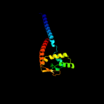
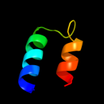
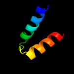
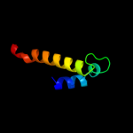
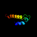
Image coloured by rainbow N → C terminus
Model (left) based on template d1dvoa_
Top template information
Fold:FinO-like
Superfamily:FinO-like
Family:FinO-like
Confidence and coverage
Confidence:100.0% Coverage: 82%
152 residues ( 82% of your sequence) have been modelled with 100.0% confidence by the single highest scoring template.
3D viewing
Interactive 3D view in Jmol

Sequence analysis 

Secondary structure and disorder prediction 

   1........10.........20.........30.........40.........50.........60
Sequence  MTEQKRPVLTLKRKTEGETLVRSRKTIINVTTPPKWKVKKQKLAEKAAREAELAAKKAQA
Secondary structure 


SS confidence 


Disorder  ????????


?????????????????????????????????????????????????
Disorder confidence 


   .........70.........80.........90.........100.........110.........120
Sequence  RQALSIYLNLPTLDDAVNTLKPWWPGLFDGDTPRLLACGIRDVLLEDVAQRNIPLSHKKL
Secondary structure 


SS confidence 


Disorder  ??????????


??
?


Disorder confidence 


   .........130.........140.........150.........160.........170.........180
Sequence  RRALKAITRSESYLCAMKAGACRYDTEGYVTEHISQEEEAYAAERLDKIRRQNRIKAELQ
Secondary structure 


SS confidence 


Disorder 


?


????


Disorder confidence 


   ......
Sequence  AVLDEK
Secondary structure 

SS confidence 


Disorder 


???
Disorder confidence 


Confidence Key
High(9)                    Low (0)
?Disordered
Alpha helix
Beta strand

Domain analysis 

Hover over an aligned region to see model and summary info

Please note, only up to the top 20 hits are modelled to reduce computer load

RankAligned region

PDB 1dvo chain A

3D model

Region: 33 - 184
Aligned: 152
Modelled: 152
Confidence: 100.0%
Identity: 97%
Fold: FinO-like
Superfamily: FinO-like
Family: FinO-like

Phyre2

PDB 2hxj chain A domain 1

3D model

Region: 63 - 165
Aligned: 99
Modelled: 103
Confidence: 100.0%
Identity: 23%
Fold: FinO-like
Superfamily: FinO-like
Family: FinO-like

Phyre2

PDB 2hxj chain F

3D model

Region: 62 - 165
Aligned: 100
Modelled: 104
Confidence: 100.0%
Identity: 23%
PDB header:structural genomics, unknown function
Chain: F: PDB Molecule:hypothetical protein;
PDBTitle: crystal structure of a protein of unknown function nmb1681 from2 neisseria meningitidis mc58, possible nucleic acid binding protein

Phyre2

PDB 2k19 chain A

3D model

Region: 88 - 127
Aligned: 37
Modelled: 40
Confidence: 53.1%
Identity: 30%
PDB header:antimicrobial protein
Chain: A: PDB Molecule:putative piscicolin 126 immunity protein;
PDBTitle: nmr solution structure of pisi

Phyre2

PDB 2zrr chain A

3D model

Region: 97 - 127
Aligned: 28
Modelled: 31
Confidence: 37.4%
Identity: 36%
PDB header:antimicrobial protein
Chain: A: PDB Molecule:mundticin ks immunity protein;
PDBTitle: crystal structure of an immunity protein that contributes2 to the self-protection of bacteriocin-producing3 enterococcus mundtii 15-1a

Phyre2

PDB 2jdq chain D domain 1

3D model

Region: 4 - 22
Aligned: 19
Modelled: 19
Confidence: 26.1%
Identity: 37%
Fold: PB2 C-terminal domain-like
Superfamily: PB2 C-terminal domain-like
Family: PB2 C-terminal domain-like

Phyre2

PDB 2yic chain C

3D model

Region: 68 - 148
Aligned: 81
Modelled: 81
Confidence: 21.3%
Identity: 17%
PDB header:lyase
Chain: C: PDB Molecule:2-oxoglutarate decarboxylase;
PDBTitle: crystal structure of the suca domain of mycobacterium smegmatis2 alpha-ketoglutarate decarboxylase (triclinic form)

Phyre2

PDB 2i5u chain A domain 1

3D model

Region: 86 - 137
Aligned: 52
Modelled: 52
Confidence: 14.1%
Identity: 15%
Fold: DnaD domain-like
Superfamily: DnaD domain-like
Family: DnaD domain

Phyre2

PDB 2w50 chain B

3D model

Region: 96 - 130
Aligned: 35
Modelled: 35
Confidence: 13.4%
Identity: 9%
PDB header:hormone
Chain: B: PDB Molecule:armet-like protein 1;
PDBTitle: n-terminal domain of human conserved dopamine neurotrophic2 factor (cdnf)

Phyre2

PDB 2xt6 chain B

3D model

Region: 72 - 148
Aligned: 77
Modelled: 77
Confidence: 12.4%
Identity: 17%
PDB header:lyase
Chain: B: PDB Molecule:2-oxoglutarate decarboxylase;
PDBTitle: crystal structure of mycobacterium smegmatis alpha-ketoglutarate2 decarboxylase homodimer (orthorhombic form)

Phyre2

PDB 2w51 chain A

3D model

Region: 96 - 130
Aligned: 35
Modelled: 35
Confidence: 12.4%
Identity: 14%
PDB header:hormone
Chain: A: PDB Molecule:protein armet;
PDBTitle: human mesencephalic astrocyte-derived neurotrophic factor (2 manf)

Phyre2

PDB 3pp5 chain A

3D model

Region: 97 - 137
Aligned: 41
Modelled: 41
Confidence: 12.3%
Identity: 12%
PDB header:structural protein
Chain: A: PDB Molecule:brk1;
PDBTitle: high-resolution structure of the trimeric scar/wave complex precursor2 brk1

Phyre2

PDB 1b33 chain A

3D model

Region: 73 - 140
Aligned: 48
Modelled: 48
Confidence: 12.1%
Identity: 17%
Fold: Globin-like
Superfamily: Globin-like
Family: Phycocyanin-like phycobilisome proteins

Phyre2

PDB 3mix chain A

3D model

Region: 74 - 130
Aligned: 57
Modelled: 57
Confidence: 12.1%
Identity: 18%
PDB header:protein transport
Chain: A: PDB Molecule:flagellar biosynthesis protein flha;
PDBTitle: crystal structure of the cytosolic domain of b. subtilis flha

Phyre2

PDB 3a5i chain B

3D model

Region: 74 - 115
Aligned: 42
Modelled: 42
Confidence: 11.2%
Identity: 12%
PDB header:protein transport
Chain: B: PDB Molecule:flagellar biosynthesis protein flha;
PDBTitle: structure of the cytoplasmic domain of flha

Phyre2

PDB 2kq6 chain A

3D model

Region: 103 - 170
Aligned: 63
Modelled: 68
Confidence: 11.1%
Identity: 25%
PDB header:transport protein
Chain: A: PDB Molecule:polycystin-2;
PDBTitle: the structure of the ef-hand domain of polycystin-2 suggests a2 mechanism for ca2+-dependent regulation of polycystin-2 channel3 activity

Phyre2

PDB 1all chain A

3D model

Region: 73 - 140
Aligned: 48
Modelled: 48
Confidence: 11.0%
Identity: 17%
Fold: Globin-like
Superfamily: Globin-like
Family: Phycocyanin-like phycobilisome proteins

Phyre2

PDB 1us2 chain A

3D model

Region: 84 - 105
Aligned: 22
Modelled: 22
Confidence: 10.9%
Identity: 36%
PDB header:hydrolase
Chain: A: PDB Molecule:endo-beta-1,4-xylanase;
PDBTitle: xylanase10c (mutant e385a) from cellvibrio japonicus in2 complex with xylopentaose

Phyre2

PDB 1qnt chain A domain 2

3D model

Region: 72 - 84
Aligned: 13
Modelled: 13
Confidence: 10.7%
Identity: 15%
Fold: Ribonuclease H-like motif
Superfamily: Methylated DNA-protein cysteine methyltransferase domain
Family: Methylated DNA-protein cysteine methyltransferase domain

Phyre2

PDB 1v4v chain A

3D model

Region: 98 - 157
Aligned: 59
Modelled: 60
Confidence: 10.7%
Identity: 12%
Fold: UDP-Glycosyltransferase/glycogen phosphorylase
Superfamily: UDP-Glycosyltransferase/glycogen phosphorylase
Family: UDP-N-acetylglucosamine 2-epimerase

Phyre2
1

d1dvoa_
2

d2hxja1
3

c2hxjF_
4

c2k19A_
5

c2zrrA_
6

d2jdqd1
7

c2yicC_
8

d2i5ua1
9

c2w50B_
10

c2xt6B_
11

c2w51A_
12

c3pp5A_
13

d1b33a_
14

c3mixA_
15

c3a5iB_
16

c2kq6A_
17

d1alla_
18

c1us2A_
19

d1qnta2
20

d1v4va_
21


22


23


24


25


26


27


28


29


30


31


32


33


34


35


36


37


38


39


40


41


42


43


44


45


46


47


48


49


Detailed template information 

#
Template Alignment Coverage3D Model Confidence
% i.d. Template Information
1d1dvoa_


100.0 97 Fold:FinO-like
Superfamily:FinO-like
Family:FinO-like
2d2hxja1


100.0 23 Fold:FinO-like
Superfamily:FinO-like
Family:FinO-like
3c2hxjF_


100.0 23 PDB header:structural genomics, unknown function
Chain: F: PDB Molecule:hypothetical protein;
PDBTitle: crystal structure of a protein of unknown function nmb1681 from2 neisseria meningitidis mc58, possible nucleic acid binding protein
4c2k19A_


53.1 30 PDB header:antimicrobial protein
Chain: A: PDB Molecule:putative piscicolin 126 immunity protein;
PDBTitle: nmr solution structure of pisi
5c2zrrA_


37.4 36 PDB header:antimicrobial protein
Chain: A: PDB Molecule:mundticin ks immunity protein;
PDBTitle: crystal structure of an immunity protein that contributes2 to the self-protection of bacteriocin-producing3 enterococcus mundtii 15-1a
6d2jdqd1


26.1 37 Fold:PB2 C-terminal domain-like
Superfamily:PB2 C-terminal domain-like
Family:PB2 C-terminal domain-like
7c2yicC_


21.3 17 PDB header:lyase
Chain: C: PDB Molecule:2-oxoglutarate decarboxylase;
PDBTitle: crystal structure of the suca domain of mycobacterium smegmatis2 alpha-ketoglutarate decarboxylase (triclinic form)
8d2i5ua1


14.1 15 Fold:DnaD domain-like
Superfamily:DnaD domain-like
Family:DnaD domain
9c2w50B_


13.4 9 PDB header:hormone
Chain: B: PDB Molecule:armet-like protein 1;
PDBTitle: n-terminal domain of human conserved dopamine neurotrophic2 factor (cdnf)
10c2xt6B_


12.4 17 PDB header:lyase
Chain: B: PDB Molecule:2-oxoglutarate decarboxylase;
PDBTitle: crystal structure of mycobacterium smegmatis alpha-ketoglutarate2 decarboxylase homodimer (orthorhombic form)
11c2w51A_


12.4 14 PDB header:hormone
Chain: A: PDB Molecule:protein armet;
PDBTitle: human mesencephalic astrocyte-derived neurotrophic factor (2 manf)
12c3pp5A_


12.3 12 PDB header:structural protein
Chain: A: PDB Molecule:brk1;
PDBTitle: high-resolution structure of the trimeric scar/wave complex precursor2 brk1
13d1b33a_


12.1 17 Fold:Globin-like
Superfamily:Globin-like
Family:Phycocyanin-like phycobilisome proteins
14c3mixA_


12.1 18 PDB header:protein transport
Chain: A: PDB Molecule:flagellar biosynthesis protein flha;
PDBTitle: crystal structure of the cytosolic domain of b. subtilis flha
15c3a5iB_


11.2 12 PDB header:protein transport
Chain: B: PDB Molecule:flagellar biosynthesis protein flha;
PDBTitle: structure of the cytoplasmic domain of flha
16c2kq6A_


11.1 25 PDB header:transport protein
Chain: A: PDB Molecule:polycystin-2;
PDBTitle: the structure of the ef-hand domain of polycystin-2 suggests a2 mechanism for ca2+-dependent regulation of polycystin-2 channel3 activity
17d1alla_


11.0 17 Fold:Globin-like
Superfamily:Globin-like
Family:Phycocyanin-like phycobilisome proteins
18c1us2A_


10.9 36 PDB header:hydrolase
Chain: A: PDB Molecule:endo-beta-1,4-xylanase;
PDBTitle: xylanase10c (mutant e385a) from cellvibrio japonicus in2 complex with xylopentaose
19d1qnta2


10.7 15 Fold:Ribonuclease H-like motif
Superfamily:Methylated DNA-protein cysteine methyltransferase domain
Family:Methylated DNA-protein cysteine methyltransferase domain
20d1v4va_


10.7 12 Fold:UDP-Glycosyltransferase/glycogen phosphorylase
Superfamily:UDP-Glycosyltransferase/glycogen phosphorylase
Family:UDP-N-acetylglucosamine 2-epimerase
21c3dv9A_


not modelled 9.7 19 PDB header:isomerase
Chain: A: PDB Molecule:beta-phosphoglucomutase;
PDBTitle: putative beta-phosphoglucomutase from bacteroides vulgatus.
22d1qh8b_


not modelled 9.6 15 Fold:Chelatase-like
Superfamily:"Helical backbone" metal receptor
Family:Nitrogenase iron-molybdenum protein
23d1kn1a_


not modelled 9.4 15 Fold:Globin-like
Superfamily:Globin-like
Family:Phycocyanin-like phycobilisome proteins
24d1cpcb_


not modelled 9.1 23 Fold:Globin-like
Superfamily:Globin-like
Family:Phycocyanin-like phycobilisome proteins
25c3aicC_


not modelled 8.9 24 PDB header:transferase
Chain: C: PDB Molecule:glucosyltransferase-si;
PDBTitle: crystal structure of glucansucrase from streptococcus mutans
26d1gjja2


not modelled 8.9 29 Fold:LEM/SAP HeH motif
Superfamily:LEM domain
Family:LEM domain
27d1xg0c_


not modelled 8.8 21 Fold:Globin-like
Superfamily:Globin-like
Family:Phycocyanin-like phycobilisome proteins
28c3mydA_


not modelled 8.4 15 PDB header:protein transport
Chain: A: PDB Molecule:flagellar biosynthesis protein flha;
PDBTitle: structure of the cytoplasmic domain of flha from helicobacter pylori
29d1ka8a_


not modelled 7.9 15 Fold:DNA/RNA-binding 3-helical bundle
Superfamily:"Winged helix" DNA-binding domain
Family:P4 origin-binding domain-like
30c3hz3A_


not modelled 7.6 35 PDB header:transferase
Chain: A: PDB Molecule:glucansucrase;
PDBTitle: lactobacillus reuteri n-terminally truncated glucansucrase2 gtf180(d1025n)-sucrose complex
31c4ktqA_


not modelled 7.2 25 PDB header:transferase/dna
Chain: A: PDB Molecule:protein (large fragment of dna polymerase i);
PDBTitle: binary complex of the large fragment of dna polymerase i2 from t. aquaticus bound to a primer/template dna
32d1s9aa_


not modelled 6.8 3 Fold:Prealbumin-like
Superfamily:Aromatic compound dioxygenase
Family:Aromatic compound dioxygenase
33c3hj8A_


not modelled 6.8 10 PDB header:oxidoreductase
Chain: A: PDB Molecule:catechol 1,2-dioxygenase;
PDBTitle: crystal structure determination of catechol 1,2-dioxygenase from2 rhodococcus opacus 1cp in complex with 4-chlorocatechol
34c3s2xB_


not modelled 6.7 12 PDB header:transferase
Chain: B: PDB Molecule:acetyl-coa synthase subunit alpha;
PDBTitle: structure of acetyl-coenzyme a synthase alpha subunit c-terminal2 domain
35c1tmxA_


not modelled 6.5 13 PDB header:oxidoreductase
Chain: A: PDB Molecule:hydroxyquinol 1,2-dioxygenase;
PDBTitle: crystal structure of hydroxyquinol 1,2-dioxygenase from2 nocardioides simplex 3e
36c2boyC_


not modelled 6.4 7 PDB header:oxidoreductase
Chain: C: PDB Molecule:3-chlorocatechol 1,2-dioxygenase;
PDBTitle: crystal structure of 3-chlorocatechol 1,2-dioxygenase from2 rhodococcus opacus 1cp
37c2c7jB_


not modelled 6.4 21 PDB header:electron transport
Chain: B: PDB Molecule:phycoerythrocyanin beta chain;
PDBTitle: phycoerythrocyanin from mastigocladus laminosus, 295 k,2 3.0 a
38d1dmha_


not modelled 6.4 10 Fold:Prealbumin-like
Superfamily:Aromatic compound dioxygenase
Family:Aromatic compound dioxygenase
39c2azqA_


not modelled 6.1 20 PDB header:oxidoreductase
Chain: A: PDB Molecule:catechol 1,2-dioxygenase;
PDBTitle: crystal structure of catechol 1,2-dioxygenase from pseudomonas arvilla2 c-1
40d2c7fa2


not modelled 6.0 25 Fold:TIM beta/alpha-barrel
Superfamily:(Trans)glycosidases
Family:beta-glycanases
41d2b1xa1


not modelled 6.0 17 Fold:ISP domain
Superfamily:ISP domain
Family:Ring hydroxylating alpha subunit ISP domain
42d2ijqa1


not modelled 5.9 11 Fold:Hyaluronidase domain-like
Superfamily:TTHA0068-like
Family:TTHA0068-like
43c2kpqA_


not modelled 5.9 21 PDB header:structural genomics, unknown function
Chain: A: PDB Molecule:uncharacterized protein;
PDBTitle: nmr structure of agrobacterium tumefaciens protein atu1219:2 northeast structural genomics consortium target att14
44c2zc2A_


not modelled 5.5 22 PDB header:replication
Chain: A: PDB Molecule:dnad-like replication protein;
PDBTitle: crystal structure of dnad-like replication protein from2 streptococcus mutans ua159, gi 24377835, residues 127-199
45c2vmlA_


not modelled 5.5 17 PDB header:photosynthesis
Chain: A: PDB Molecule:phycocyanin alpha chain;
PDBTitle: the monoclinic structure of phycocyanin from gloeobacter2 violaceus
46d1ezfa_


not modelled 5.5 26 Fold:Terpenoid synthases
Superfamily:Terpenoid synthases
Family:Squalene synthase
47d1liab_


not modelled 5.4 23 Fold:Globin-like
Superfamily:Globin-like
Family:Phycocyanin-like phycobilisome proteins
48d2ghra1


not modelled 5.2 33 Fold:Flavodoxin-like
Superfamily:Class I glutamine amidotransferase-like
Family:HTS-like
49d1b8da_


not modelled 5.2 15 Fold:Globin-like
Superfamily:Globin-like
Family:Phycocyanin-like phycobilisome proteins

Binding site prediction 

Due to computational demand, binding site predictions are not run for batch jobs

If you want to predict binding sites, please manually submit your model of choice to 3DLigandSite


Phyre is for academic use only

Please cite: Protein structure prediction on the web: a case study using the Phyre server
Kelley LA and Sternberg MJE. Nature Protocols 4, 363 - 371 (2009) [pdf] [Import into BibTeX]
 
If you use the binding site predictions from 3DLigandSite, please also cite:
3DLigandSite: predicting ligand-binding sites using similar structures.
Wass MN, Kelley LA and Sternberg MJ Nucleic Acids Research 38, W469-73 (2010) [PubMed]
 
© Structural Bioinformatics Group
Imperial College London
Lawrence Kelley, Benjamin Jefferys
Disclaimer
Terms and Conditions
Component software
Template detection: HHpred 1.51
Secondary structure prediction: Psi-pred 2.5
Disorder prediction: Disopred 2.4
Transmembrane prediction: Memsat_SVM
Multi-template modelling and ab initio: Poing 1.0