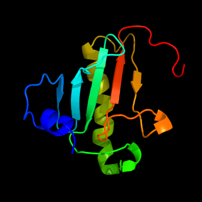
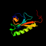
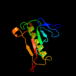
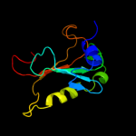
1 c3n1tE_
100.0
99
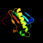
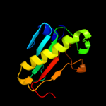
PDB header: hydrolaseChain: E: PDB Molecule: hit-like protein hint;PDBTitle: crystal structure of the h101a mutant echint gmp complex
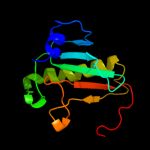
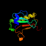
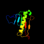
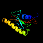
2 d1xqua_
100.0
48
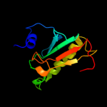
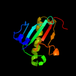
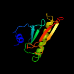
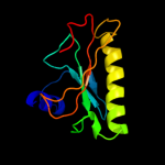
Fold: HIT-likeSuperfamily: HIT-likeFamily: HIT (HINT, histidine triad) family of protein kinase-interacting proteins3 c1xquA_
100.0
48
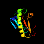
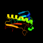
PDB header: hydrolaseChain: A: PDB Molecule: hit family hydrolase;PDBTitle: hit family hydrolase from clostridium thermocellum cth-393
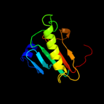
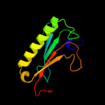
4 d1kpfa_
100.0
47
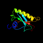
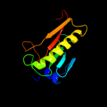
Fold: HIT-likeSuperfamily: HIT-likeFamily: HIT (HINT, histidine triad) family of protein kinase-interacting proteins5 c3l7xA_
100.0
29
PDB header: cell cycleChain: A: PDB Molecule: putative hit-like protein involved in cell-cyclePDBTitle: the crystal structure of smu.412c from streptococcus mutans ua159
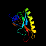
6 c3ksvA_
100.0
27
PDB header: unknown functionChain: A: PDB Molecule: uncharacterized protein;PDBTitle: hypothetical protein from leishmania major
7 d1rzya_
100.0
46
Fold: HIT-likeSuperfamily: HIT-likeFamily: HIT (HINT, histidine triad) family of protein kinase-interacting proteins8 c3o0mB_
100.0
25
PDB header: hydrolaseChain: B: PDB Molecule: hit family protein;PDBTitle: crystal structure of a zn-bound histidine triad family protein from2 mycobacterium smegmatis
9 c3imiB_
100.0
29
PDB header: structural genomics, unknown functionChain: B: PDB Molecule: hit family protein;PDBTitle: 2.01 angstrom resolution crystal structure of a hit family protein2 from bacillus anthracis str. 'ames ancestor'
10 c3lb5B_
100.0
35
PDB header: cell cycleChain: B: PDB Molecule: hit-like protein involved in cell-cycle regulation;PDBTitle: crystal structure of hit-like protein involved in cell-cycle2 regulation from bartonella henselae with unknown ligand
11 c2eo4A_
100.0
25
PDB header: hydrolaseChain: A: PDB Molecule: 150aa long hypothetical histidine triad nucleotide-bindingPDBTitle: crystal structure of hypothetical histidine triad nucleotide-binding2 protein st2152 from sulfolobus tokodaii strain7
12 c3anoA_
100.0
19
PDB header: transferaseChain: A: PDB Molecule: ap-4-a phosphorylase;PDBTitle: crystal structure of a novel diadenosine 5',5'''-p1,p4-tetraphosphate2 phosphorylase from mycobacterium tuberculosis h37rv
13 c3oj7A_
100.0
44
PDB header: metal binding proteinChain: A: PDB Molecule: putative histidine triad family protein;PDBTitle: crystal structure of a histidine triad family protein from entamoeba2 histolytica, bound to sulfate
14 d1y23a_
100.0
34
Fold: HIT-likeSuperfamily: HIT-likeFamily: HIT (HINT, histidine triad) family of protein kinase-interacting proteins15 c1emsB_
99.9
25
PDB header: antitumor proteinChain: B: PDB Molecule: nit-fragile histidine triad fusion protein;PDBTitle: crystal structure of the c. elegans nitfhit protein
16 d1emsa1
99.9
22
Fold: HIT-likeSuperfamily: HIT-likeFamily: HIT (HINT, histidine triad) family of protein kinase-interacting proteins17 c3p0tB_
99.9
31
PDB header: unknown functionChain: B: PDB Molecule: uncharacterized protein;PDBTitle: crystal structure of an hit-like protein from mycobacterium2 paratuberculosis
18 d2oika1
99.9
15
Fold: HIT-likeSuperfamily: HIT-likeFamily: HIT (HINT, histidine triad) family of protein kinase-interacting proteins19 c3r6fA_
99.9
25
PDB header: hydrolaseChain: A: PDB Molecule: hit family protein;PDBTitle: crystal structure of a zinc-containing hit family protein from2 encephalitozoon cuniculi
20 d1guqa2
99.9
12
Fold: HIT-likeSuperfamily: HIT-likeFamily: Hexose-1-phosphate uridylyltransferase21 d1z84a2
not modelled
99.9
10
Fold: HIT-likeSuperfamily: HIT-likeFamily: Hexose-1-phosphate uridylyltransferase22 d1fita_
not modelled
99.9
19
Fold: HIT-likeSuperfamily: HIT-likeFamily: HIT (HINT, histidine triad) family of protein kinase-interacting proteins23 c1gupC_
not modelled
99.9
13
PDB header: nucleotidyltransferaseChain: C: PDB Molecule: galactose-1-phosphate uridylyltransferase;PDBTitle: structure of nucleotidyltransferase complexed with udp-2 galactose
24 c1zwjA_
not modelled
99.9
10
PDB header: structural genomics, unknown functionChain: A: PDB Molecule: putative galactose-1-phosphate uridyl transferase;PDBTitle: x-ray structure of galt-like protein from arabidopsis thaliana2 at5g18200
25 c3i24B_
not modelled
99.8
14
PDB header: hydrolaseChain: B: PDB Molecule: hit family hydrolase;PDBTitle: crystal structure of a hit family hydrolase protein from2 vibrio fischeri. northeast structural genomics consortium3 target id vfr176
26 c3i4sB_
not modelled
99.8
13
PDB header: hydrolaseChain: B: PDB Molecule: histidine triad protein;PDBTitle: crystal structure of histidine triad protein blr8122 from2 bradyrhizobium japonicum
27 c3nrdB_
not modelled
99.8
15
PDB header: nucleotide binding proteinChain: B: PDB Molecule: histidine triad (hit) protein;PDBTitle: crystal structure of a histidine triad (hit) protein (smc02904) from2 sinorhizobium meliloti 1021 at 2.06 a resolution
28 c3oheA_
not modelled
99.7
15
PDB header: hydrolaseChain: A: PDB Molecule: histidine triad (hit) protein;PDBTitle: crystal structure of a histidine triad protein (maqu_1709) from2 marinobacter aquaeolei vt8 at 1.20 a resolution
29 d1z84a1
not modelled
99.4
10
Fold: HIT-likeSuperfamily: HIT-likeFamily: Hexose-1-phosphate uridylyltransferase30 d3bl9a1
not modelled
99.4
17
Fold: HIT-likeSuperfamily: HIT-likeFamily: mRNA decapping enzyme DcpS C-terminal domain31 c3bl9B_
not modelled
99.3
17
PDB header: hydrolaseChain: B: PDB Molecule: scavenger mrna-decapping enzyme dcps;PDBTitle: synthetic gene encoded dcps bound to inhibitor dg157493
32 d1vlra1
not modelled
99.2
17
Fold: HIT-likeSuperfamily: HIT-likeFamily: mRNA decapping enzyme DcpS C-terminal domain33 c1xmlA_
not modelled
99.0
18
PDB header: chaperoneChain: A: PDB Molecule: heat shock-like protein 1;PDBTitle: structure of human dcps
34 d1guqa1
not modelled
98.1
10
Fold: HIT-likeSuperfamily: HIT-likeFamily: Hexose-1-phosphate uridylyltransferase35 d2pofa1
not modelled
95.9
17
Fold: HIT-likeSuperfamily: HIT-likeFamily: CDH-like36 c3csqC_
not modelled
40.3
23
PDB header: hydrolaseChain: C: PDB Molecule: morphogenesis protein 1;PDBTitle: crystal and cryoem structural studies of a cell wall2 degrading enzyme in the bacteriophage phi29 tail
37 c3hfnA_
not modelled
33.4
19
PDB header: rna binding proteinChain: A: PDB Molecule: asl2047 protein;PDBTitle: crystal structure of an hfq protein from anabaena sp.
38 c1qyuA_
not modelled
18.8
41
PDB header: lyaseChain: A: PDB Molecule: ribosomal large subunit pseudouridine synthase d;PDBTitle: structure of the catalytic domain of 23s rrna pseudouridine2 synthase rlud
39 d1v9ka_
not modelled
17.1
33
Fold: Pseudouridine synthaseSuperfamily: Pseudouridine synthaseFamily: Pseudouridine synthase RsuA/RluD40 d2cs7a1
not modelled
15.4
25
Fold: IL8-likeSuperfamily: PhtA domain-likeFamily: PhtA domain-like41 c2i82D_
not modelled
15.1
33
PDB header: lyase/rnaChain: D: PDB Molecule: ribosomal large subunit pseudouridine synthase a;PDBTitle: crystal structure of pseudouridine synthase rlua: indirect2 sequence readout through protein-induced rna structure
42 d2at2a1
not modelled
14.4
9
Fold: ATC-likeSuperfamily: Aspartate/ornithine carbamoyltransferaseFamily: Aspartate/ornithine carbamoyltransferase43 d1v9fa_
not modelled
14.1
41
Fold: Pseudouridine synthaseSuperfamily: Pseudouridine synthaseFamily: Pseudouridine synthase RsuA/RluD44 c1v9fA_
not modelled
14.1
41
PDB header: lyaseChain: A: PDB Molecule: ribosomal large subunit pseudouridine synthase d;PDBTitle: crystal structure of catalytic domain of pseudouridine2 synthase rlud from escherichia coli
45 c3hfoC_
not modelled
13.5
19
PDB header: rna binding proteinChain: C: PDB Molecule: ssr3341 protein;PDBTitle: crystal structure of an hfq protein from synechocystis sp.
46 c3sdsA_
not modelled
12.8
13
PDB header: transferaseChain: A: PDB Molecule: ornithine carbamoyltransferase, mitochondrial;PDBTitle: crystal structure of a mitochondrial ornithine carbamoyltransferase2 from coccidioides immitis
47 d2hnga1
not modelled
12.6
12
Fold: SecB-likeSuperfamily: SecB-likeFamily: SP1558-like48 d1ryba_
not modelled
12.3
8
Fold: Phosphorylase/hydrolase-likeSuperfamily: Peptidyl-tRNA hydrolase-likeFamily: Peptidyl-tRNA hydrolase-like49 c1t3bA_
not modelled
12.2
11
PDB header: isomeraseChain: A: PDB Molecule: thiol:disulfide interchange protein dsbc;PDBTitle: x-ray structure of dsbc from haemophilus influenzae
50 c2qwwB_
not modelled
12.1
8
PDB header: transcriptionChain: B: PDB Molecule: transcriptional regulator, marr family;PDBTitle: crystal structure of multiple antibiotic-resistance repressor (marr)2 (yp_013417.1) from listeria monocytogenes 4b f2365 at 2.07 a3 resolution
51 c2ef0A_
not modelled
11.1
11
PDB header: transferaseChain: A: PDB Molecule: ornithine carbamoyltransferase;PDBTitle: crystal structure of ornithine carbamoyltransferase from thermus2 thermophilus
52 d1tuga1
not modelled
11.1
11
Fold: ATC-likeSuperfamily: Aspartate/ornithine carbamoyltransferaseFamily: Aspartate/ornithine carbamoyltransferase53 d1vlva1
not modelled
10.9
17
Fold: ATC-likeSuperfamily: Aspartate/ornithine carbamoyltransferaseFamily: Aspartate/ornithine carbamoyltransferase54 d1ekxa1
not modelled
10.6
11
Fold: ATC-likeSuperfamily: Aspartate/ornithine carbamoyltransferaseFamily: Aspartate/ornithine carbamoyltransferase55 c3gd5D_
not modelled
10.5
9
PDB header: transferaseChain: D: PDB Molecule: ornithine carbamoyltransferase;PDBTitle: crystal structure of ornithine carbamoyltransferase from gloeobacter2 violaceus
56 d1pg5a1
not modelled
10.3
10
Fold: ATC-likeSuperfamily: Aspartate/ornithine carbamoyltransferaseFamily: Aspartate/ornithine carbamoyltransferase57 d1pvva1
not modelled
9.6
9
Fold: ATC-likeSuperfamily: Aspartate/ornithine carbamoyltransferaseFamily: Aspartate/ornithine carbamoyltransferase58 c3lxmC_
not modelled
9.4
17
PDB header: transferaseChain: C: PDB Molecule: aspartate carbamoyltransferase;PDBTitle: 2.00 angstrom resolution crystal structure of a catalytic2 subunit of an aspartate carbamoyltransferase (pyrb) from3 yersinia pestis co92
59 c3l6tB_
not modelled
9.4
25
PDB header: hydrolaseChain: B: PDB Molecule: mobilization protein trai;PDBTitle: crystal structure of an n-terminal mutant of the plasmid pcu1 trai2 relaxase domain
60 d1otha1
not modelled
9.4
10
Fold: ATC-likeSuperfamily: Aspartate/ornithine carbamoyltransferaseFamily: Aspartate/ornithine carbamoyltransferase61 d1dxha1
not modelled
9.3
6
Fold: ATC-likeSuperfamily: Aspartate/ornithine carbamoyltransferaseFamily: Aspartate/ornithine carbamoyltransferase62 c2qgpA_
not modelled
8.4
17
PDB header: hydrolaseChain: A: PDB Molecule: hnh endonuclease;PDBTitle: x-ray structure of the nhn endonuclease from geobacter2 metallireducens. northeast structural genomics consortium3 target gmr87.
63 c3vg8F_
not modelled
8.4
22
PDB header: unknown functionChain: F: PDB Molecule: hypothetical protein tthb210;PDBTitle: crystal structure of hypothetical protein tthb210 from thermus2 thermophilus hb8
64 d1ex4a1
not modelled
8.3
19
Fold: SH3-like barrelSuperfamily: DNA-binding domain of retroviral integraseFamily: DNA-binding domain of retroviral integrase65 d1ml4a1
not modelled
8.1
14
Fold: ATC-likeSuperfamily: Aspartate/ornithine carbamoyltransferaseFamily: Aspartate/ornithine carbamoyltransferase66 c3py7A_
not modelled
8.0
7
PDB header: viral proteinChain: A: PDB Molecule: maltose-binding periplasmic protein,paxillin ld1,protein e6PDBTitle: crystal structure of full-length bovine papillomavirus oncoprotein e62 in complex with ld1 motif of paxillin at 2.3a resolution
67 c2elpA_
not modelled
7.9
13
PDB header: transcriptionChain: A: PDB Molecule: zinc finger protein 406;PDBTitle: solution structure of the 13th c2h2 zinc finger of human2 zinc finger protein 406
68 d2jz6a1
not modelled
7.9
25
Fold: L28p-likeSuperfamily: L28p-likeFamily: Ribosomal protein L2869 d2d8ya1
not modelled
7.4
11
Fold: Glucocorticoid receptor-like (DNA-binding domain)Superfamily: Glucocorticoid receptor-like (DNA-binding domain)Family: LIM domain70 c1jzdA_
not modelled
7.1
7
PDB header: oxidoreductaseChain: A: PDB Molecule: thiol:disulfide interchange protein dsbc;PDBTitle: dsbc-dsbdalpha complex
71 d1j0ga_
not modelled
6.9
8
Fold: beta-Grasp (ubiquitin-like)Superfamily: Ubiquitin-likeFamily: BM-002-like72 c1fvoB_
not modelled
6.6
10
PDB header: transferaseChain: B: PDB Molecule: ornithine transcarbamylase;PDBTitle: crystal structure of human ornithine transcarbamylase complexed with2 carbamoyl phosphate
73 c3fmbA_
not modelled
6.4
13
PDB header: structural genomics, unknown functionChain: A: PDB Molecule: dimeric protein of unknown function and ferredoxin-likePDBTitle: crystal structure of dimeric protein of unknown function and2 ferredoxin-like fold (yp_212648.1) from bacteroides fragilis nctc3 9343 at 1.85 a resolution
74 c2dnfA_
not modelled
6.3
17
PDB header: protein bindingChain: A: PDB Molecule: doublecortin domain-containing protein 2;PDBTitle: solution structure of rsgi ruh-062, a dcx domain from human
75 c3d6nB_
not modelled
6.2
8
PDB header: hydrolase/transferaseChain: B: PDB Molecule: aspartate carbamoyltransferase;PDBTitle: crystal structure of aquifex dihydroorotase activated by aspartate2 transcarbamoylase
76 c2at2B_
not modelled
6.2
9
PDB header: PDB COMPND: 77 c2rgwD_
not modelled
6.1
6
PDB header: transferaseChain: D: PDB Molecule: aspartate carbamoyltransferase;PDBTitle: catalytic subunit of m. jannaschii aspartate2 transcarbamoylase
78 d1u2ma_
not modelled
6.1
11
Fold: OmpH-likeSuperfamily: OmpH-likeFamily: OmpH-like79 c2k6pA_
not modelled
6.1
10
PDB header: unknown functionChain: A: PDB Molecule: uncharacterized protein hp_1423;PDBTitle: solution structure of hypothetical protein, hp1423
80 d1p4da_
not modelled
6.1
8
Fold: Origin of replication-binding domain, RBD-likeSuperfamily: Origin of replication-binding domain, RBD-likeFamily: Relaxase domain81 c2zaeC_
not modelled
6.0
8
PDB header: hydrolaseChain: C: PDB Molecule: ribonuclease p protein component 1;PDBTitle: crystal structure of protein ph1601p in complex with protein ph1771p2 of archaeal ribonuclease p from pyrococcus horikoshii ot3
82 d1duvg1
not modelled
5.8
6
Fold: ATC-likeSuperfamily: Aspartate/ornithine carbamoyltransferaseFamily: Aspartate/ornithine carbamoyltransferase83 c1zq2A_
not modelled
5.7
15
PDB header: transferaseChain: A: PDB Molecule: ornithine carbamoyltransferase;PDBTitle: crystal structure of n-acetyl-l-ornithine transcarbamylase2 complexed with cp
84 d1wira_
not modelled
5.4
20
Fold: beta-beta-alpha zinc fingersSuperfamily: beta-beta-alpha zinc fingersFamily: variant C2H2 finger85 d1ihwa_
not modelled
5.2
19
Fold: SH3-like barrelSuperfamily: DNA-binding domain of retroviral integraseFamily: DNA-binding domain of retroviral integrase86 d1abaa_
not modelled
5.2
0
Fold: Thioredoxin foldSuperfamily: Thioredoxin-likeFamily: Thioltransferase87 c1vlvA_
not modelled
5.2
17
PDB header: transferaseChain: A: PDB Molecule: ornithine carbamoyltransferase;PDBTitle: crystal structure of ornithine carbamoyltransferase (tm1097) from2 thermotoga maritima at 2.25 a resolution
88 d1oqka_
not modelled
5.2
10
Fold: Rof/RNase P subunit-likeSuperfamily: Rof/RNase P subunit-likeFamily: RNase P subunit p29-like89 d1uf0a_
not modelled
5.2
15
Fold: beta-Grasp (ubiquitin-like)Superfamily: Doublecortin (DC)Family: Doublecortin (DC)90 d1wiia_
not modelled
5.2
17
Fold: Rubredoxin-likeSuperfamily: Zinc beta-ribbonFamily: Putative zinc binding domain91 c3tpfF_
not modelled
5.1
23
PDB header: transferaseChain: F: PDB Molecule: ornithine carbamoyltransferase;PDBTitle: crystal structure of anabolic ornithine carbamoyltransferase from2 campylobacter jejuni subsp. jejuni nctc 11168
92 d2bo9b2
not modelled
5.1
23
Fold: Cystatin-likeSuperfamily: Cystatin/monellinFamily: Latexin-like93 c2p2gD_
not modelled
5.1
12
PDB header: transferaseChain: D: PDB Molecule: ornithine carbamoyltransferase;PDBTitle: crystal structure of ornithine carbamoyltransferase from mycobacterium2 tuberculosis (rv1656): orthorhombic form