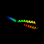
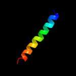
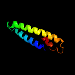
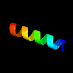
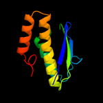
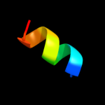
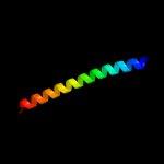
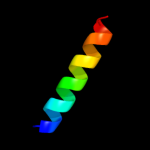
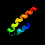
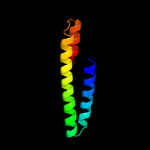
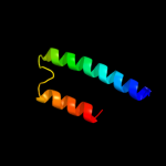
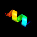
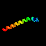
1 d1nt2b_
35.9
22
Fold: Nop domainSuperfamily: Nop domainFamily: Nop domain2 c1junB_
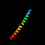
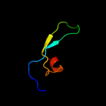
26.9
26
PDB header: transcription regulationChain: B: PDB Molecule: c-jun homodimer;PDBTitle: nmr study of c-jun homodimer
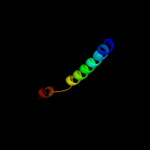
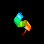
3 d1vcsa1
25.4
14
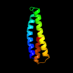
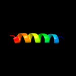
Fold: STAT-likeSuperfamily: t-snare proteinsFamily: t-snare proteins4 d2gtsa1
24.9
22
Fold: Ferritin-likeSuperfamily: HP0062-likeFamily: HP0062-like5 c2xzmI_
24.5
12
PDB header: ribosomeChain: I: PDB Molecule: rps16e;PDBTitle: crystal structure of the eukaryotic 40s ribosomal2 subunit in complex with initiation factor 1. this file3 contains the 40s subunit and initiation factor for4 molecule 1
6 c3n4xB_
23.7
17
PDB header: replicationChain: B: PDB Molecule: monopolin complex subunit csm1;PDBTitle: structure of csm1 full-length
7 c3swfA_
22.9
16
PDB header: transport proteinChain: A: PDB Molecule: cgmp-gated cation channel alpha-1;PDBTitle: cnga1 621-690 containing clz domain
8 d1lvfa_
22.7
15
Fold: STAT-likeSuperfamily: t-snare proteinsFamily: t-snare proteins9 c2f3yB_
18.1
40
PDB header: metal binding proteinChain: B: PDB Molecule: voltage-dependent l-type calcium channel alpha-PDBTitle: calmodulin/iq domain complex
10 c1t3jA_
17.1
13
PDB header: membrane proteinChain: A: PDB Molecule: mitofusin 1;PDBTitle: mitofusin domain hr2 v686m/i708m mutant
11 d1wmib1
13.9
35
Fold: Non-globular all-alpha subunits of globular proteinsSuperfamily: RelB-likeFamily: RelB-like12 c2rpaA_
13.7
11
PDB header: hydrolaseChain: A: PDB Molecule: katanin p60 atpase-containing subunit a1;PDBTitle: the solution structure of n-terminal domain of microtubule severing2 enzyme
13 c3onjA_
13.5
13
PDB header: protein transportChain: A: PDB Molecule: t-snare vti1;PDBTitle: crystal structure of yeast vti1p_habc domain
14 d1wgda_
12.7
13
Fold: beta-Grasp (ubiquitin-like)Superfamily: Ubiquitin-likeFamily: Ubiquitin-related15 d3saka_
12.0
57
Fold: p53 tetramerization domainSuperfamily: p53 tetramerization domainFamily: p53 tetramerization domain16 c3q9dB_
11.9
30
PDB header: unknown functionChain: B: PDB Molecule: protein cpn_0803/cp_1068/cpj0803/cpb0832;PDBTitle: crystal structure of cpn0803 from c. pneumoniae.
17 c3n1bA_
11.7
9
PDB header: transport proteinChain: A: PDB Molecule: vacuolar protein sorting-associated protein 54;PDBTitle: c-terminal domain of vps54 subunit of the garp complex
18 c2vayB_
10.4
40
PDB header: metal transportChain: B: PDB Molecule: voltage-dependent l-type calcium channel subunitPDBTitle: calmodulin complexed with cav1.1 iq peptide
19 c3f42A_
10.1
14
PDB header: structural genomics, unknown functionChain: A: PDB Molecule: protein hp0035;PDBTitle: crystal structure of uncharacterized protein hp0035 from helicobacter2 pylori
20 d1a9xb1
9.8
80
Fold: The "swivelling" beta/beta/alpha domainSuperfamily: Carbamoyl phosphate synthetase, small subunit N-terminal domainFamily: Carbamoyl phosphate synthetase, small subunit N-terminal domain21 c2jroA_
not modelled
9.2
17
PDB header: structural genomics, unknown functionChain: A: PDB Molecule: uncharacterized protein;PDBTitle: solution nmr structure of so0334 from shewanella oneidensis. northeast2 structural genomics target sor75
22 d2elba1
not modelled
9.1
12
Fold: BAR/IMD domain-likeSuperfamily: BAR/IMD domain-likeFamily: BAR domain23 c2j11D_
not modelled
8.9
57
PDB header: transcriptionChain: D: PDB Molecule: cellular tumor antigen p53;PDBTitle: p53 tetramerization domain mutant y327s t329g q331g
24 c1gngX_
not modelled
8.8
35
PDB header: transferaseChain: X: PDB Molecule: frattide;PDBTitle: glycogen synthase kinase-3 beta (gsk3) complex with frattide2 peptide
25 c3r2cJ_
not modelled
8.7
21
PDB header: transcription/rnaChain: J: PDB Molecule: 30s ribosomal protein s10;PDBTitle: crystal structure of antitermination factors nusb and nuse in complex2 with boxa rna
26 c2ywfA_
not modelled
8.4
17
PDB header: translationChain: A: PDB Molecule: gtp-binding protein lepa;PDBTitle: crystal structure of gmppnp-bound lepa from aquifex aeolicus
27 d1aiea_
not modelled
8.1
57
Fold: p53 tetramerization domainSuperfamily: p53 tetramerization domainFamily: p53 tetramerization domain28 c1mpgB_
not modelled
7.9
26
PDB header: hydrolaseChain: B: PDB Molecule: 3-methyladenine dna glycosylase ii;PDBTitle: 3-methyladenine dna glycosylase ii from escherichia coli
29 d1we3a2
not modelled
7.9
18
Fold: The "swivelling" beta/beta/alpha domainSuperfamily: GroEL apical domain-likeFamily: GroEL-like chaperone, apical domain30 c2j10D_
not modelled
7.7
57
PDB header: transcriptionChain: D: PDB Molecule: cellular tumor antigen p53;PDBTitle: p53 tetramerization domain mutant t329f q331k
31 c2j10B_
not modelled
7.7
57
PDB header: transcriptionChain: B: PDB Molecule: cellular tumor antigen p53;PDBTitle: p53 tetramerization domain mutant t329f q331k
32 c2j10A_
not modelled
7.7
57
PDB header: transcriptionChain: A: PDB Molecule: cellular tumor antigen p53;PDBTitle: p53 tetramerization domain mutant t329f q331k
33 d3bypa1
not modelled
7.5
15
Fold: Alpha-lytic protease prodomain-likeSuperfamily: Cation efflux protein cytoplasmic domain-likeFamily: Cation efflux protein cytoplasmic domain-like34 c2zkqi_
not modelled
7.5
22
PDB header: ribosomal protein/rnaChain: I: PDB Molecule: PDBTitle: structure of a mammalian ribosomal 40s subunit within an2 80s complex obtained by docking homology models of the rna3 and proteins into an 8.7 a cryo-em map
35 c4a4kI_
not modelled
7.5
12
PDB header: hydrolaseChain: I: PDB Molecule: antiviral helicase ski2;PDBTitle: crystal structure of the s. cerevisiae ski2 insertion domain
36 d1jjcb2
not modelled
7.5
33
Fold: Putative DNA-binding domainSuperfamily: Putative DNA-binding domainFamily: Domains B1 and B5 of PheRS-beta, PheT37 c3n6sA_
not modelled
7.5
24
PDB header: transcription, replication/dnaChain: A: PDB Molecule: transcription termination factor, mitochondrial;PDBTitle: crystal structure of human mitochondrial mterf in complex with a 15-2 mer dna encompassing the trnaleu(uur) binding sequence
38 c1keeH_
not modelled
7.4
80
PDB header: ligaseChain: H: PDB Molecule: carbamoyl-phosphate synthetase small chain;PDBTitle: inactivation of the amidotransferase activity of carbamoyl phosphate2 synthetase by the antibiotic acivicin
39 d1z1va1
not modelled
7.4
50
Fold: SAM domain-likeSuperfamily: SAM/Pointed domainFamily: SAM (sterile alpha motif) domain40 d1g3nc2
not modelled
7.2
50
Fold: Cyclin-likeSuperfamily: Cyclin-likeFamily: Cyclin41 c2it3B_
not modelled
7.2
15
PDB header: structural genomics, unknown functionChain: B: PDB Molecule: upf0130 protein ph1069;PDBTitle: structure of ph1069 protein from pyrococcus horikoshii
42 c3eabK_
not modelled
7.1
31
PDB header: cell cycleChain: K: PDB Molecule: chmp1b;PDBTitle: crystal structure of spastin mit in complex with escrt iii
43 c1lj2C_
not modelled
7.0
50
PDB header: viral protein/ translationChain: C: PDB Molecule: eukaryotic protein synthesis initiation factor;PDBTitle: recognition of eif4g by rotavirus nsp3 reveals a basis for2 mrna circularization
44 c2kdbA_
not modelled
6.9
13
PDB header: protein bindingChain: A: PDB Molecule: homocysteine-responsive endoplasmic reticulum-PDBTitle: solution structure of human ubiquitin-like domain of2 herpud2_9_85, northeast structural genomics consortium3 (nesg) target ht53a
45 d1k1fa_
not modelled
6.8
22
Fold: Bcr-Abl oncoprotein oligomerization domainSuperfamily: Bcr-Abl oncoprotein oligomerization domainFamily: Bcr-Abl oncoprotein oligomerization domain46 c3n5aA_
not modelled
6.8
31
PDB header: protein transportChain: A: PDB Molecule: synaptotagmin-7;PDBTitle: synaptotagmin-7, c2b-domain, calcium bound
47 c3kizA_
not modelled
6.7
16
PDB header: ligaseChain: A: PDB Molecule: phosphoribosylformylglycinamidine cyclo-ligase;PDBTitle: crystal structure of putative phosphoribosylformylglycinamidine cyclo-2 ligase (yp_676759.1) from cytophaga hutchinsonii atcc 33406 at 1.50 a3 resolution
48 d1kida_
not modelled
6.7
19
Fold: The "swivelling" beta/beta/alpha domainSuperfamily: GroEL apical domain-likeFamily: GroEL-like chaperone, apical domain49 d1sjpa2
not modelled
6.7
19
Fold: The "swivelling" beta/beta/alpha domainSuperfamily: GroEL apical domain-likeFamily: GroEL-like chaperone, apical domain50 c3p8cD_
not modelled
6.6
10
PDB header: protein bindingChain: D: PDB Molecule: wiskott-aldrich syndrome protein family member 1;PDBTitle: structure and control of the actin regulatory wave complex
51 c1lj2D_
not modelled
6.6
50
PDB header: viral protein/ translationChain: D: PDB Molecule: eukaryotic protein synthesis initiation factor;PDBTitle: recognition of eif4g by rotavirus nsp3 reveals a basis for2 mrna circularization
52 c2elxA_
not modelled
6.6
44
PDB header: transcriptionChain: A: PDB Molecule: zinc finger protein 406;PDBTitle: solution structure of the 8th c2h2 zinc finger of mouse2 zinc finger protein 406
53 d2oufa1
not modelled
6.4
18
Fold: HP0242-likeSuperfamily: HP0242-likeFamily: HP0242-like54 c3ks7D_
not modelled
6.4
22
PDB header: hydrolaseChain: D: PDB Molecule: putative putative pngase f;PDBTitle: crystal structure of putative peptide:n-glycosidase f (pngase f)2 (yp_210507.1) from bacteroides fragilis nctc 9343 at 2.30 a3 resolution
55 d1g73a_
not modelled
6.4
14
Fold: Spectrin repeat-likeSuperfamily: Smac/diabloFamily: Smac/diablo56 d1igqa_
not modelled
6.1
14
Fold: SH3-like barrelSuperfamily: C-terminal domain of transcriptional repressorsFamily: Transcriptional repressor protein KorB57 d3efza1
not modelled
6.1
8
Fold: alpha-alpha superhelixSuperfamily: 14-3-3 proteinFamily: 14-3-3 protein58 c3efzA_
not modelled
6.1
8
PDB header: signaling proteinChain: A: PDB Molecule: 14-3-3 protein;PDBTitle: crystal structure of a 14-3-3 protein from cryptosporidium parvum2 (cgd1_2980)
59 c3swyB_
not modelled
6.0
12
PDB header: transport proteinChain: B: PDB Molecule: cyclic nucleotide-gated cation channel alpha-3;PDBTitle: cnga3 626-672 containing clz domain
60 c3c19A_
not modelled
6.0
6
PDB header: structural genomics, unknown functionChain: A: PDB Molecule: uncharacterized protein mk0293;PDBTitle: crystal structure of protein mk0293 from methanopyrus kandleri av19
61 c1gw4A_
not modelled
6.0
31
PDB header: high density lipoproteinsChain: A: PDB Molecule: apoa-i;PDBTitle: the helix-hinge-helix structural motif in human2 apolipoprotein a-i determined by nmr spectroscopy, 13 structure
62 d1dula_
not modelled
6.0
50
Fold: Signal peptide-binding domainSuperfamily: Signal peptide-binding domainFamily: Signal peptide-binding domain63 d1igub_
not modelled
5.9
14
Fold: SH3-like barrelSuperfamily: C-terminal domain of transcriptional repressorsFamily: Transcriptional repressor protein KorB64 d1fewa_
not modelled
5.8
14
Fold: Spectrin repeat-likeSuperfamily: Smac/diabloFamily: Smac/diablo65 c1zvaA_
not modelled
5.8
13
PDB header: viral proteinChain: A: PDB Molecule: e2 glycoprotein;PDBTitle: a structure-based mechanism of sars virus membrane fusion
66 c1jqmA_
not modelled
5.8
5
PDB header: ribosomeChain: A: PDB Molecule: 50s ribosomal protein l11;PDBTitle: fitting of l11 protein and elongation factor g (ef-g) in2 the cryo-em map of e. coli 70s ribosome bound with ef-g,3 gdp and fusidic acid
67 c1rrqA_
not modelled
5.8
26
PDB header: hydrolase/dnaChain: A: PDB Molecule: muty;PDBTitle: muty adenine glycosylase in complex with dna containing an2 a:oxog pair
68 d1ip9a_
not modelled
5.8
14
Fold: beta-Grasp (ubiquitin-like)Superfamily: CAD & PB1 domainsFamily: PB1 domain69 d1orja_
not modelled
5.8
14
Fold: Four-helical up-and-down bundleSuperfamily: Flagellar export chaperone FliSFamily: Flagellar export chaperone FliS70 c3bboK_
not modelled
5.7
13
PDB header: ribosomeChain: K: PDB Molecule: ribosomal protein l11;PDBTitle: homology model for the spinach chloroplast 50s subunit2 fitted to 9.4a cryo-em map of the 70s chlororibosome
71 c3degC_
not modelled
5.7
17
PDB header: ribosomeChain: C: PDB Molecule: gtp-binding protein lepa;PDBTitle: complex of elongating escherichia coli 70s ribosome and ef4(lepa)-2 gmppnp
72 c3cb4D_
not modelled
5.6
17
PDB header: translationChain: D: PDB Molecule: gtp-binding protein lepa;PDBTitle: the crystal structure of lepa
73 d1a1ua_
not modelled
5.3
57
Fold: p53 tetramerization domainSuperfamily: p53 tetramerization domainFamily: p53 tetramerization domain74 d1dfca1
not modelled
5.3
56
Fold: beta-TrefoilSuperfamily: Actin-crosslinking proteinsFamily: Fascin75 c2v8sV_
not modelled
5.2
11
PDB header: protein transportChain: V: PDB Molecule: vesicle transport through interaction withPDBTitle: vti1b habc domain - epsinr enth domain complex
76 c2be6F_
not modelled
5.2
40
PDB header: membrane proteinChain: F: PDB Molecule: voltage-dependent l-type calcium channel alpha-1c subunit;PDBTitle: 2.0 a crystal structure of the cav1.2 iq domain-ca/cam complex
77 d1js8a2
not modelled
5.2
5
Fold: C-terminal domain of mollusc hemocyaninSuperfamily: C-terminal domain of mollusc hemocyaninFamily: C-terminal domain of mollusc hemocyanin78 c1bf5A_
not modelled
5.2
16
PDB header: gene regulation/dnaChain: A: PDB Molecule: signal transducer and activator of transcriptionPDBTitle: tyrosine phosphorylated stat-1/dna complex
79 c3okqA_
not modelled
5.1
21
PDB header: protein bindingChain: A: PDB Molecule: bud site selection protein 6;PDBTitle: crystal structure of a core domain of yeast actin nucleation cofactor2 bud6


