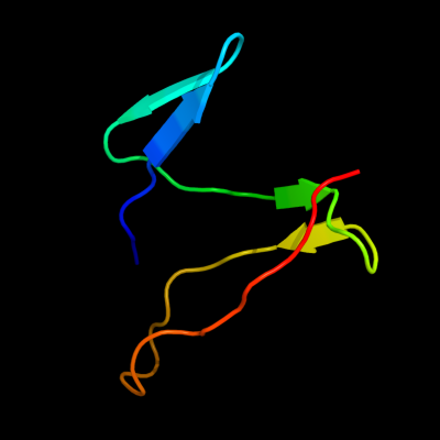
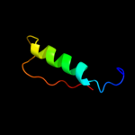
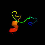
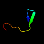
1 d1aqta2
37.3
21
Fold: Epsilon subunit of F1F0-ATP synthase N-terminal domainSuperfamily: Epsilon subunit of F1F0-ATP synthase N-terminal domainFamily: Epsilon subunit of F1F0-ATP synthase N-terminal domain2 c1s3rA_
35.3
27
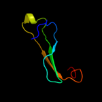
PDB header: toxinChain: A: PDB Molecule: intermedilysin;PDBTitle: crystal structure of the human-specific toxin intermedilysin
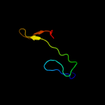
3 c1pfoA_
33.1
20
PDB header: toxinChain: A: PDB Molecule: perfringolysin o;PDBTitle: perfringolysin o
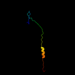
4 d1pfoa_
33.1
20
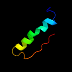
Fold: PerfringolysinSuperfamily: PerfringolysinFamily: Perfringolysin5 c1fs0E_
30.3
13
PDB header: hydrolaseChain: E: PDB Molecule: atp synthase epsilon subunit;PDBTitle: complex of gamma/epsilon atp synthase from e.coli
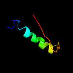
6 c2rq7A_
29.8
7
PDB header: hydrolaseChain: A: PDB Molecule: atp synthase epsilon chain;PDBTitle: solution structure of the epsilon subunit chimera combining2 the n-terminal beta-sandwich domain from t. elongatus bp-13 f1 and the c-terminal alpha-helical domain from spinach4 chloroplast f1
7 d2p02a3
29.2
30
Fold: S-adenosylmethionine synthetaseSuperfamily: S-adenosylmethionine synthetaseFamily: S-adenosylmethionine synthetase8 d1g94a1
28.2
20
Fold: Glycosyl hydrolase domainSuperfamily: Glycosyl hydrolase domainFamily: alpha-Amylases, C-terminal beta-sheet domain9 d1qm4a3
27.6
30
Fold: S-adenosylmethionine synthetaseSuperfamily: S-adenosylmethionine synthetaseFamily: S-adenosylmethionine synthetase10 d3dhpa1
27.2
24
Fold: Glycosyl hydrolase domainSuperfamily: Glycosyl hydrolase domainFamily: alpha-Amylases, C-terminal beta-sheet domain11 c3b5wE_
25.2
19
PDB header: membrane proteinChain: E: PDB Molecule: lipid a export atp-binding/permease protein msba;PDBTitle: crystal structure of eschericia coli msba
12 c2xu8B_
22.5
27
PDB header: structural genomicsChain: B: PDB Molecule: pa1645;PDBTitle: structure of pa1645
13 d2e74d1
22.5
23
Fold: ISP domainSuperfamily: ISP domainFamily: Rieske iron-sulfur protein (ISP)14 c3so4C_
21.0
27
PDB header: transferaseChain: C: PDB Molecule: methionine-adenosyltransferase;PDBTitle: methionine-adenosyltransferase from entamoeba histolytica
15 c3hvnA_
20.2
20
PDB header: toxinChain: A: PDB Molecule: hemolysin;PDBTitle: crystal structure of cytotoxin protein suilysin from2 streptococcus suis
16 c2e5yA_
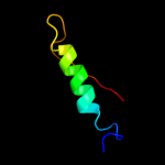
19.9
18
PDB header: hydrolaseChain: A: PDB Molecule: atp synthase epsilon chain;PDBTitle: epsilon subunit and atp complex of f1f0-atp synthase from2 the thermophilic bacillus ps3
17 d1v58a2
19.8
44
Fold: Cystatin-likeSuperfamily: DsbC/DsbG N-terminal domain-likeFamily: DsbC/DsbG N-terminal domain-like18 c2obvA_
19.7
30
PDB header: transferaseChain: A: PDB Molecule: s-adenosylmethionine synthetase isoform type-1;PDBTitle: crystal structure of the human s-adenosylmethionine synthetase 1 in2 complex with the product
19 c1rg9D_
19.2
34
PDB header: transferaseChain: D: PDB Molecule: s-adenosylmethionine synthetase;PDBTitle: s-adenosylmethionine synthetase complexed with sam and ppnp
20 c3imlB_
18.9
24
PDB header: transferaseChain: B: PDB Molecule: s-adenosylmethionine synthetase;PDBTitle: crystal structure of s-adenosylmethionine synthetase from burkholderia2 pseudomallei
21 c3rv2B_
not modelled
17.6
27
PDB header: transferaseChain: B: PDB Molecule: s-adenosylmethionine synthase;PDBTitle: crystal structure of s-adenosylmethionine synthetase from2 mycobacterium marinum
22 c2wanA_
not modelled
16.2
25
PDB header: hydrolaseChain: A: PDB Molecule: pullulanase;PDBTitle: pullulanase from bacillus acidopullulyticus
23 c2l92A_
not modelled
13.6
50
PDB header: dna binding proteinChain: A: PDB Molecule: histone family protein nucleoid-structuring protein h-ns;PDBTitle: solution structure of the c-terminal domain of h-ns like protein bv3f
24 c1h8eH_
not modelled
13.3
14
PDB header: hydrolaseChain: H: PDB Molecule: bovine mitochondrial f1-atpase;PDBTitle: (adp.alf4)2(adp.so4) bovine f1-atpase2 (all three catalytic sites occupied)
25 c3ebeA_
not modelled
11.8
24
PDB header: replicationChain: A: PDB Molecule: protein mcm10 homolog;PDBTitle: crystal structure of xenopus laevis replication initiation factor2 mcm10 internal domain
26 d2j07a1
not modelled
11.8
23
Fold: Cryptochrome/photolyase FAD-binding domainSuperfamily: Cryptochrome/photolyase FAD-binding domainFamily: Cryptochrome/photolyase FAD-binding domain27 d1ig4a_
not modelled
10.6
50
Fold: DNA-binding domainSuperfamily: DNA-binding domainFamily: Methyl-CpG-binding domain, MBD28 d1e6vb2
not modelled
10.5
18
Fold: Ferredoxin-likeSuperfamily: Methyl-coenzyme M reductase subunitsFamily: Methyl-coenzyme M reductase alpha and beta chain N-terminal domain29 c2qe7H_
not modelled
10.3
27
PDB header: hydrolaseChain: H: PDB Molecule: atp synthase subunit epsilon;PDBTitle: crystal structure of the f1-atpase from the thermoalkaliphilic2 bacterium bacillus sp. ta2.a1
30 d1ik0a_
not modelled
9.9
35
Fold: 4-helical cytokinesSuperfamily: 4-helical cytokinesFamily: Short-chain cytokines31 d1hbnb2
not modelled
9.4
45
Fold: Ferredoxin-likeSuperfamily: Methyl-coenzyme M reductase subunitsFamily: Methyl-coenzyme M reductase alpha and beta chain N-terminal domain32 d1mxaa3
not modelled
9.2
30
Fold: S-adenosylmethionine synthetaseSuperfamily: S-adenosylmethionine synthetaseFamily: S-adenosylmethionine synthetase33 d1jaea1
not modelled
9.1
26
Fold: Glycosyl hydrolase domainSuperfamily: Glycosyl hydrolase domainFamily: alpha-Amylases, C-terminal beta-sheet domain34 c1dvaY_
not modelled
8.9
40
PDB header: hydrolase/hydrolase inhibitorChain: Y: PDB Molecule: peptide e-76;PDBTitle: crystal structure of the complex between the peptide exosite inhibitor2 e-76 and coagulation factor viia
35 c1dvaX_
not modelled
8.9
40
PDB header: hydrolase/hydrolase inhibitorChain: X: PDB Molecule: peptide e-76;PDBTitle: crystal structure of the complex between the peptide exosite inhibitor2 e-76 and coagulation factor viia
36 d1r44a_
not modelled
8.8
20
Fold: Hedgehog/DD-peptidaseSuperfamily: Hedgehog/DD-peptidaseFamily: VanX-like37 d2j2ja1
not modelled
8.6
33
Fold: Virus attachment protein globular domainSuperfamily: Virus attachment protein globular domainFamily: Adenovirus fiber protein "knob" domain38 c2e76D_
not modelled
8.6
25
PDB header: photosynthesisChain: D: PDB Molecule: cytochrome b6-f complex iron-sulfur subunit;PDBTitle: crystal structure of the cytochrome b6f complex with tridecyl-2 stigmatellin (tds) from m.laminosus
39 c3ideD_
not modelled
8.5
21
PDB header: virus like particleChain: D: PDB Molecule: capsid protein vp2;PDBTitle: structure of ipnv subviral particle
40 c2df7H_
not modelled
8.5
43
PDB header: virus like particleChain: H: PDB Molecule: structural polyprotein vp2;PDBTitle: crystal structure of infectious bursal disease virus vp2 subviral2 particle
41 c2wstE_
not modelled
8.4
50
PDB header: viral proteinChain: E: PDB Molecule: putative fiber protein;PDBTitle: head domain of porcine adenovirus type 4 nadc-1 isolate2 fibre
42 d2df7a1
not modelled
8.3
43
Fold: Nucleoplasmin-like/VP (viral coat and capsid proteins)Superfamily: Positive stranded ssRNA virusesFamily: Birnaviridae-like VP43 c2rkcA_
not modelled
8.2
20
PDB header: viral proteinChain: A: PDB Molecule: hemagglutinin;PDBTitle: crystal structure of the measles virus hemagglutinin
44 c3m05A_
not modelled
8.0
29
PDB header: structural genomics, unknown functionChain: A: PDB Molecule: uncharacterized protein pepe_1480;PDBTitle: the crystal structure of a functionally unknown protein2 pepe_1480 from pediococcus pentosaceus atcc 25745
45 c2ky8A_
not modelled
7.7
60
PDB header: transcription/dnaChain: A: PDB Molecule: methyl-cpg-binding domain protein 2;PDBTitle: solution structure and dynamic analysis of chicken mbd2 methyl binding2 domain bound to a target methylated dna sequence
46 d1v1ha1
not modelled
7.6
25
Fold: Triple beta-spiralSuperfamily: Fibre shaft of virus attachment proteinsFamily: Adenovirus47 c2yxhB_
not modelled
7.4
75
PDB header: structural genomics, unknown functionChain: B: PDB Molecule: mazg-related protein;PDBTitle: crystal structure of mazg-related protein from thermotoga maritima
48 d1ua7a1
not modelled
7.4
17
Fold: Glycosyl hydrolase domainSuperfamily: Glycosyl hydrolase domainFamily: alpha-Amylases, C-terminal beta-sheet domain49 d1wi9a_
not modelled
7.4
24
Fold: DNA/RNA-binding 3-helical bundleSuperfamily: "Winged helix" DNA-binding domainFamily: PCI domain (PINT motif)50 c3oqcB_
not modelled
7.3
25
PDB header: hydrolaseChain: B: PDB Molecule: ufm1-specific protease 2;PDBTitle: ubiquitin-fold modifier 1 specific protease, ufsp2
51 c1z8rA_
not modelled
7.3
34
PDB header: hydrolaseChain: A: PDB Molecule: coxsackievirus b4 polyprotein;PDBTitle: 2a cysteine proteinase from human coxsackievirus b4 (strain2 jvb / benschoten / new york / 51)
52 c2z84A_
not modelled
7.2
67
PDB header: hydrolaseChain: A: PDB Molecule: ufm1-specific protease 1;PDBTitle: insights from crystal and solution structures of mouse ufsp1
53 c3crcB_
not modelled
7.0
25
PDB header: hydrolaseChain: B: PDB Molecule: protein mazg;PDBTitle: crystal structure of escherichia coli mazg, the regulator2 of nutritional stress response
54 d2j4wd1
not modelled
6.7
80
Fold: Apical membrane antigen 1Superfamily: Apical membrane antigen 1Family: Apical membrane antigen 155 c2j4wD_
not modelled
6.7
80
PDB header: immune systemChain: D: PDB Molecule: apical membrane antigen 1;PDBTitle: structure of a plasmodium vivax apical membrane antigen 1-2 fab f8.12.19 complex
56 d2ysca1
not modelled
6.0
71
Fold: WW domain-likeSuperfamily: WW domainFamily: WW domain57 c2i5pO_
not modelled
5.9
21
PDB header: oxidoreductaseChain: O: PDB Molecule: glyceraldehyde-3-phosphate dehydrogenase 1;PDBTitle: crystal structure of glyceraldehyde-3-phosphate2 dehydrogenase isoform 1 from k. marxianus
58 c2l1qA_
not modelled
5.9
42
PDB header: antimicrobial proteinChain: A: PDB Molecule: liver-expressed antimicrobial peptide 2;PDBTitle: solution structure of human liver expressed antimicrobial peptide 2
59 c3obhA_
not modelled
5.9
28
PDB header: structural genomics, unknown functionChain: A: PDB Molecule: uncharacterized protein;PDBTitle: x-ray crystal structure of protein sp_0782 (7-79) from streptococcus2 pneumoniae. northeast structural genomics consortium target spr104
60 c2pbdV_
not modelled
5.9
75
PDB header: structural proteinChain: V: PDB Molecule: vasodilator-stimulated phosphoprotein;PDBTitle: ternary complex of profilin-actin with the poly-pro-gab2 domain of vasp*
61 c2yyyB_
not modelled
5.9
23
PDB header: oxidoreductaseChain: B: PDB Molecule: glyceraldehyde-3-phosphate dehydrogenase;PDBTitle: crystal structure of glyceraldehyde-3-phosphate2 dehydrogenase
62 c2j5lA_
not modelled
5.7
100
PDB header: immune systemChain: A: PDB Molecule: apical membrane antigen 1;PDBTitle: structure of a plasmodium falciparum apical membrane2 antigen 1-fab f8.12.19 complex
63 d2j5la1
not modelled
5.7
100
Fold: Apical membrane antigen 1Superfamily: Apical membrane antigen 1Family: Apical membrane antigen 164 d1obfo2
not modelled
5.7
21
Fold: FwdE/GAPDH domain-likeSuperfamily: Glyceraldehyde-3-phosphate dehydrogenase-like, C-terminal domainFamily: GAPDH-like65 d1rm4a2
not modelled
5.2
27
Fold: FwdE/GAPDH domain-likeSuperfamily: Glyceraldehyde-3-phosphate dehydrogenase-like, C-terminal domainFamily: GAPDH-like