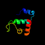
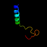
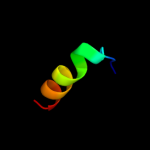
| 1 |
|
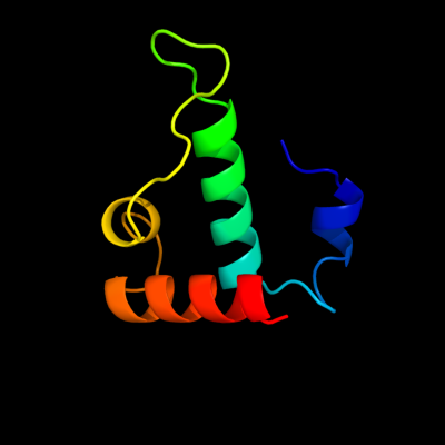
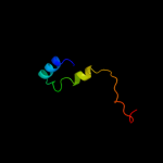
PDB 2v79 chain B
Region: 114 - 181
Aligned: 62
Modelled: 68
Confidence: 76.1%
Identity: 11%
PDB header:dna-binding protein
Chain: B: PDB Molecule:dna replication protein dnad;
PDBTitle: crystal structure of the n-terminal domain of dnad from2 bacillus subtilis
Phyre2
| 2 |
|
PDB 3h8d chain E
Region: 52 - 62
Aligned: 11
Modelled: 11
Confidence: 34.8%
Identity: 73%
PDB header:motor protein/signaling protein
Chain: E: PDB Molecule:disabled homolog 2;
PDBTitle: crystal structure of myosin vi in complex with dab2 peptide
Phyre2
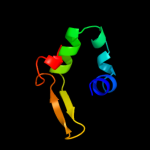
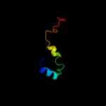
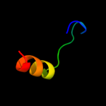
| 3 |
|
PDB 1vje chain A
Region: 170 - 203
Aligned: 34
Modelled: 34
Confidence: 33.6%
Identity: 24%
Fold: LuxS/MPP-like metallohydrolase
Superfamily: LuxS/MPP-like metallohydrolase
Family: Autoinducer-2 production protein LuxS
Phyre2
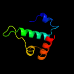
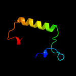
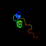
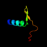
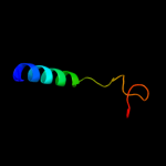
| 4 |
|
PDB 2vn2 chain B
Region: 114 - 180
Aligned: 61
Modelled: 67
Confidence: 32.4%
Identity: 20%
PDB header:replication
Chain: B: PDB Molecule:chromosome replication initiation protein;
PDBTitle: crystal structure of the n-terminal domain of dnad protein2 from geobacillus kaustophilus hta426
Phyre2
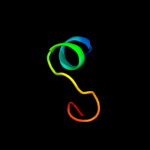
| 5 |
|
PDB 2r7c chain A domain 2
Region: 7 - 53
Aligned: 47
Modelled: 47
Confidence: 23.6%
Identity: 28%
Fold: Rotavirus NSP2 fragment, N-terminal domain
Superfamily: Rotavirus NSP2 fragment, N-terminal domain
Family: Rotavirus NSP2 fragment, N-terminal domain
Phyre2
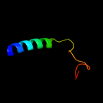
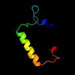
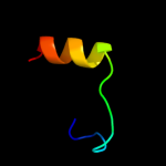
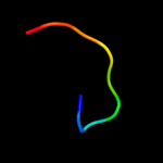
| 6 |
|
PDB 2k5c chain A
Region: 34 - 85
Aligned: 52
Modelled: 52
Confidence: 14.4%
Identity: 31%
PDB header:metal binding protein
Chain: A: PDB Molecule:uncharacterized protein pf0385;
PDBTitle: nmr structure for pf0385
Phyre2
| 7 |
|
PDB 2r7c chain A
Region: 7 - 53
Aligned: 47
Modelled: 47
Confidence: 10.2%
Identity: 28%
PDB header:rna binding protein
Chain: A: PDB Molecule:non-structural rna-binding protein 35;
PDBTitle: crystallographic and biochemical analysis of rotavirus nsp22 with nucleotides reveals an ndp kinase like activity
Phyre2
| 8 |
|
PDB 1j6x chain A
Region: 170 - 206
Aligned: 36
Modelled: 37
Confidence: 8.8%
Identity: 19%
Fold: LuxS/MPP-like metallohydrolase
Superfamily: LuxS/MPP-like metallohydrolase
Family: Autoinducer-2 production protein LuxS
Phyre2
| 9 |
|
PDB 3zqu chain A
Region: 103 - 129
Aligned: 22
Modelled: 27
Confidence: 8.7%
Identity: 41%
PDB header:lyase
Chain: A: PDB Molecule:probable aromatic acid decarboxylase;
PDBTitle: structure of a probable aromatic acid decarboxylase
Phyre2
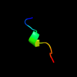
| 10 |
|
PDB 2fmm chain E domain 1
Region: 129 - 171
Aligned: 41
Modelled: 43
Confidence: 7.9%
Identity: 32%
Fold: ENT-like
Superfamily: ENT-like
Family: Emsy N terminal (ENT) domain-like
Phyre2
| 11 |
|
PDB 3iyd chain E
Region: 170 - 185
Aligned: 16
Modelled: 16
Confidence: 7.4%
Identity: 38%
PDB header:transcription/dna
Chain: E: PDB Molecule:dna-directed rna polymerase subunit omega;
PDBTitle: three-dimensional em structure of an intact activator-dependent2 transcription initiation complex
Phyre2
| 12 |
|
PDB 2fmm chain E
Region: 129 - 171
Aligned: 41
Modelled: 43
Confidence: 7.1%
Identity: 32%
PDB header:transcription
Chain: E: PDB Molecule:protein emsy;
PDBTitle: crystal structure of emsy-hp1 complex
Phyre2
| 13 |
|
PDB 1p6r chain A
Region: 166 - 204
Aligned: 39
Modelled: 39
Confidence: 6.9%
Identity: 31%
Fold: DNA/RNA-binding 3-helical bundle
Superfamily: "Winged helix" DNA-binding domain
Family: Penicillinase repressor
Phyre2
| 14 |
|
PDB 2j9w chain B
Region: 39 - 60
Aligned: 22
Modelled: 22
Confidence: 6.8%
Identity: 32%
PDB header:protein transport
Chain: B: PDB Molecule:vps28-prov protein;
PDBTitle: structural insight into the escrt-i-ii link and its role in2 mvb trafficking
Phyre2
| 15 |
|
PDB 2j9u chain A domain 1
Region: 39 - 60
Aligned: 22
Modelled: 22
Confidence: 6.8%
Identity: 14%
Fold: Four-helical up-and-down bundle
Superfamily: VPS28 C-terminal domain-like
Family: VPS28 C-terminal domain-like
Phyre2
| 16 |
|
PDB 3dom chain C
Region: 42 - 59
Aligned: 18
Modelled: 18
Confidence: 5.6%
Identity: 22%
PDB header:transcription
Chain: C: PDB Molecule:rna polymerase ii transcription factor b subunit 2;
PDBTitle: crystal structure of the complex between tfb5 and the c-terminal2 domain of tfb2
Phyre2
| 17 |
|
PDB 1j6w chain A
Region: 170 - 203
Aligned: 34
Modelled: 34
Confidence: 5.5%
Identity: 18%
Fold: LuxS/MPP-like metallohydrolase
Superfamily: LuxS/MPP-like metallohydrolase
Family: Autoinducer-2 production protein LuxS
Phyre2
| 18 |
|
PDB 9wga chain A domain 2
Region: 148 - 156
Aligned: 9
Modelled: 9
Confidence: 5.5%
Identity: 56%
Fold: Knottins (small inhibitors, toxins, lectins)
Superfamily: Plant lectins/antimicrobial peptides
Family: Hevein-like agglutinin (lectin) domain
Phyre2
| 19 |
|
PDB 2cwg chain A domain 2
Region: 148 - 156
Aligned: 9
Modelled: 9
Confidence: 5.3%
Identity: 56%
Fold: Knottins (small inhibitors, toxins, lectins)
Superfamily: Plant lectins/antimicrobial peptides
Family: Hevein-like agglutinin (lectin) domain
Phyre2
| 20 |
|
PDB 2gu0 chain A
Region: 7 - 16
Aligned: 10
Modelled: 10
Confidence: 5.0%
Identity: 70%
PDB header:viral protein
Chain: A: PDB Molecule:nonstructural protein 2;
PDBTitle: crystal structure of human rotavirus nsp2 (group c /2 bristol strain)
Phyre2