Phyre Home Retrieve Phyre Job Id
Emaill.a.kelley@imperial.ac.uk
DescriptionP64548
DateThu Jan 5 12:09:21 GMT 2012
Unique Job IDa3b6bf063340b2f9

Summary 

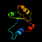
Top model
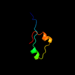
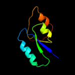
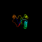
Image coloured by rainbow N → C terminus
Model (left) based on template c2qh8A_
Top template information
PDB header:structural genomics, unknown function
Chain: A: PDB Molecule:uncharacterized protein;
PDBTitle: crystal structure of conserved domain protein from vibrio2 cholerae o1 biovar eltor str. n16961
Confidence and coverage
Confidence: 38.9% Coverage: 20%
35 residues ( 20% of your sequence) have been modelled with 38.9% confidence by the single highest scoring template.
You may wish to submit your sequence to Phyrealarm. This will automatically scan your sequence every week for new potential templates as they appear in the Phyre2 library.
Please note: You must be registered and logged in to use Phyrealarm.
3D viewing
Interactive 3D view in Jmol

Sequence analysis 

Secondary structure and disorder prediction 

   1........10.........20.........30.........40.........50.........60
Sequence  MRFSHRLFLLLILLLTGAPILAQEPSDVAKNVRMMVSGIVSYTRWPALSGPPKLCIFSSS
Secondary structure 


SS confidence 


Disorder  ????


?????????


Disorder confidence 


   .........70.........80.........90.........100.........110.........120
Sequence  RFSTALQENAATSLPYLPVIIHTQQEAMISGCNGFYFGNESPTFQMELTEQYPSKALLLI
Secondary structure 


SS confidence 


Disorder 


??????????


??????


????


Disorder confidence 


   .........130.........140.........150.........160.........170..
Sequence  AEQNTECIIGSAFCLIIHNNDVRFAVNLDALSRSGVKVNPDVLMLARKKNDG
Secondary structure 


SS confidence 


Disorder 


?


????

?


???????
Disorder confidence 


Confidence Key
High(9)                    Low (0)
?Disordered
Alpha helix
Beta strand

Domain analysis 

Hover over an aligned region to see model and summary info

Please note, only up to the top 20 hits are modelled to reduce computer load

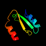
RankAligned region

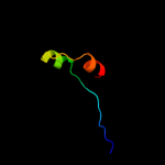
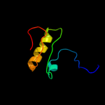
PDB 2qh8 chain A

3D model

Region: 136 - 170
Aligned: 35
Modelled: 35
Confidence: 38.9%
Identity: 17%
PDB header:structural genomics, unknown function
Chain: A: PDB Molecule:uncharacterized protein;
PDBTitle: crystal structure of conserved domain protein from vibrio2 cholerae o1 biovar eltor str. n16961

Phyre2

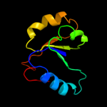
PDB 3lft chain A

3D model

Region: 136 - 166
Aligned: 31
Modelled: 31
Confidence: 34.6%
Identity: 16%
PDB header:structure genomics, unknown function
Chain: A: PDB Molecule:uncharacterized protein;
PDBTitle: the crystal structure of the abc domain in complex with l-trp from2 streptococcus pneumonia to 1.35a

Phyre2

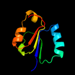
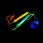
PDB 1iib chain A

3D model

Region: 52 - 131
Aligned: 79
Modelled: 80
Confidence: 29.9%
Identity: 10%
Fold: Phosphotyrosine protein phosphatases I-like
Superfamily: PTS system IIB component-like
Family: PTS system, Lactose/Cellobiose specific IIB subunit

Phyre2

PDB 2hjs chain A domain 1

3D model

Region: 50 - 126
Aligned: 76
Modelled: 77
Confidence: 26.5%
Identity: 11%
Fold: NAD(P)-binding Rossmann-fold domains
Superfamily: NAD(P)-binding Rossmann-fold domains
Family: Glyceraldehyde-3-phosphate dehydrogenase-like, N-terminal domain

Phyre2

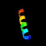
PDB 2ljc chain A

3D model

Region: 28 - 45
Aligned: 18
Modelled: 18
Confidence: 22.5%
Identity: 22%
PDB header:transport protein/inhibitor
Chain: A: PDB Molecule:m2 protein, bm2 protein chimera;
PDBTitle: structure of the influenza am2-bm2 chimeric channel bound to2 rimantadine

Phyre2

PDB 1pjq chain A domain 1

3D model

Region: 73 - 128
Aligned: 56
Modelled: 56
Confidence: 19.9%
Identity: 9%
Fold: NAD(P)-binding Rossmann-fold domains
Superfamily: NAD(P)-binding Rossmann-fold domains
Family: Siroheme synthase N-terminal domain-like

Phyre2

PDB 2hu9 chain B

3D model

Region: 90 - 101
Aligned: 12
Modelled: 12
Confidence: 19.0%
Identity: 25%
PDB header:metal transport
Chain: B: PDB Molecule:mercuric transport protein periplasmic component;
PDBTitle: x-ray structure of the archaeoglobus fulgidus copz n-2 terminal domain

Phyre2

PDB 2hwk chain A

3D model

Region: 106 - 149
Aligned: 42
Modelled: 44
Confidence: 18.3%
Identity: 14%
PDB header:hydrolase
Chain: A: PDB Molecule:helicase nsp2;
PDBTitle: crystal structure of venezuelan equine encephalitis2 alphavirus nsp2 protease domain

Phyre2

PDB 2abw chain A domain 1

3D model

Region: 50 - 121
Aligned: 72
Modelled: 72
Confidence: 17.8%
Identity: 11%
Fold: Flavodoxin-like
Superfamily: Class I glutamine amidotransferase-like
Family: Class I glutamine amidotransferases (GAT)

Phyre2

PDB 3cu0 chain A domain 1

3D model

Region: 94 - 163
Aligned: 64
Modelled: 70
Confidence: 16.3%
Identity: 25%
Fold: Nucleotide-diphospho-sugar transferases
Superfamily: Nucleotide-diphospho-sugar transferases
Family: 1,3-glucuronyltransferase

Phyre2

PDB 1k9v chain F

3D model

Region: 52 - 121
Aligned: 70
Modelled: 70
Confidence: 15.3%
Identity: 6%
Fold: Flavodoxin-like
Superfamily: Class I glutamine amidotransferase-like
Family: Class I glutamine amidotransferases (GAT)

Phyre2

PDB 2bs3 chain A

3D model

Region: 88 - 132
Aligned: 45
Modelled: 45
Confidence: 11.1%
Identity: 9%
PDB header:oxidoreductase
Chain: A: PDB Molecule:quinol-fumarate reductase flavoprotein subunit a;
PDBTitle: glu c180 -> gln variant quinol:fumarate reductase from2 wolinella succinogenes

Phyre2

PDB 2l2q chain A

3D model

Region: 52 - 131
Aligned: 79
Modelled: 80
Confidence: 11.1%
Identity: 11%
PDB header:transferase
Chain: A: PDB Molecule:pts system, cellobiose-specific iib component (cela);
PDBTitle: solution structure of cellobiose-specific phosphotransferase iib2 component protein from borrelia burgdorferi

Phyre2

PDB 1wvh chain A domain 1

3D model

Region: 90 - 101
Aligned: 12
Modelled: 12
Confidence: 10.4%
Identity: 17%
Fold: PH domain-like barrel
Superfamily: PH domain-like
Family: Phosphotyrosine-binding domain (PTB)

Phyre2

PDB 2h1q chain A domain 1

3D model

Region: 53 - 127
Aligned: 75
Modelled: 75
Confidence: 10.3%
Identity: 13%
Fold: PLP-dependent transferase-like
Superfamily: Dhaf3308-like
Family: Dhaf3308-like

Phyre2

PDB 2fwt chain A

3D model

Region: 90 - 115
Aligned: 26
Modelled: 26
Confidence: 10.0%
Identity: 19%
PDB header:electron transport
Chain: A: PDB Molecule:dhc, diheme cytochrome c;
PDBTitle: crystal structure of dhc purified from rhodobacter2 sphaeroides

Phyre2

PDB 3c85 chain A

3D model

Region: 46 - 124
Aligned: 79
Modelled: 76
Confidence: 9.1%
Identity: 16%
PDB header:transport protein
Chain: A: PDB Molecule:putative glutathione-regulated potassium-efflux system
PDBTitle: crystal structure of trka domain of putative glutathione-regulated2 potassium-efflux kefb from vibrio parahaemolyticus

Phyre2

PDB 2ioj chain A domain 1

3D model

Region: 95 - 170
Aligned: 61
Modelled: 61
Confidence: 8.9%
Identity: 23%
Fold: MurF and HprK N-domain-like
Superfamily: HprK N-terminal domain-like
Family: DRTGG domain

Phyre2

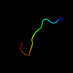
PDB 2x77 chain B

3D model

Region: 34 - 63
Aligned: 29
Modelled: 30
Confidence: 8.0%
Identity: 7%
PDB header:gtp-binding protein
Chain: B: PDB Molecule:adp-ribosylation factor;
PDBTitle: crystal structure of leishmania major adp ribosylation2 factor-like 1.

Phyre2

PDB 2qn6 chain A domain 3

3D model

Region: 45 - 63
Aligned: 19
Modelled: 19
Confidence: 8.0%
Identity: 11%
Fold: P-loop containing nucleoside triphosphate hydrolases
Superfamily: P-loop containing nucleoside triphosphate hydrolases
Family: G proteins

Phyre2
1

c2qh8A_
2

c3lftA_
3

d1iiba_
4

d2hjsa1
5

c2ljcA_
6

d1pjqa1
7

c2hu9B_
8

c2hwkA_
9

d2abwa1
10

d3cu0a1
11

d1k9vf_
12

c2bs3A_
13

c2l2qA_
14

d1wvha1
15

d2h1qa1
16

c2fwtA_
17

c3c85A_
18

d2ioja1
19

c2x77B_
20

d2qn6a3
21


22


23


24


25


26


27


28


29


30


31


32


33


34


Detailed template information 

#
Template Alignment Coverage3D Model Confidence
% i.d. Template Information
1c2qh8A_


38.9 17 PDB header:structural genomics, unknown function
Chain: A: PDB Molecule:uncharacterized protein;
PDBTitle: crystal structure of conserved domain protein from vibrio2 cholerae o1 biovar eltor str. n16961
2c3lftA_


34.6 16 PDB header:structure genomics, unknown function
Chain: A: PDB Molecule:uncharacterized protein;
PDBTitle: the crystal structure of the abc domain in complex with l-trp from2 streptococcus pneumonia to 1.35a
3d1iiba_


29.9 10 Fold:Phosphotyrosine protein phosphatases I-like
Superfamily:PTS system IIB component-like
Family:PTS system, Lactose/Cellobiose specific IIB subunit
4d2hjsa1


26.5 11 Fold:NAD(P)-binding Rossmann-fold domains
Superfamily:NAD(P)-binding Rossmann-fold domains
Family:Glyceraldehyde-3-phosphate dehydrogenase-like, N-terminal domain
5c2ljcA_


22.5 22 PDB header:transport protein/inhibitor
Chain: A: PDB Molecule:m2 protein, bm2 protein chimera;
PDBTitle: structure of the influenza am2-bm2 chimeric channel bound to2 rimantadine
6d1pjqa1


19.9 9 Fold:NAD(P)-binding Rossmann-fold domains
Superfamily:NAD(P)-binding Rossmann-fold domains
Family:Siroheme synthase N-terminal domain-like
7c2hu9B_


19.0 25 PDB header:metal transport
Chain: B: PDB Molecule:mercuric transport protein periplasmic component;
PDBTitle: x-ray structure of the archaeoglobus fulgidus copz n-2 terminal domain
8c2hwkA_


18.3 14 PDB header:hydrolase
Chain: A: PDB Molecule:helicase nsp2;
PDBTitle: crystal structure of venezuelan equine encephalitis2 alphavirus nsp2 protease domain
9d2abwa1


17.8 11 Fold:Flavodoxin-like
Superfamily:Class I glutamine amidotransferase-like
Family:Class I glutamine amidotransferases (GAT)
10d3cu0a1


16.3 25 Fold:Nucleotide-diphospho-sugar transferases
Superfamily:Nucleotide-diphospho-sugar transferases
Family:1,3-glucuronyltransferase
11d1k9vf_


15.3 6 Fold:Flavodoxin-like
Superfamily:Class I glutamine amidotransferase-like
Family:Class I glutamine amidotransferases (GAT)
12c2bs3A_


11.1 9 PDB header:oxidoreductase
Chain: A: PDB Molecule:quinol-fumarate reductase flavoprotein subunit a;
PDBTitle: glu c180 -> gln variant quinol:fumarate reductase from2 wolinella succinogenes
13c2l2qA_


11.1 11 PDB header:transferase
Chain: A: PDB Molecule:pts system, cellobiose-specific iib component (cela);
PDBTitle: solution structure of cellobiose-specific phosphotransferase iib2 component protein from borrelia burgdorferi
14d1wvha1


10.4 17 Fold:PH domain-like barrel
Superfamily:PH domain-like
Family:Phosphotyrosine-binding domain (PTB)
15d2h1qa1


10.3 13 Fold:PLP-dependent transferase-like
Superfamily:Dhaf3308-like
Family:Dhaf3308-like
16c2fwtA_


10.0 19 PDB header:electron transport
Chain: A: PDB Molecule:dhc, diheme cytochrome c;
PDBTitle: crystal structure of dhc purified from rhodobacter2 sphaeroides
17c3c85A_


9.1 16 PDB header:transport protein
Chain: A: PDB Molecule:putative glutathione-regulated potassium-efflux system
PDBTitle: crystal structure of trka domain of putative glutathione-regulated2 potassium-efflux kefb from vibrio parahaemolyticus
18d2ioja1


8.9 23 Fold:MurF and HprK N-domain-like
Superfamily:HprK N-terminal domain-like
Family:DRTGG domain
19c2x77B_


8.0 7 PDB header:gtp-binding protein
Chain: B: PDB Molecule:adp-ribosylation factor;
PDBTitle: crystal structure of leishmania major adp ribosylation2 factor-like 1.
20d2qn6a3


8.0 11 Fold:P-loop containing nucleoside triphosphate hydrolases
Superfamily:P-loop containing nucleoside triphosphate hydrolases
Family:G proteins
21c3doeA_


not modelled 7.9 8 PDB header:signaling protein/hydrolase
Chain: A: PDB Molecule:adp-ribosylation factor-like protein 2;
PDBTitle: complex of arl2 and bart, crystal form 1
22d2imra1


not modelled 7.0 45 Fold:Composite domain of metallo-dependent hydrolases
Superfamily:Composite domain of metallo-dependent hydrolases
Family:DR0824-like
23d2gp4a1


not modelled 6.8 15 Fold:The "swivelling" beta/beta/alpha domain
Superfamily:LeuD/IlvD-like
Family:IlvD/EDD C-terminal domain-like
24c2d0jD_


not modelled 6.7 22 PDB header:transferase
Chain: D: PDB Molecule:galactosylgalactosylxylosylprotein 3-beta-
PDBTitle: crystal structure of human glcat-s apo form
25c2dc1A_


not modelled 6.6 6 PDB header:oxidoreductase
Chain: A: PDB Molecule:l-aspartate dehydrogenase;
PDBTitle: crystal structure of l-aspartate dehydrogenase from2 hyperthermophilic archaeon archaeoglobus fulgidus
26c3c1aB_


not modelled 6.3 14 PDB header:oxidoreductase
Chain: B: PDB Molecule:putative oxidoreductase;
PDBTitle: crystal structure of a putative oxidoreductase (zp_00056571.1) from2 magnetospirillum magnetotacticum ms-1 at 1.85 a resolution
27c4a1eF_


not modelled 6.2 33 PDB header:ribosome
Chain: F: PDB Molecule:rpl7a;
PDBTitle: t.thermophila 60s ribosomal subunit in complex with2 initiation factor 6. this file contains 5s rrna, 5.8s rrna3 and proteins of molecule 1
28d1cjba_


not modelled 6.2 15 Fold:PRTase-like
Superfamily:PRTase-like
Family:Phosphoribosyltransferases (PRTases)
29c1yq4A_


not modelled 5.8 12 PDB header:oxidoreductase
Chain: A: PDB Molecule:succinate dehydrogenase flavoprotein subunit;
PDBTitle: avian respiratory complex ii with 3-nitropropionate and ubiquinone
30c3db0B_


not modelled 5.8 7 PDB header:oxidoreductase
Chain: B: PDB Molecule:lin2891 protein;
PDBTitle: crystal structure of putative pyridoxamine 5'-phosphate oxidase2 (np_472219.1) from listeria innocua at 2.00 a resolution
31c2ywjA_


not modelled 5.8 9 PDB header:transferase
Chain: A: PDB Molecule:glutamine amidotransferase subunit pdxt;
PDBTitle: crystal structure of uncharacterized conserved protein from2 methanocaldococcus jannaschii
32c2yueA_


not modelled 5.6 16 PDB header:rna binding protein
Chain: A: PDB Molecule:protein neuralized;
PDBTitle: solution structure of the neuz (nhr) domain in neuralized2 from drosophila melanogaster
33c3k6jA_


not modelled 5.6 21 PDB header:oxidoreductase
Chain: A: PDB Molecule:protein f01g10.3, confirmed by transcript evidence;
PDBTitle: crystal structure of the dehydrogenase part of multifuctional enzyme 12 from c.elegans
34d1gq2a2


not modelled 5.1 7 Fold:Aminoacid dehydrogenase-like, N-terminal domain
Superfamily:Aminoacid dehydrogenase-like, N-terminal domain
Family:Malic enzyme N-domain

Binding site prediction 

Due to computational demand, binding site predictions are not run for batch jobs

If you want to predict binding sites, please manually submit your model of choice to 3DLigandSite


Phyre is for academic use only

Please cite: Protein structure prediction on the web: a case study using the Phyre server
Kelley LA and Sternberg MJE. Nature Protocols 4, 363 - 371 (2009) [pdf] [Import into BibTeX]
 
If you use the binding site predictions from 3DLigandSite, please also cite:
3DLigandSite: predicting ligand-binding sites using similar structures.
Wass MN, Kelley LA and Sternberg MJ Nucleic Acids Research 38, W469-73 (2010) [PubMed]
 
© Structural Bioinformatics Group
Imperial College London
Lawrence Kelley, Benjamin Jefferys
Disclaimer
Terms and Conditions
Component software
Template detection: HHpred 1.51
Secondary structure prediction: Psi-pred 2.5
Disorder prediction: Disopred 2.4
Transmembrane prediction: Memsat_SVM
Multi-template modelling and ab initio: Poing 1.0