74 residues ( 24% of your sequence) have been modelled with 97.7% confidence by the single highest scoring template.
You may wish to submit your sequence to Phyrealarm. This will automatically scan your sequence every week for new potential templates as they appear in the Phyre2 library.
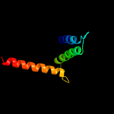
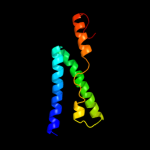
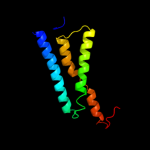
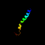
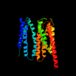
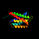
Region: 228 - 291 Aligned: 58 Modelled: 64 Confidence: 84.2% Identity: 22% PDB header:transport protein Chain: B: PDB Molecule:protein emre; PDBTitle: cryo-em based theoretical model structure of transmembrane2 domain of the multidrug-resistance antiporter from e. coli3 emre
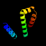
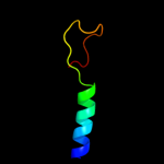
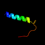
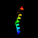
Region: 276 - 307 Aligned: 32 Modelled: 32 Confidence: 22.8% Identity: 16% PDB header:transcription Chain: A: PDB Molecule:fxyd domain-containing ion transport regulator 4; PDBTitle: solution structure of the human fxyd4 (chif) protein in sds2 micelles
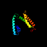
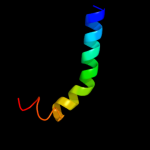
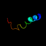
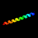
Region: 275 - 307 Aligned: 33 Modelled: 33 Confidence: 9.5% Identity: 3% PDB header:membrane protein Chain: D: PDB Molecule:kcsa channel; PDBTitle: cs+ complex of a k channel with an amide to ester substitution in the2 selectivity filter
Phyre2
21
22
23
24
25
26
27
28
29
Detailed template information
Binding site prediction
Due to computational demand, binding site predictions are not run for batch jobs
If you want to predict binding sites, please manually submit your model of choice to 3DLigandSite
Phyre is for academic use only
Please cite: Protein structure prediction on
the web: a case study using the Phyre server
Kelley LA and Sternberg MJE. Nature Protocols
4, 363 - 371 (2009) [pdf] [Import into BibTeX]
If you use the binding site
predictions from 3DLigandSite, please also cite:
3DLigandSite: predicting ligand-binding sites using similar structures.
Wass MN, Kelley LA and Sternberg
MJ Nucleic Acids Research 38, W469-73 (2010) [PubMed]