Phyre Home Retrieve Phyre Job Id
Emaill.a.kelley@imperial.ac.uk
DescriptionQ9S4X3
DateThu Jan 5 12:38:27 GMT 2012
Unique Job ID9b6a37a703d5ebff

Summary 

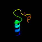
Top model
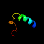
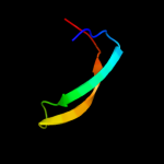
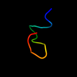
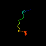
Image coloured by rainbow N → C terminus
Model (left) based on template d1keaa_
Top template information
Fold:DNA-glycosylase
Superfamily:DNA-glycosylase
Family:Mismatch glycosylase
Confidence and coverage
Confidence: 38.2% Coverage: 30%
30 residues ( 30% of your sequence) have been modelled with 38.2% confidence by the single highest scoring template.
You may wish to submit your sequence to Phyrealarm. This will automatically scan your sequence every week for new potential templates as they appear in the Phyre2 library.
Please note: You must be registered and logged in to use Phyrealarm.
3D viewing
Interactive 3D view in Jmol

Sequence analysis 

Secondary structure and disorder prediction 

   1........10.........20.........30.........40.........50.........60
Sequence  MTETGGQPPVSFPVKDVAGLLFLLRRLTRRGRNAACGQCPASGWARDGRFYRSRLASVTV
Secondary structure 


SS confidence 


Disorder  ???????


??

?????
???????


Disorder confidence 


   .........70.........80.........90.........100.
Sequence  YASPSPFSDERPSSRFRGIFSPSKRRRLRYSTVGLTRYRTR
Secondary structure 


SS confidence 


Disorder 


?????


?

??


??


???????
Disorder confidence 


Confidence Key
High(9)                    Low (0)
?Disordered
Alpha helix
Beta strand

Domain analysis 

Hover over an aligned region to see model and summary info

Please note, only up to the top 20 hits are modelled to reduce computer load

RankAligned region

PDB 1kea chain A

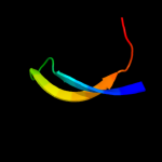
3D model

Region: 15 - 44
Aligned: 30
Modelled: 30
Confidence: 38.2%
Identity: 20%
Fold: DNA-glycosylase
Superfamily: DNA-glycosylase
Family: Mismatch glycosylase

Phyre2

PDB 1u61 chain A

3D model

Region: 25 - 43
Aligned: 19
Modelled: 19
Confidence: 23.2%
Identity: 42%
Fold: RNase III domain-like
Superfamily: RNase III domain-like
Family: RNase III catalytic domain-like

Phyre2

PDB 1rrq chain A

3D model

Region: 15 - 44
Aligned: 30
Modelled: 30
Confidence: 21.9%
Identity: 23%
PDB header:hydrolase/dna
Chain: A: PDB Molecule:muty;
PDBTitle: muty adenine glycosylase in complex with dna containing an2 a:oxog pair

Phyre2

PDB 2qvt chain A

3D model

Region: 49 - 60
Aligned: 12
Modelled: 12
Confidence: 20.9%
Identity: 75%
PDB header:unknown function
Chain: A: PDB Molecule:avrl567-d;
PDBTitle: structure of melampsora lini avirulence protein, avrl567-d

Phyre2

PDB 1mhn chain A

3D model

Region: 32 - 59
Aligned: 28
Modelled: 28
Confidence: 19.7%
Identity: 25%
Fold: SH3-like barrel
Superfamily: Tudor/PWWP/MBT
Family: Tudor domain

Phyre2

PDB 1orn chain A

3D model

Region: 15 - 49
Aligned: 35
Modelled: 35
Confidence: 18.3%
Identity: 17%
Fold: DNA-glycosylase
Superfamily: DNA-glycosylase
Family: Endonuclease III

Phyre2

PDB 2y8s chain E

3D model

Region: 1 - 11
Aligned: 11
Modelled: 11
Confidence: 18.1%
Identity: 64%
PDB header:membrane protein
Chain: E: PDB Molecule:rhoptry neck protein 2;
PDBTitle: co-structure of an ama1 mutant (y230a) with a surface2 exposed region of ron2 from toxoplasma gondii

Phyre2

PDB 2abk chain A

3D model

Region: 15 - 43
Aligned: 29
Modelled: 29
Confidence: 17.2%
Identity: 31%
Fold: DNA-glycosylase
Superfamily: DNA-glycosylase
Family: Endonuclease III

Phyre2

PDB 3n5n chain X

3D model

Region: 15 - 46
Aligned: 32
Modelled: 32
Confidence: 17.1%
Identity: 22%
PDB header:hydrolase
Chain: X: PDB Molecule:a/g-specific adenine dna glycosylase;
PDBTitle: crystal structure analysis of the catalytic domain and interdomain2 connector of human muty homologue

Phyre2

PDB 4a4f chain A

3D model

Region: 37 - 59
Aligned: 23
Modelled: 23
Confidence: 14.3%
Identity: 22%
PDB header:rna binding protein
Chain: A: PDB Molecule:survival of motor neuron-related-splicing factor 30;
PDBTitle: solution structure of spf30 tudor domain in complex with2 symmetrically dimethylated arginine

Phyre2

PDB 1g5v chain A

3D model

Region: 37 - 59
Aligned: 23
Modelled: 23
Confidence: 14.2%
Identity: 30%
PDB header:translation
Chain: A: PDB Molecule:survival motor neuron protein 1;
PDBTitle: solution structure of the tudor domain of the human smn2 protein

Phyre2

PDB 1kg2 chain A

3D model

Region: 14 - 43
Aligned: 30
Modelled: 30
Confidence: 12.6%
Identity: 17%
Fold: DNA-glycosylase
Superfamily: DNA-glycosylase
Family: Mismatch glycosylase

Phyre2

PDB 3ush chain B

3D model

Region: 34 - 48
Aligned: 15
Modelled: 15
Confidence: 12.0%
Identity: 47%
PDB header:structural genomics, unknown function
Chain: B: PDB Molecule:uncharacterized protein;
PDBTitle: crystal structure of the q2s0r5 protein from salinibacter ruber,2 northeast structural genomics consortium target srr207

Phyre2

PDB 2d9t chain A domain 1

3D model

Region: 37 - 59
Aligned: 23
Modelled: 23
Confidence: 11.4%
Identity: 22%
Fold: SH3-like barrel
Superfamily: Tudor/PWWP/MBT
Family: Tudor domain

Phyre2

PDB 2d9t chain A

3D model

Region: 44 - 59
Aligned: 16
Modelled: 16
Confidence: 10.6%
Identity: 25%
PDB header:structural genomics, unknown function
Chain: A: PDB Molecule:tudor domain-containing protein 3;
PDBTitle: solution structure of the tudor domain of tudor domain2 containing protein 3 from mouse

Phyre2

PDB 2aqa chain A domain 1

3D model

Region: 57 - 80
Aligned: 24
Modelled: 24
Confidence: 10.5%
Identity: 33%
Fold: Rubredoxin-like
Superfamily: Nop10-like SnoRNP
Family: Nucleolar RNA-binding protein Nop10-like

Phyre2

PDB 2ey4 chain E domain 1

3D model

Region: 57 - 77
Aligned: 21
Modelled: 21
Confidence: 10.4%
Identity: 38%
Fold: Rubredoxin-like
Superfamily: Nop10-like SnoRNP
Family: Nucleolar RNA-binding protein Nop10-like

Phyre2

PDB 2jnv chain A

3D model

Region: 35 - 42
Aligned: 8
Modelled: 8
Confidence: 10.2%
Identity: 75%
PDB header:metal transport
Chain: A: PDB Molecule:nifu-like protein 1, chloroplast;
PDBTitle: solution structure of c-terminal domain of nifu-like2 protein from oryza sativa

Phyre2

PDB 3qii chain A

3D model

Region: 37 - 59
Aligned: 22
Modelled: 23
Confidence: 10.2%
Identity: 27%
PDB header:transcription regulator
Chain: A: PDB Molecule:phd finger protein 20;
PDBTitle: crystal structure of tudor domain 2 of human phd finger protein 20

Phyre2

PDB 2b5l chain C

3D model

Region: 35 - 41
Aligned: 7
Modelled: 7
Confidence: 9.8%
Identity: 57%
PDB header:protein binding/viral protein
Chain: C: PDB Molecule:nonstructural protein v;
PDBTitle: crystal structure of ddb1 in complex with simian virus 5 v2 protein

Phyre2
1

d1keaa_
2

d1u61a_
3

c1rrqA_
4

c2qvtA_
5

d1mhna_
6

d1orna_
7

c2y8sE_
8

d2abka_
9

c3n5nX_
10

c4a4fA_
11

c1g5vA_
12

d1kg2a_
13

c3ushB_
14

d2d9ta1
15

c2d9tA_
16

d2aqaa1
17

d2ey4e1
18

c2jnvA_
19

c3qiiA_
20

c2b5lC_
21


22


23


24


25


26


27


28


29


30


31


32


33


34


35


36


37


38


39


40


41


42


Detailed template information 

#
Template Alignment Coverage3D Model Confidence
% i.d. Template Information
1d1keaa_


38.2 20 Fold:DNA-glycosylase
Superfamily:DNA-glycosylase
Family:Mismatch glycosylase
2d1u61a_


23.2 42 Fold:RNase III domain-like
Superfamily:RNase III domain-like
Family:RNase III catalytic domain-like
3c1rrqA_


21.9 23 PDB header:hydrolase/dna
Chain: A: PDB Molecule:muty;
PDBTitle: muty adenine glycosylase in complex with dna containing an2 a:oxog pair
4c2qvtA_


20.9 75 PDB header:unknown function
Chain: A: PDB Molecule:avrl567-d;
PDBTitle: structure of melampsora lini avirulence protein, avrl567-d
5d1mhna_


19.7 25 Fold:SH3-like barrel
Superfamily:Tudor/PWWP/MBT
Family:Tudor domain
6d1orna_


18.3 17 Fold:DNA-glycosylase
Superfamily:DNA-glycosylase
Family:Endonuclease III
7c2y8sE_


18.1 64 PDB header:membrane protein
Chain: E: PDB Molecule:rhoptry neck protein 2;
PDBTitle: co-structure of an ama1 mutant (y230a) with a surface2 exposed region of ron2 from toxoplasma gondii
8d2abka_


17.2 31 Fold:DNA-glycosylase
Superfamily:DNA-glycosylase
Family:Endonuclease III
9c3n5nX_


17.1 22 PDB header:hydrolase
Chain: X: PDB Molecule:a/g-specific adenine dna glycosylase;
PDBTitle: crystal structure analysis of the catalytic domain and interdomain2 connector of human muty homologue
10c4a4fA_


14.3 22 PDB header:rna binding protein
Chain: A: PDB Molecule:survival of motor neuron-related-splicing factor 30;
PDBTitle: solution structure of spf30 tudor domain in complex with2 symmetrically dimethylated arginine
11c1g5vA_


14.2 30 PDB header:translation
Chain: A: PDB Molecule:survival motor neuron protein 1;
PDBTitle: solution structure of the tudor domain of the human smn2 protein
12d1kg2a_


12.6 17 Fold:DNA-glycosylase
Superfamily:DNA-glycosylase
Family:Mismatch glycosylase
13c3ushB_


12.0 47 PDB header:structural genomics, unknown function
Chain: B: PDB Molecule:uncharacterized protein;
PDBTitle: crystal structure of the q2s0r5 protein from salinibacter ruber,2 northeast structural genomics consortium target srr207
14d2d9ta1


11.4 22 Fold:SH3-like barrel
Superfamily:Tudor/PWWP/MBT
Family:Tudor domain
15c2d9tA_


10.6 25 PDB header:structural genomics, unknown function
Chain: A: PDB Molecule:tudor domain-containing protein 3;
PDBTitle: solution structure of the tudor domain of tudor domain2 containing protein 3 from mouse
16d2aqaa1


10.5 33 Fold:Rubredoxin-like
Superfamily:Nop10-like SnoRNP
Family:Nucleolar RNA-binding protein Nop10-like
17d2ey4e1


10.4 38 Fold:Rubredoxin-like
Superfamily:Nop10-like SnoRNP
Family:Nucleolar RNA-binding protein Nop10-like
18c2jnvA_


10.2 75 PDB header:metal transport
Chain: A: PDB Molecule:nifu-like protein 1, chloroplast;
PDBTitle: solution structure of c-terminal domain of nifu-like2 protein from oryza sativa
19c3qiiA_


10.2 27 PDB header:transcription regulator
Chain: A: PDB Molecule:phd finger protein 20;
PDBTitle: crystal structure of tudor domain 2 of human phd finger protein 20
20c2b5lC_


9.8 57 PDB header:protein binding/viral protein
Chain: C: PDB Molecule:nonstructural protein v;
PDBTitle: crystal structure of ddb1 in complex with simian virus 5 v2 protein
21d1xhja_


not modelled 9.0 75 Fold:Alpha-lytic protease prodomain-like
Superfamily:Fe-S cluster assembly (FSCA) domain-like
Family:NifU C-terminal domain-like
22c3pnwX_


not modelled 8.9 22 PDB header:protein binding/immune system
Chain: X: PDB Molecule:tudor domain-containing protein 3;
PDBTitle: crystal structure of the tudor domain of human tdrd3 in complex with2 an anti-tdrd3 fab
23c2equA_


not modelled 8.8 21 PDB header:protein binding
Chain: A: PDB Molecule:phd finger protein 20-like 1;
PDBTitle: solution structure of the tudor domain of phd finger2 protein 20-like 1
24d1en2a2


not modelled 8.6 48 Fold:Knottins (small inhibitors, toxins, lectins)
Superfamily:Plant lectins/antimicrobial peptides
Family:Hevein-like agglutinin (lectin) domain
25d1ehda2


not modelled 8.5 48 Fold:Knottins (small inhibitors, toxins, lectins)
Superfamily:Plant lectins/antimicrobial peptides
Family:Hevein-like agglutinin (lectin) domain
26d1rrqa1


not modelled 8.4 24 Fold:DNA-glycosylase
Superfamily:DNA-glycosylase
Family:Mismatch glycosylase
27d2apob1


not modelled 8.4 33 Fold:Rubredoxin-like
Superfamily:Nop10-like SnoRNP
Family:Nucleolar RNA-binding protein Nop10-like
28c2gslE_


not modelled 8.1 22 PDB header:structural genomics, unknown function
Chain: E: PDB Molecule:hypothetical protein;
PDBTitle: x-ray crystal structure of protein fn1578 from fusobacterium2 nucleatum. northeast structural genomics consortium target nr1.
29d1veha_


not modelled 8.0 50 Fold:Alpha-lytic protease prodomain-like
Superfamily:Fe-S cluster assembly (FSCA) domain-like
Family:NifU C-terminal domain-like
30d1khia2


not modelled 7.7 39 Fold:OB-fold
Superfamily:Nucleic acid-binding proteins
Family:Cold shock DNA-binding domain-like
31c2z51A_


not modelled 7.5 75 PDB header:metal transport
Chain: A: PDB Molecule:nifu-like protein 2, chloroplast;
PDBTitle: crystal structure of arabidopsis cnfu involved in iron-2 sulfur cluster biosynthesis
32d1eysh2


not modelled 7.0 46 Fold:Single transmembrane helix
Superfamily:Photosystem II reaction centre subunit H, transmembrane region
Family:Photosystem II reaction centre subunit H, transmembrane region
33c3p8dB_


not modelled 6.8 27 PDB header:protein binding
Chain: B: PDB Molecule:medulloblastoma antigen mu-mb-50.72;
PDBTitle: crystal structure of the second tudor domain of human phf20 (homodimer2 form)
34c2h2rB_


not modelled 6.0 44 PDB header:immune system
Chain: B: PDB Molecule:low affinity immunoglobulin epsilon fc receptor
PDBTitle: crystal structure of the human cd23 lectin domain, apo form
35d1v7wa2


not modelled 5.8 19 Fold:Supersandwich
Superfamily:Galactose mutarotase-like
Family:Glycosyltransferase family 36 N-terminal domain
36c1xniI_


not modelled 5.7 25 PDB header:cell cycle
Chain: I: PDB Molecule:tumor suppressor p53-binding protein 1;
PDBTitle: tandem tudor domain of 53bp1
37d1vqoi1


not modelled 5.6 50 Fold:DNA/RNA-binding 3-helical bundle
Superfamily:Ribosomal protein L11, C-terminal domain
Family:Ribosomal protein L11, C-terminal domain
38d2fug11


not modelled 5.4 29 Fold:Bromodomain-like
Superfamily:Nqo1C-terminal domain-like
Family:Nqo1C-terminal domain-like
39c3i0uA_


not modelled 5.2 27 PDB header:lyase
Chain: A: PDB Molecule:phosphothreonine lyase ospf;
PDBTitle: structure of the type iii effector/phosphothreonine lyase ospf from2 shigella flexneri
40c1vq8I_


not modelled 5.1 50 PDB header:ribosome
Chain: I: PDB Molecule:50s ribosomal protein l11p;
PDBTitle: the structure of ccda-phe-cap-bio and the antibiotic sparsomycin bound2 to the large ribosomal subunit of haloarcula marismortui
41d2p8ta1


not modelled 5.1 43 Fold:DNA/RNA-binding 3-helical bundle
Superfamily:"Winged helix" DNA-binding domain
Family:PH0730 N-terminal domain-like
42d1twfi1


not modelled 5.1 29 Fold:Rubredoxin-like
Superfamily:Zinc beta-ribbon
Family:Transcriptional factor domain

Binding site prediction 

Due to computational demand, binding site predictions are not run for batch jobs

If you want to predict binding sites, please manually submit your model of choice to 3DLigandSite


Phyre is for academic use only

Please cite: Protein structure prediction on the web: a case study using the Phyre server
Kelley LA and Sternberg MJE. Nature Protocols 4, 363 - 371 (2009) [pdf] [Import into BibTeX]
 
If you use the binding site predictions from 3DLigandSite, please also cite:
3DLigandSite: predicting ligand-binding sites using similar structures.
Wass MN, Kelley LA and Sternberg MJ Nucleic Acids Research 38, W469-73 (2010) [PubMed]
 
© Structural Bioinformatics Group
Imperial College London
Lawrence Kelley, Benjamin Jefferys
Disclaimer
Terms and Conditions
Component software
Template detection: HHpred 1.51
Secondary structure prediction: Psi-pred 2.5
Disorder prediction: Disopred 2.4
Transmembrane prediction: Memsat_SVM
Multi-template modelling and ab initio: Poing 1.0