Phyre Home Retrieve Phyre Job Id
Emaill.a.kelley@imperial.ac.uk
DescriptionC3UPD1
DateThu Jan 5 10:56:22 GMT 2012
Unique Job ID9481017d6fde861c

Summary 

Top model
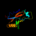
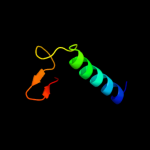
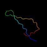
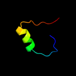
Image coloured by rainbow N → C terminus
Model (left) based on template d1p6va_
Top template information
Fold:Small protein B (SmpB)
Superfamily:Small protein B (SmpB)
Family:Small protein B (SmpB)
Confidence and coverage
Confidence:100.0% Coverage: 81%
122 residues ( 81% of your sequence) have been modelled with 100.0% confidence by the single highest scoring template.
3D viewing
Interactive 3D view in Jmol

Sequence analysis 

Secondary structure and disorder prediction 

   1........10.........20.........30.........40.........50.........60
Sequence  GSATIALNKRARHEYFIEEEFEAGLALQGWEVKSLRAGKANISDSYVLLRDGEAFLFGAN
Secondary structure 


SS confidence 


Disorder  ???????


???


?


Disorder confidence 


   .........70.........80.........90.........100.........110.........120
Sequence  ITPMAVASTHVVCDPTRTRKLLLNQRELDSLYGRVNREGYTVVALSLYWKNAWCKVKIGV
Secondary structure 


SS confidence 


Disorder 


??

?


Disorder confidence 


   .........130.........140.........150.
Sequence  AKGKKQHDKRSDIKEREWQVDKARIMKNAHR
Secondary structure 


SS confidence 


Disorder 


?


?


???
Disorder confidence 


Confidence Key
High(9)                    Low (0)
?Disordered
Alpha helix
Beta strand

Domain analysis 

Hover over an aligned region to see model and summary info

Please note, only up to the top 20 hits are modelled to reduce computer load

RankAligned region

PDB 1p6v chain A

3D model

Region: 1 - 126
Aligned: 122
Modelled: 126
Confidence: 100.0%
Identity: 50%
Fold: Small protein B (SmpB)
Superfamily: Small protein B (SmpB)
Family: Small protein B (SmpB)

Phyre2

PDB 1p6v chain C

3D model

Region: 1 - 126
Aligned: 122
Modelled: 126
Confidence: 100.0%
Identity: 50%
PDB header:rna binding protein/rna
Chain: C: PDB Molecule:ssra-binding protein;
PDBTitle: crystal structure of the trna domain of transfer-messenger2 rna in complex with smpb

Phyre2

PDB 1j1h chain A

3D model

Region: 3 - 124
Aligned: 121
Modelled: 122
Confidence: 100.0%
Identity: 46%
PDB header:rna binding protein
Chain: A: PDB Molecule:small protein b;
PDBTitle: solution structure of a tmrna-binding protein, smpb, from2 thermus thermophilus

Phyre2

PDB 1wjx chain A

3D model

Region: 5 - 124
Aligned: 111
Modelled: 120
Confidence: 100.0%
Identity: 51%
Fold: Small protein B (SmpB)
Superfamily: Small protein B (SmpB)
Family: Small protein B (SmpB)

Phyre2

PDB 2ipq chain X domain 1

3D model

Region: 18 - 57
Aligned: 40
Modelled: 40
Confidence: 34.1%
Identity: 28%
Fold: DNA/RNA-binding 3-helical bundle
Superfamily: "Winged helix" DNA-binding domain
Family: STY4665 C-terminal domain-like

Phyre2

PDB 2qnk chain A

3D model

Region: 11 - 80
Aligned: 66
Modelled: 70
Confidence: 32.5%
Identity: 33%
PDB header:oxidoreductase
Chain: A: PDB Molecule:3-hydroxyanthranilate 3,4-dioxygenase;
PDBTitle: crystal structure of human 3-hydroxyanthranilate 3,4-dioxygenase

Phyre2

PDB 2aga chain A

3D model

Region: 51 - 106
Aligned: 56
Modelled: 56
Confidence: 23.3%
Identity: 14%
PDB header:transcription
Chain: A: PDB Molecule:machado-joseph disease protein 1;
PDBTitle: de-ubiquitinating function of ataxin-3: insights from the2 solution structure of the josephin domain

Phyre2

PDB 1yfu chain A domain 1

3D model

Region: 11 - 66
Aligned: 53
Modelled: 56
Confidence: 18.0%
Identity: 19%
Fold: Double-stranded beta-helix
Superfamily: RmlC-like cupins
Family: 3-hydroxyanthranilic acid dioxygenase-like

Phyre2

PDB 1sdw chain A domain 2

3D model

Region: 21 - 59
Aligned: 39
Modelled: 39
Confidence: 17.0%
Identity: 15%
Fold: Nucleoplasmin-like/VP (viral coat and capsid proteins)
Superfamily: PHM/PNGase F
Family: Peptidylglycine alpha-hydroxylating monooxygenase, PHM

Phyre2

PDB 1wqs chain A

3D model

Region: 22 - 69
Aligned: 48
Modelled: 48
Confidence: 16.5%
Identity: 21%
PDB header:hydrolase
Chain: A: PDB Molecule:3c-like protease;
PDBTitle: crystal structure of norovirus 3c-like protease

Phyre2

PDB 1g31 chain A

3D model

Region: 10 - 34
Aligned: 22
Modelled: 25
Confidence: 14.1%
Identity: 36%
Fold: GroES-like
Superfamily: GroES-like
Family: GroES

Phyre2

PDB 3jsb chain A

3D model

Region: 88 - 105
Aligned: 18
Modelled: 18
Confidence: 11.9%
Identity: 22%
PDB header:rna binding protein
Chain: A: PDB Molecule:rna-directed rna polymerase;
PDBTitle: crystal structure of the n-terminal domain of the lymphocytic2 choriomeningitis virus l protein

Phyre2

PDB 1hf9 chain B

3D model

Region: 84 - 97
Aligned: 14
Modelled: 14
Confidence: 11.5%
Identity: 21%
PDB header:atpase inhibitor
Chain: B: PDB Molecule:atpase inhibitor (mitochondrial);
PDBTitle: c-terminal coiled-coil domain from bovine if1

Phyre2

PDB 2qfa chain A domain 1

3D model

Region: 10 - 31
Aligned: 22
Modelled: 22
Confidence: 11.4%
Identity: 27%
Fold: Inhibitor of apoptosis (IAP) repeat
Superfamily: Inhibitor of apoptosis (IAP) repeat
Family: Inhibitor of apoptosis (IAP) repeat

Phyre2

PDB 1zvf chain A domain 1

3D model

Region: 4 - 62
Aligned: 59
Modelled: 59
Confidence: 10.0%
Identity: 15%
Fold: Double-stranded beta-helix
Superfamily: RmlC-like cupins
Family: 3-hydroxyanthranilic acid dioxygenase-like

Phyre2

PDB 2quo chain A

3D model

Region: 102 - 127
Aligned: 26
Modelled: 26
Confidence: 10.0%
Identity: 27%
PDB header:toxin
Chain: A: PDB Molecule:heat-labile enterotoxin b chain;
PDBTitle: crystal structure of c terminal fragment of clostridium2 perfringens enterotoxin

Phyre2

PDB 2w3z chain A

3D model

Region: 81 - 106
Aligned: 26
Modelled: 26
Confidence: 8.9%
Identity: 23%
PDB header:hydrolase
Chain: A: PDB Molecule:putative deacetylase;
PDBTitle: structure of a streptococcus mutans ce4 esterase

Phyre2

PDB 2oa9 chain B

3D model

Region: 27 - 47
Aligned: 21
Modelled: 21
Confidence: 8.7%
Identity: 10%
PDB header:hydrolase
Chain: B: PDB Molecule:r.mvai;
PDBTitle: restriction endonuclease mvai in the absence of dna

Phyre2

PDB 2rax chain A domain 1

3D model

Region: 10 - 31
Aligned: 22
Modelled: 22
Confidence: 8.3%
Identity: 27%
Fold: Inhibitor of apoptosis (IAP) repeat
Superfamily: Inhibitor of apoptosis (IAP) repeat
Family: Inhibitor of apoptosis (IAP) repeat

Phyre2

PDB 2nly chain A domain 1

3D model

Region: 81 - 106
Aligned: 26
Modelled: 26
Confidence: 7.8%
Identity: 19%
Fold: 7-stranded beta/alpha barrel
Superfamily: Glycoside hydrolase/deacetylase
Family: Divergent polysaccharide deacetylase

Phyre2
1

d1p6va_
2

c1p6vC_
3

c1j1hA_
4

d1wjxa_
5

d2ipqx1
6

c2qnkA_
7

c2agaA_
8

d1yfua1
9

d1sdwa2
10

c1wqsA_
11

d1g31a_
12

c3jsbA_
13

c1hf9B_
14

d2qfaa1
15

d1zvfa1
16

c2quoA_
17

c2w3zA_
18

c2oa9B_
19

d2raxa1
20

d2nlya1
21


22


23


24


25


26


27


28


29


30


31


32


33


34


35


36


37


38


39


40


41


42


Detailed template information 

#
Template Alignment Coverage3D Model Confidence
% i.d. Template Information
1d1p6va_


100.0 50 Fold:Small protein B (SmpB)
Superfamily:Small protein B (SmpB)
Family:Small protein B (SmpB)
2c1p6vC_


100.0 50 PDB header:rna binding protein/rna
Chain: C: PDB Molecule:ssra-binding protein;
PDBTitle: crystal structure of the trna domain of transfer-messenger2 rna in complex with smpb
3c1j1hA_


100.0 46 PDB header:rna binding protein
Chain: A: PDB Molecule:small protein b;
PDBTitle: solution structure of a tmrna-binding protein, smpb, from2 thermus thermophilus
4d1wjxa_


100.0 51 Fold:Small protein B (SmpB)
Superfamily:Small protein B (SmpB)
Family:Small protein B (SmpB)
5d2ipqx1


34.1 28 Fold:DNA/RNA-binding 3-helical bundle
Superfamily:"Winged helix" DNA-binding domain
Family:STY4665 C-terminal domain-like
6c2qnkA_


32.5 33 PDB header:oxidoreductase
Chain: A: PDB Molecule:3-hydroxyanthranilate 3,4-dioxygenase;
PDBTitle: crystal structure of human 3-hydroxyanthranilate 3,4-dioxygenase
7c2agaA_


23.3 14 PDB header:transcription
Chain: A: PDB Molecule:machado-joseph disease protein 1;
PDBTitle: de-ubiquitinating function of ataxin-3: insights from the2 solution structure of the josephin domain
8d1yfua1


18.0 19 Fold:Double-stranded beta-helix
Superfamily:RmlC-like cupins
Family:3-hydroxyanthranilic acid dioxygenase-like
9d1sdwa2


17.0 15 Fold:Nucleoplasmin-like/VP (viral coat and capsid proteins)
Superfamily:PHM/PNGase F
Family:Peptidylglycine alpha-hydroxylating monooxygenase, PHM
10c1wqsA_


16.5 21 PDB header:hydrolase
Chain: A: PDB Molecule:3c-like protease;
PDBTitle: crystal structure of norovirus 3c-like protease
11d1g31a_


14.1 36 Fold:GroES-like
Superfamily:GroES-like
Family:GroES
12c3jsbA_


11.9 22 PDB header:rna binding protein
Chain: A: PDB Molecule:rna-directed rna polymerase;
PDBTitle: crystal structure of the n-terminal domain of the lymphocytic2 choriomeningitis virus l protein
13c1hf9B_


11.5 21 PDB header:atpase inhibitor
Chain: B: PDB Molecule:atpase inhibitor (mitochondrial);
PDBTitle: c-terminal coiled-coil domain from bovine if1
14d2qfaa1


11.4 27 Fold:Inhibitor of apoptosis (IAP) repeat
Superfamily:Inhibitor of apoptosis (IAP) repeat
Family:Inhibitor of apoptosis (IAP) repeat
15d1zvfa1


10.0 15 Fold:Double-stranded beta-helix
Superfamily:RmlC-like cupins
Family:3-hydroxyanthranilic acid dioxygenase-like
16c2quoA_


10.0 27 PDB header:toxin
Chain: A: PDB Molecule:heat-labile enterotoxin b chain;
PDBTitle: crystal structure of c terminal fragment of clostridium2 perfringens enterotoxin
17c2w3zA_


8.9 23 PDB header:hydrolase
Chain: A: PDB Molecule:putative deacetylase;
PDBTitle: structure of a streptococcus mutans ce4 esterase
18c2oa9B_


8.7 10 PDB header:hydrolase
Chain: B: PDB Molecule:r.mvai;
PDBTitle: restriction endonuclease mvai in the absence of dna
19d2raxa1


8.3 27 Fold:Inhibitor of apoptosis (IAP) repeat
Superfamily:Inhibitor of apoptosis (IAP) repeat
Family:Inhibitor of apoptosis (IAP) repeat
20d2nlya1


7.8 19 Fold:7-stranded beta/alpha barrel
Superfamily:Glycoside hydrolase/deacetylase
Family:Divergent polysaccharide deacetylase
21d2c1ia1


not modelled 7.7 24 Fold:7-stranded beta/alpha barrel
Superfamily:Glycoside hydrolase/deacetylase
Family:NodB-like polysaccharide deacetylase
22d1m4ma_


not modelled 7.7 27 Fold:Inhibitor of apoptosis (IAP) repeat
Superfamily:Inhibitor of apoptosis (IAP) repeat
Family:Inhibitor of apoptosis (IAP) repeat
23d2qqra1


not modelled 7.6 31 Fold:SH3-like barrel
Superfamily:Tudor/PWWP/MBT
Family:Tudor domain
24d1d1na_


not modelled 7.4 20 Fold:Reductase/isomerase/elongation factor common domain
Superfamily:Translation proteins
Family:Elongation factors
25d2cc0a1


not modelled 7.4 15 Fold:7-stranded beta/alpha barrel
Superfamily:Glycoside hydrolase/deacetylase
Family:NodB-like polysaccharide deacetylase
26c3mjdA_


not modelled 6.9 22 PDB header:transferase
Chain: A: PDB Molecule:orotate phosphoribosyltransferase;
PDBTitle: 1.9 angstrom crystal structure of orotate2 phosphoribosyltransferase (pyre) francisella tularensis.
27c2ylaA_


not modelled 6.4 15 PDB header:hydrolase
Chain: A: PDB Molecule:beta-n-acetylhexosaminidase;
PDBTitle: inhibition of the pneumococcal virulence factor strh and2 molecular insights into n-glycan recognition and3 hydrolysis
28d1im3d_


not modelled 6.3 28 Fold:Immunoglobulin-like beta-sandwich
Superfamily:E set domains
Family:Cytomegalovirus protein US2
29d8ohma2


not modelled 6.2 24 Fold:P-loop containing nucleoside triphosphate hydrolases
Superfamily:P-loop containing nucleoside triphosphate hydrolases
Family:RNA helicase
30d1kk1a1


not modelled 6.1 36 Fold:Reductase/isomerase/elongation factor common domain
Superfamily:Translation proteins
Family:Elongation factors
31c2z59A_


not modelled 6.0 29 PDB header:protein transport
Chain: A: PDB Molecule:protein adrm1;
PDBTitle: complex structures of mouse rpn13 (22-130aa) and ubiquitin
32c2r2yA_


not modelled 6.0 29 PDB header:protein binding
Chain: A: PDB Molecule:protein adrm1;
PDBTitle: crystal structure of the proteasomal rpn13 pru-domain
33d1jd5a_


not modelled 5.9 26 Fold:Inhibitor of apoptosis (IAP) repeat
Superfamily:Inhibitor of apoptosis (IAP) repeat
Family:Inhibitor of apoptosis (IAP) repeat
34d2ieca1


not modelled 5.7 71 Fold:MK0786-like
Superfamily:MK0786-like
Family:MK0786-like
35c2crvA_


not modelled 5.5 29 PDB header:translation
Chain: A: PDB Molecule:translation initiation factor if-2;
PDBTitle: solution structure of c-terminal domain of mitochondrial2 translational initiationfactor 2
36d2gjxa1


not modelled 5.4 15 Fold:TIM beta/alpha-barrel
Superfamily:(Trans)glycosidases
Family:beta-N-acetylhexosaminidase catalytic domain
37d2i52a1


not modelled 5.4 71 Fold:MK0786-like
Superfamily:MK0786-like
Family:MK0786-like
38d1jaka1


not modelled 5.4 15 Fold:TIM beta/alpha-barrel
Superfamily:(Trans)glycosidases
Family:beta-N-acetylhexosaminidase catalytic domain
39d1yhta1


not modelled 5.3 20 Fold:TIM beta/alpha-barrel
Superfamily:(Trans)glycosidases
Family:beta-N-acetylhexosaminidase catalytic domain
40d1mkya3


not modelled 5.2 9 Fold:Alpha-lytic protease prodomain-like
Superfamily:Probable GTPase Der, C-terminal domain
Family:Probable GTPase Der, C-terminal domain
41c2elvA_


not modelled 5.2 21 PDB header:transcription
Chain: A: PDB Molecule:zinc finger protein 406;
PDBTitle: solution structure of the 6th c2h2 zinc finger of human2 zinc finger protein 406
42c2ogfD_


not modelled 5.1 57 PDB header:structural genomics, unknown function
Chain: D: PDB Molecule:hypothetical protein mj0408;
PDBTitle: crystal structure of protein mj0408 from methanococcus jannaschii,2 pfam duf372

Binding site prediction 

Due to computational demand, binding site predictions are not run for batch jobs

If you want to predict binding sites, please manually submit your model of choice to 3DLigandSite


Phyre is for academic use only

Please cite: Protein structure prediction on the web: a case study using the Phyre server
Kelley LA and Sternberg MJE. Nature Protocols 4, 363 - 371 (2009) [pdf] [Import into BibTeX]
 
If you use the binding site predictions from 3DLigandSite, please also cite:
3DLigandSite: predicting ligand-binding sites using similar structures.
Wass MN, Kelley LA and Sternberg MJ Nucleic Acids Research 38, W469-73 (2010) [PubMed]
 
© Structural Bioinformatics Group
Imperial College London
Lawrence Kelley, Benjamin Jefferys
Disclaimer
Terms and Conditions
Component software
Template detection: HHpred 1.51
Secondary structure prediction: Psi-pred 2.5
Disorder prediction: Disopred 2.4
Transmembrane prediction: Memsat_SVM
Multi-template modelling and ab initio: Poing 1.0