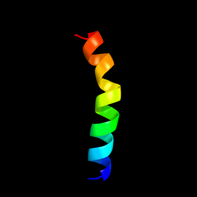
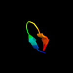
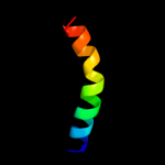
PDB header:electron transport Chain: Y: PDB Molecule:cytochrome c oxidase subunit 7c; PDBTitle: bovine heart cytochrome c oxidase re-refined with molecular2 oxygen
Confidence and coverage
Confidence:
31.2%
Coverage:
6%
23 residues ( 6% of your sequence) have been modelled with 31.2% confidence by the single highest scoring template.
You may wish to submit your sequence to Phyrealarm. This will automatically scan your sequence every week for new potential templates as they appear in the Phyre2 library.
Please note: You must be registered and logged in to use Phyrealarm.
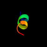
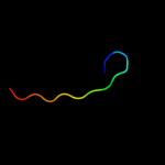
Region: 36 - 45 Aligned: 10 Modelled: 10 Confidence: 19.3% Identity: 40% Fold: Single transmembrane helix Superfamily: Photosystem II reaction centre subunit H, transmembrane region Family: Photosystem II reaction centre subunit H, transmembrane region
Region: 36 - 45 Aligned: 10 Modelled: 10 Confidence: 18.4% Identity: 40% Fold: Single transmembrane helix Superfamily: Photosystem II reaction centre subunit H, transmembrane region Family: Photosystem II reaction centre subunit H, transmembrane region
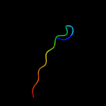
Region: 36 - 45 Aligned: 10 Modelled: 10 Confidence: 16.3% Identity: 40% Fold: Single transmembrane helix Superfamily: Photosystem II reaction centre subunit H, transmembrane region Family: Photosystem II reaction centre subunit H, transmembrane region
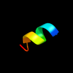
Region: 50 - 60 Aligned: 11 Modelled: 11 Confidence: 12.2% Identity: 36% Fold: DNA topoisomerase I domain Superfamily: DNA topoisomerase I domain Family: Vaccinia DNA topoisomerase I, 9 kDa N-terminal fragment
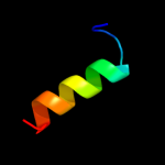
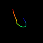
Region: 62 - 71 Aligned: 10 Modelled: 10 Confidence: 6.8% Identity: 40% PDB header:structural genomics, unknown function Chain: A: PDB Molecule:ntf2-like protein of unknown function; PDBTitle: crystal structure of ntf2-like protein of unknown function mn2a_05052 from prochlorococcus marinus (yp_291699.1) from prochlorococcus sp.3 natl2a at 1.40 a resolution
Phyre2
21
22
23
24
25
26
27
28
29
30
31
32
33
34
35
36
37
Detailed template information
Binding site prediction
Due to computational demand, binding site predictions are not run for batch jobs
If you want to predict binding sites, please manually submit your model of choice to 3DLigandSite
Phyre is for academic use only
Please cite: Protein structure prediction on
the web: a case study using the Phyre server
Kelley LA and Sternberg MJE. Nature Protocols
4, 363 - 371 (2009) [pdf] [Import into BibTeX]
If you use the binding site
predictions from 3DLigandSite, please also cite:
3DLigandSite: predicting ligand-binding sites using similar structures.
Wass MN, Kelley LA and Sternberg
MJ Nucleic Acids Research 38, W469-73 (2010) [PubMed]