Phyre Home Retrieve Phyre Job Id
Emaill.a.kelley@imperial.ac.uk
DescriptionP77307
DateThu Jan 5 12:27:34 GMT 2012
Unique Job ID806ffb3b4e4c7347

Summary 

Top model
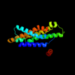
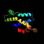
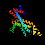
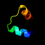
Image coloured by rainbow N → C terminus
Model (left) based on template c3k07A_
Top template information
PDB header:transport protein
Chain: A: PDB Molecule:cation efflux system protein cusa;
PDBTitle: crystal structure of cusa
Confidence and coverage
Confidence: 95.6% Coverage: 73%
189 residues ( 73% of your sequence) have been modelled with 95.6% confidence by the single highest scoring template.
Additional confident templates have been detected (see Domain analysis) which cover other regions of your sequence.
249 residues ( 96%) could be modelled at >90% confidence using multiple-templates.
You may wish to try resubmitting your sequence in "intensive" mode to model more of your sequence.
3D viewing
Interactive 3D view in Jmol

Sequence analysis 

Secondary structure and disorder prediction 

   1........10.........20.........30.........40.........50.........60
Sequence  MNSHNITNESLALALMLVVVAILISHKEKLALEKDILWSVGRAIIQLIIVGYVLKYIFSV
Secondary structure 


SS confidence 


Disorder  ???????


?
?
?


Disorder confidence 


   .........70.........80.........90.........100.........110.........120
Sequence  DDASLTLLMVLFICFNAAWNAQKRSKYIAKAFISSFIAITVGAGITLAVLILSGSIEFIP
Secondary structure 


SS confidence 


Disorder 


????
???


Disorder confidence 


   .........130.........140.........150.........160.........170.........180
Sequence  MQVIPIAGMIAGNAMVAVGLCYNNLGQRVISEQQQIQEKLSLGATPKQASAILIRDSIRA
Secondary structure 


SS confidence 


Disorder 


Disorder confidence 


   .........190.........200.........210.........220.........230.........240
Sequence  ALIPTVDSAKTVGLVSLPGMMSGLIFAGIDPVKAIKYQIMVTFMLLSTASLSTIIACYLT
Secondary structure 


SS confidence 


Disorder 


?

?


Disorder confidence 


   .........250.........
Sequence  YRKFYNSRHQLVVTQLKKK
Secondary structure 


SS confidence 


Disorder 


???????
Disorder confidence 


Confidence Key
High(9)                    Low (0)
?Disordered
Alpha helix
Beta strand

Domain analysis 

Hover over an aligned region to see model and summary info

Please note, only up to the top 20 hits are modelled to reduce computer load

RankAligned region

PDB 3k07 chain A

3D model

Region: 61 - 259
Aligned: 189
Modelled: 199
Confidence: 95.6%
Identity: 16%
PDB header:transport protein
Chain: A: PDB Molecule:cation efflux system protein cusa;
PDBTitle: crystal structure of cusa

Phyre2

PDB 3d31 chain C domain 1

3D model

Region: 11 - 237
Aligned: 221
Modelled: 224
Confidence: 93.7%
Identity: 12%
Fold: MetI-like
Superfamily: MetI-like
Family: MetI-like

Phyre2

PDB 3d31 chain D

3D model

Region: 11 - 237
Aligned: 221
Modelled: 224
Confidence: 93.7%
Identity: 12%
PDB header:transport protein
Chain: D: PDB Molecule:sulfate/molybdate abc transporter, permease
PDBTitle: modbc from methanosarcina acetivorans

Phyre2

PDB 3dhw chain A domain 1

3D model

Region: 36 - 234
Aligned: 185
Modelled: 185
Confidence: 92.9%
Identity: 12%
Fold: MetI-like
Superfamily: MetI-like
Family: MetI-like

Phyre2

PDB 2r6g chain F

3D model

Region: 63 - 186
Aligned: 124
Modelled: 124
Confidence: 88.0%
Identity: 13%
PDB header:hydrolase/transport protein
Chain: F: PDB Molecule:maltose transport system permease protein malf;
PDBTitle: the crystal structure of the e. coli maltose transporter

Phyre2

PDB 2r6g chain F domain 2

3D model

Region: 63 - 186
Aligned: 124
Modelled: 124
Confidence: 83.2%
Identity: 12%
Fold: MetI-like
Superfamily: MetI-like
Family: MetI-like

Phyre2

PDB 3fh6 chain F

3D model

Region: 63 - 186
Aligned: 124
Modelled: 124
Confidence: 80.5%
Identity: 12%
PDB header:transport protein
Chain: F: PDB Molecule:maltose transport system permease protein malf;
PDBTitle: crystal structure of the resting state maltose transporter from e.2 coli

Phyre2

PDB 2onk chain C

3D model

Region: 11 - 240
Aligned: 215
Modelled: 218
Confidence: 69.7%
Identity: 12%
PDB header:membrane protein
Chain: C: PDB Molecule:molybdate/tungstate abc transporter, permease
PDBTitle: abc transporter modbc in complex with its binding protein2 moda

Phyre2

PDB 2onk chain C domain 1

3D model

Region: 11 - 240
Aligned: 215
Modelled: 218
Confidence: 69.7%
Identity: 12%
Fold: MetI-like
Superfamily: MetI-like
Family: MetI-like

Phyre2

PDB 3aqp chain B

3D model

Region: 65 - 242
Aligned: 153
Modelled: 174
Confidence: 65.8%
Identity: 15%
PDB header:membrane protein
Chain: B: PDB Molecule:probable secdf protein-export membrane protein;
PDBTitle: crystal structure of secdf, a translocon-associated membrane protein,2 from thermus thrmophilus

Phyre2

PDB 2r6g chain G domain 1

3D model

Region: 55 - 186
Aligned: 132
Modelled: 132
Confidence: 59.3%
Identity: 11%
Fold: MetI-like
Superfamily: MetI-like
Family: MetI-like

Phyre2

PDB 1iwg chain A domain 8

3D model

Region: 59 - 201
Aligned: 123
Modelled: 129
Confidence: 39.5%
Identity: 19%
Fold: Multidrug efflux transporter AcrB transmembrane domain
Superfamily: Multidrug efflux transporter AcrB transmembrane domain
Family: Multidrug efflux transporter AcrB transmembrane domain

Phyre2

PDB 2job chain A

3D model

Region: 167 - 183
Aligned: 17
Modelled: 17
Confidence: 35.0%
Identity: 18%
PDB header:lipid binding protein
Chain: A: PDB Molecule:antilipopolysaccharide factor;
PDBTitle: solution structure of an antilipopolysaccharide factor from2 shrimp and its possible lipid a binding site

Phyre2

PDB 2gy9 chain P domain 1

3D model

Region: 152 - 176
Aligned: 25
Modelled: 25
Confidence: 34.5%
Identity: 28%
Fold: Ribosomal protein S16
Superfamily: Ribosomal protein S16
Family: Ribosomal protein S16

Phyre2

PDB 2uub chain P domain 1

3D model

Region: 152 - 177
Aligned: 26
Modelled: 26
Confidence: 32.7%
Identity: 27%
Fold: Ribosomal protein S16
Superfamily: Ribosomal protein S16
Family: Ribosomal protein S16

Phyre2

PDB 1cuk chain A domain 1

3D model

Region: 150 - 175
Aligned: 26
Modelled: 26
Confidence: 31.5%
Identity: 27%
Fold: RuvA C-terminal domain-like
Superfamily: DNA helicase RuvA subunit, C-terminal domain
Family: DNA helicase RuvA subunit, C-terminal domain

Phyre2

PDB 3bn0 chain A domain 1

3D model

Region: 152 - 177
Aligned: 26
Modelled: 26
Confidence: 27.1%
Identity: 8%
Fold: Ribosomal protein S16
Superfamily: Ribosomal protein S16
Family: Ribosomal protein S16

Phyre2

PDB 2akc chain C

3D model

Region: 126 - 162
Aligned: 37
Modelled: 37
Confidence: 24.2%
Identity: 16%
PDB header:hydrolase
Chain: C: PDB Molecule:class a nonspecific acid phosphatase phon;
PDBTitle: crystal structure of tungstate complex of the phon protein2 from s. typhimurium

Phyre2

PDB 3i5d chain C

3D model

Region: 44 - 62
Aligned: 19
Modelled: 19
Confidence: 22.1%
Identity: 21%
PDB header:transport protein
Chain: C: PDB Molecule:p2x purinoceptor;
PDBTitle: crystal structure of the atp-gated p2x4 ion channel in the closed, apo2 state at 3.5 angstroms (r3)

Phyre2

PDB 2amw chain A

3D model

Region: 181 - 196
Aligned: 16
Modelled: 16
Confidence: 21.5%
Identity: 31%
PDB header:structural genomics, unknown function
Chain: A: PDB Molecule:hypothetical protein ne2163;
PDBTitle: solution nmr structure of protein ne2163 from nitrosomonas europaea.2 northeast structural genomics consortium target net1.

Phyre2
1

c3k07A_
2

d3d31c1
3

c3d31D_
4

d3dhwa1
5

c2r6gF_
6

d2r6gf2
7

c3fh6F_
8

c2onkC_
9

d2onkc1
10

c3aqpB_
11

d2r6gg1
12

d1iwga8
13

c2jobA_
14

d2gy9p1
15

d2uubp1
16

d1cuka1
17

d3bn0a1
18

c2akcC_
19

c3i5dC_
20

c2amwA_
21


22


23


24


25


26


27


28


29


30


31


32


33


34


35


36


37


Detailed template information 

#
Template Alignment Coverage3D Model Confidence
% i.d. Template Information
1c3k07A_


95.6 16 PDB header:transport protein
Chain: A: PDB Molecule:cation efflux system protein cusa;
PDBTitle: crystal structure of cusa
2d3d31c1


93.7 12 Fold:MetI-like
Superfamily:MetI-like
Family:MetI-like
3c3d31D_


93.7 12 PDB header:transport protein
Chain: D: PDB Molecule:sulfate/molybdate abc transporter, permease
PDBTitle: modbc from methanosarcina acetivorans
4d3dhwa1


92.9 12 Fold:MetI-like
Superfamily:MetI-like
Family:MetI-like
5c2r6gF_


88.0 13 PDB header:hydrolase/transport protein
Chain: F: PDB Molecule:maltose transport system permease protein malf;
PDBTitle: the crystal structure of the e. coli maltose transporter
6d2r6gf2


83.2 12 Fold:MetI-like
Superfamily:MetI-like
Family:MetI-like
7c3fh6F_


80.5 12 PDB header:transport protein
Chain: F: PDB Molecule:maltose transport system permease protein malf;
PDBTitle: crystal structure of the resting state maltose transporter from e.2 coli
8c2onkC_


69.7 12 PDB header:membrane protein
Chain: C: PDB Molecule:molybdate/tungstate abc transporter, permease
PDBTitle: abc transporter modbc in complex with its binding protein2 moda
9d2onkc1


69.7 12 Fold:MetI-like
Superfamily:MetI-like
Family:MetI-like
10c3aqpB_


65.8 15 PDB header:membrane protein
Chain: B: PDB Molecule:probable secdf protein-export membrane protein;
PDBTitle: crystal structure of secdf, a translocon-associated membrane protein,2 from thermus thrmophilus
11d2r6gg1


59.3 11 Fold:MetI-like
Superfamily:MetI-like
Family:MetI-like
12d1iwga8


39.5 19 Fold:Multidrug efflux transporter AcrB transmembrane domain
Superfamily:Multidrug efflux transporter AcrB transmembrane domain
Family:Multidrug efflux transporter AcrB transmembrane domain
13c2jobA_


35.0 18 PDB header:lipid binding protein
Chain: A: PDB Molecule:antilipopolysaccharide factor;
PDBTitle: solution structure of an antilipopolysaccharide factor from2 shrimp and its possible lipid a binding site
14d2gy9p1


34.5 28 Fold:Ribosomal protein S16
Superfamily:Ribosomal protein S16
Family:Ribosomal protein S16
15d2uubp1


32.7 27 Fold:Ribosomal protein S16
Superfamily:Ribosomal protein S16
Family:Ribosomal protein S16
16d1cuka1


31.5 27 Fold:RuvA C-terminal domain-like
Superfamily:DNA helicase RuvA subunit, C-terminal domain
Family:DNA helicase RuvA subunit, C-terminal domain
17d3bn0a1


27.1 8 Fold:Ribosomal protein S16
Superfamily:Ribosomal protein S16
Family:Ribosomal protein S16
18c2akcC_


24.2 16 PDB header:hydrolase
Chain: C: PDB Molecule:class a nonspecific acid phosphatase phon;
PDBTitle: crystal structure of tungstate complex of the phon protein2 from s. typhimurium
19c3i5dC_


22.1 21 PDB header:transport protein
Chain: C: PDB Molecule:p2x purinoceptor;
PDBTitle: crystal structure of the atp-gated p2x4 ion channel in the closed, apo2 state at 3.5 angstroms (r3)
20c2amwA_


21.5 31 PDB header:structural genomics, unknown function
Chain: A: PDB Molecule:hypothetical protein ne2163;
PDBTitle: solution nmr structure of protein ne2163 from nitrosomonas europaea.2 northeast structural genomics consortium target net1.
21c3idwA_


not modelled 19.7 21 PDB header:endocytosis
Chain: A: PDB Molecule:actin cytoskeleton-regulatory complex protein sla1;
PDBTitle: crystal structure of sla1 homology domain 2
22c2lkiA_


not modelled 19.5 31 PDB header:lipid transport
Chain: A: PDB Molecule:putative uncharacterized protein;
PDBTitle: solution nmr structure of holo acyl carrier protein ne2163 from2 nitrosomonas europaea. northeast structural genomics consortium3 target net1.
23c3h4cA_


not modelled 19.5 13 PDB header:transcription
Chain: A: PDB Molecule:transcription factor tfiib-like;
PDBTitle: structure of the c-terminal domain of transcription factor iib from2 trypanosoma brucei
24d1njra_


not modelled 15.4 16 Fold:Macro domain-like
Superfamily:Macro domain-like
Family:Macro domain
25d1pn2a1


not modelled 10.7 39 Fold:Thioesterase/thiol ester dehydrase-isomerase
Superfamily:Thioesterase/thiol ester dehydrase-isomerase
Family:MaoC-like
26d1iwga7


not modelled 8.6 23 Fold:Multidrug efflux transporter AcrB transmembrane domain
Superfamily:Multidrug efflux transporter AcrB transmembrane domain
Family:Multidrug efflux transporter AcrB transmembrane domain
27c3mlgB_


not modelled 7.5 23 PDB header:unknown function
Chain: B: PDB Molecule:putative uncharacterized protein, linker, putative
PDBTitle: 2ouf-2x, a designed knotted protein
28c3beyC_


not modelled 7.5 11 PDB header:structural genomics, unknown function
Chain: C: PDB Molecule:conserved protein o27018;
PDBTitle: crystal structure of the protein o27018 from methanobacterium2 thermoautotrophicum. northeast structural genomics consortium target3 tt217
29d1u14a_


not modelled 7.5 17 Fold:Anticodon-binding domain-like
Superfamily:ITPase-like
Family:YjjX-like
30d1oqya2


not modelled 6.8 17 Fold:RuvA C-terminal domain-like
Superfamily:UBA-like
Family:UBA domain
31d1ijwc_


not modelled 6.6 33 Fold:DNA/RNA-binding 3-helical bundle
Superfamily:Homeodomain-like
Family:Recombinase DNA-binding domain
32c2k29A_


not modelled 6.1 14 PDB header:transcription
Chain: A: PDB Molecule:antitoxin relb;
PDBTitle: structure of the dbd domain of e. coli antitoxin relb
33c3rxyA_


not modelled 5.8 13 PDB header:structural genomics, unknown function
Chain: A: PDB Molecule:nif3 protein;
PDBTitle: crystal structure of nif3 superfamily protein from sphaerobacter2 thermophilus
34d1hcra_


not modelled 5.8 33 Fold:DNA/RNA-binding 3-helical bundle
Superfamily:Homeodomain-like
Family:Recombinase DNA-binding domain
35d2c4va1


not modelled 5.7 7 Fold:Flavodoxin-like
Superfamily:Type II 3-dehydroquinate dehydratase
Family:Type II 3-dehydroquinate dehydratase
36d1oqya1


not modelled 5.3 19 Fold:RuvA C-terminal domain-like
Superfamily:UBA-like
Family:UBA domain
37c1pn2D_


not modelled 5.3 39 PDB header:lyase
Chain: D: PDB Molecule:peroxisomal hydratase-dehydrogenase-epimerase;
PDBTitle: crystal structure analysis of the selenomethionine labelled2 2-enoyl-coa hydratase 2 domain of candida tropicalis3 multifunctional enzyme type 2

Binding site prediction 

Due to computational demand, binding site predictions are not run for batch jobs

If you want to predict binding sites, please manually submit your model of choice to 3DLigandSite


Phyre is for academic use only

Please cite: Protein structure prediction on the web: a case study using the Phyre server
Kelley LA and Sternberg MJE. Nature Protocols 4, 363 - 371 (2009) [pdf] [Import into BibTeX]
 
If you use the binding site predictions from 3DLigandSite, please also cite:
3DLigandSite: predicting ligand-binding sites using similar structures.
Wass MN, Kelley LA and Sternberg MJ Nucleic Acids Research 38, W469-73 (2010) [PubMed]
 
© Structural Bioinformatics Group
Imperial College London
Lawrence Kelley, Benjamin Jefferys
Disclaimer
Terms and Conditions
Component software
Template detection: HHpred 1.51
Secondary structure prediction: Psi-pred 2.5
Disorder prediction: Disopred 2.4
Transmembrane prediction: Memsat_SVM
Multi-template modelling and ab initio: Poing 1.0