Phyre Home Retrieve Phyre Job Id
Emaill.a.kelley@imperial.ac.uk
DescriptionP76065
DateThu Jan 5 12:18:05 GMT 2012
Unique Job ID79221046ff7d4389

Summary 

Top model
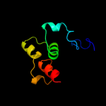
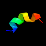
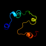
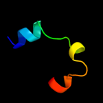
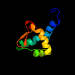
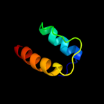
Image coloured by rainbow N → C terminus
Model (left) based on template c3knyA_
Top template information
PDB header:unknown function
Chain: A: PDB Molecule:hypothetical protein bt_3535;
PDBTitle: crystal structure of a two domain protein with unknown function2 (bt_3535) from bacteroides thetaiotaomicron vpi-5482 at 2.60 a3 resolution
Confidence and coverage
Confidence: 53.1% Coverage: 22%
62 residues ( 22% of your sequence) have been modelled with 53.1% confidence by the single highest scoring template.
You may wish to submit your sequence to Phyrealarm. This will automatically scan your sequence every week for new potential templates as they appear in the Phyre2 library.
Please note: You must be registered and logged in to use Phyrealarm.
3D viewing
Interactive 3D view in Jmol

Sequence analysis 

Secondary structure and disorder prediction 

   1........10.........20.........30.........40.........50.........60
Sequence  MLFVLILSHRAASYGAIMAALPYMQLYIADYLADTMHLSAEEHGAYLLLMFNYWQTGKPI
Secondary structure 


SS confidence 


Disorder  ???

?


Disorder confidence 


   .........70.........80.........90.........100.........110.........120
Sequence  PKNRLAKIARLTNERWADVEPSLQEFFCDNGEEWVHLRIEEDLASVREKLTKKSAAGKAS
Secondary structure 


SS confidence 


Disorder 


?????????
Disorder confidence 


   .........130.........140.........150.........160.........170.........180
Sequence  VQARRSRKEADVQTKQERNLTGVQTDVEVVFEHDVNTKATNKDTDKDLKTDPPLNPPRGN
Secondary structure 


SS confidence 


Disorder  ????????????????????????????????????????????????????????????
Disorder confidence 


   .........190.........200.........210.........220.........230.........240
Sequence  RGVKKFDPLDITLPNWISVSLWREWVEFRQALRKPIRTEQGANGAIRELEKFRQQGFSPE
Secondary structure 


SS confidence 


Disorder  ??????????
????


???????


Disorder confidence 


   .........250.........260.........270.........280.....
Sequence  QVIRHSIANEYQGLFAPKGVRPETLLRQVNTVSLPDSAIPPGFRG
Secondary structure 


SS confidence 


Disorder 


?


????????????????????
???????
Disorder confidence 


Confidence Key
High(9)                    Low (0)
?Disordered
Alpha helix
Beta strand

Domain analysis 

Hover over an aligned region to see model and summary info

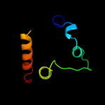
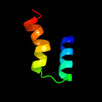
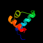
Please note, only up to the top 20 hits are modelled to reduce computer load

RankAligned region

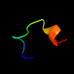
PDB 3kny chain A

3D model

Region: 176 - 250
Aligned: 62
Modelled: 73
Confidence: 53.1%
Identity: 21%
PDB header:unknown function
Chain: A: PDB Molecule:hypothetical protein bt_3535;
PDBTitle: crystal structure of a two domain protein with unknown function2 (bt_3535) from bacteroides thetaiotaomicron vpi-5482 at 2.60 a3 resolution

Phyre2

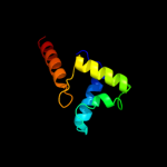
PDB 1pby chain A

3D model

Region: 31 - 91
Aligned: 61
Modelled: 61
Confidence: 48.8%
Identity: 21%
PDB header:oxidoreductase
Chain: A: PDB Molecule:quinohemoprotein amine dehydrogenase 60 kda
PDBTitle: structure of the phenylhydrazine adduct of the2 quinohemoprotein amine dehydrogenase from paracoccus3 denitrificans at 1.7 a resolution

Phyre2

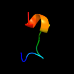
PDB 1qmg chain A domain 1

3D model

Region: 222 - 256
Aligned: 35
Modelled: 35
Confidence: 45.0%
Identity: 20%
Fold: 6-phosphogluconate dehydrogenase C-terminal domain-like
Superfamily: 6-phosphogluconate dehydrogenase C-terminal domain-like
Family: Acetohydroxy acid isomeroreductase (ketol-acid reductoisomerase, KARI)

Phyre2

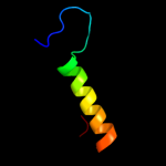
PDB 2v79 chain B

3D model

Region: 36 - 85
Aligned: 50
Modelled: 50
Confidence: 29.4%
Identity: 12%
PDB header:dna-binding protein
Chain: B: PDB Molecule:dna replication protein dnad;
PDBTitle: crystal structure of the n-terminal domain of dnad from2 bacillus subtilis

Phyre2

PDB 1khb chain A domain 1

3D model

Region: 18 - 33
Aligned: 16
Modelled: 16
Confidence: 27.5%
Identity: 31%
Fold: PEP carboxykinase-like
Superfamily: PEP carboxykinase-like
Family: PEP carboxykinase C-terminal domain

Phyre2

PDB 2vn2 chain B

3D model

Region: 36 - 113
Aligned: 77
Modelled: 78
Confidence: 24.5%
Identity: 14%
PDB header:replication
Chain: B: PDB Molecule:chromosome replication initiation protein;
PDBTitle: crystal structure of the n-terminal domain of dnad protein2 from geobacillus kaustophilus hta426

Phyre2

PDB 2qey chain A

3D model

Region: 18 - 33
Aligned: 16
Modelled: 16
Confidence: 23.8%
Identity: 31%
PDB header:lyase
Chain: A: PDB Molecule:phosphoenolpyruvate carboxykinase, cytosolic [gtp];
PDBTitle: rat cytosolic pepck in complex with gtp

Phyre2

PDB 2f5u chain A

3D model

Region: 28 - 61
Aligned: 33
Modelled: 34
Confidence: 20.6%
Identity: 30%
PDB header:viral protein
Chain: A: PDB Molecule:virion protein ul25;
PDBTitle: structural characterization of the ul25 dna packaging2 protein from herpes simplex virus type 1

Phyre2

PDB 2jrd chain A

3D model

Region: 242 - 254
Aligned: 13
Modelled: 13
Confidence: 20.6%
Identity: 31%
PDB header:viral protein
Chain: A: PDB Molecule:hemagglutinin;
PDBTitle: influenza hemagglutinin fusion domain mutant f9a

Phyre2

PDB 1bdt chain A

3D model

Region: 18 - 58
Aligned: 41
Modelled: 41
Confidence: 19.1%
Identity: 27%
Fold: Ribbon-helix-helix
Superfamily: Ribbon-helix-helix
Family: Arc/Mnt-like phage repressors

Phyre2

PDB 2zci chain D

3D model

Region: 18 - 33
Aligned: 16
Modelled: 16
Confidence: 18.5%
Identity: 38%
PDB header:signaling protein, lyase
Chain: D: PDB Molecule:phosphoenolpyruvate carboxykinase [gtp];
PDBTitle: structure of a gtp-dependent bacterial pep-carboxykinase2 from corynebacterium glutamicum

Phyre2

PDB 1t07 chain A

3D model

Region: 187 - 232
Aligned: 46
Modelled: 46
Confidence: 17.2%
Identity: 20%
Fold: YggX-like
Superfamily: YggX-like
Family: YggX-like

Phyre2

PDB 1xs8 chain A

3D model

Region: 187 - 238
Aligned: 52
Modelled: 52
Confidence: 16.1%
Identity: 17%
Fold: YggX-like
Superfamily: YggX-like
Family: YggX-like

Phyre2

PDB 1jmx chain A

3D model

Region: 31 - 90
Aligned: 59
Modelled: 60
Confidence: 15.9%
Identity: 24%
PDB header:oxidoreductase
Chain: A: PDB Molecule:amine dehydrogenase;
PDBTitle: crystal structure of a quinohemoprotein amine dehydrogenase2 from pseudomonas putida

Phyre2

PDB 1sed chain A

3D model

Region: 225 - 257
Aligned: 30
Modelled: 33
Confidence: 15.9%
Identity: 17%
Fold: Hypothetical protein YhaI
Superfamily: Hypothetical protein YhaI
Family: Hypothetical protein YhaI

Phyre2

PDB 2fah chain B

3D model

Region: 18 - 33
Aligned: 16
Modelled: 16
Confidence: 13.2%
Identity: 25%
PDB header:lyase
Chain: B: PDB Molecule:phosphoenolpyruvate carboxykinase;
PDBTitle: the structure of mitochondrial pepck, complex with mn and gdp

Phyre2

PDB 3bdr chain A

3D model

Region: 83 - 98
Aligned: 16
Modelled: 16
Confidence: 12.6%
Identity: 25%
PDB header:structural genomics, unknown function
Chain: A: PDB Molecule:ycf58 protein;
PDBTitle: crystal structure of fatty acid-binding protein-like ycf582 from thermosynecoccus elongatus. northeast structural3 genomics consortium target ter13.

Phyre2

PDB 1pby chain A domain 1

3D model

Region: 28 - 51
Aligned: 24
Modelled: 24
Confidence: 12.5%
Identity: 29%
Fold: Cytochrome c
Superfamily: Cytochrome c
Family: Quinohemoprotein amine dehydrogenase A chain, domains 1 and 2

Phyre2

PDB 1tw3 chain A

3D model

Region: 26 - 103
Aligned: 72
Modelled: 78
Confidence: 12.0%
Identity: 13%
PDB header:transferase
Chain: A: PDB Molecule:carminomycin 4-o-methyltransferase;
PDBTitle: crystal structure of carminomycin-4-o-methyltransferase2 (dnrk) in complex with s-adenosyl-l-homocystein (sah) and3 4-methoxy-e-rhodomycin t (m-et)

Phyre2

PDB 1gjj chain A domain 2

3D model

Region: 195 - 236
Aligned: 41
Modelled: 42
Confidence: 11.3%
Identity: 17%
Fold: LEM/SAP HeH motif
Superfamily: LEM domain
Family: LEM domain

Phyre2
1

c3knyA_
2

c1pbyA_
3

d1qmga1
4

c2v79B_
5

d1khba1
6

c2vn2B_
7

c2qeyA_
8

c2f5uA_
9

c2jrdA_
10

d1bdta_
11

c2zciD_
12

d1t07a_
13

d1xs8a_
14

c1jmxA_
15

d1seda_
16

c2fahB_
17

c3bdrA_
18

d1pbya1
19

c1tw3A_
20

d1gjja2
21


22


23


24


25


26


27


28


29


30


31


32


33


34


35


36


37


38


39


40


41


42


43


44


45


Detailed template information 

#
Template Alignment Coverage3D Model Confidence
% i.d. Template Information
1c3knyA_


53.1 21 PDB header:unknown function
Chain: A: PDB Molecule:hypothetical protein bt_3535;
PDBTitle: crystal structure of a two domain protein with unknown function2 (bt_3535) from bacteroides thetaiotaomicron vpi-5482 at 2.60 a3 resolution
2c1pbyA_


48.8 21 PDB header:oxidoreductase
Chain: A: PDB Molecule:quinohemoprotein amine dehydrogenase 60 kda
PDBTitle: structure of the phenylhydrazine adduct of the2 quinohemoprotein amine dehydrogenase from paracoccus3 denitrificans at 1.7 a resolution
3d1qmga1


45.0 20 Fold:6-phosphogluconate dehydrogenase C-terminal domain-like
Superfamily:6-phosphogluconate dehydrogenase C-terminal domain-like
Family:Acetohydroxy acid isomeroreductase (ketol-acid reductoisomerase, KARI)
4c2v79B_


29.4 12 PDB header:dna-binding protein
Chain: B: PDB Molecule:dna replication protein dnad;
PDBTitle: crystal structure of the n-terminal domain of dnad from2 bacillus subtilis
5d1khba1


27.5 31 Fold:PEP carboxykinase-like
Superfamily:PEP carboxykinase-like
Family:PEP carboxykinase C-terminal domain
6c2vn2B_


24.5 14 PDB header:replication
Chain: B: PDB Molecule:chromosome replication initiation protein;
PDBTitle: crystal structure of the n-terminal domain of dnad protein2 from geobacillus kaustophilus hta426
7c2qeyA_


23.8 31 PDB header:lyase
Chain: A: PDB Molecule:phosphoenolpyruvate carboxykinase, cytosolic [gtp];
PDBTitle: rat cytosolic pepck in complex with gtp
8c2f5uA_


20.6 30 PDB header:viral protein
Chain: A: PDB Molecule:virion protein ul25;
PDBTitle: structural characterization of the ul25 dna packaging2 protein from herpes simplex virus type 1
9c2jrdA_


20.6 31 PDB header:viral protein
Chain: A: PDB Molecule:hemagglutinin;
PDBTitle: influenza hemagglutinin fusion domain mutant f9a
10d1bdta_


19.1 27 Fold:Ribbon-helix-helix
Superfamily:Ribbon-helix-helix
Family:Arc/Mnt-like phage repressors
11c2zciD_


18.5 38 PDB header:signaling protein, lyase
Chain: D: PDB Molecule:phosphoenolpyruvate carboxykinase [gtp];
PDBTitle: structure of a gtp-dependent bacterial pep-carboxykinase2 from corynebacterium glutamicum
12d1t07a_


17.2 20 Fold:YggX-like
Superfamily:YggX-like
Family:YggX-like
13d1xs8a_


16.1 17 Fold:YggX-like
Superfamily:YggX-like
Family:YggX-like
14c1jmxA_


15.9 24 PDB header:oxidoreductase
Chain: A: PDB Molecule:amine dehydrogenase;
PDBTitle: crystal structure of a quinohemoprotein amine dehydrogenase2 from pseudomonas putida
15d1seda_


15.9 17 Fold:Hypothetical protein YhaI
Superfamily:Hypothetical protein YhaI
Family:Hypothetical protein YhaI
16c2fahB_


13.2 25 PDB header:lyase
Chain: B: PDB Molecule:phosphoenolpyruvate carboxykinase;
PDBTitle: the structure of mitochondrial pepck, complex with mn and gdp
17c3bdrA_


12.6 25 PDB header:structural genomics, unknown function
Chain: A: PDB Molecule:ycf58 protein;
PDBTitle: crystal structure of fatty acid-binding protein-like ycf582 from thermosynecoccus elongatus. northeast structural3 genomics consortium target ter13.
18d1pbya1


12.5 29 Fold:Cytochrome c
Superfamily:Cytochrome c
Family:Quinohemoprotein amine dehydrogenase A chain, domains 1 and 2
19c1tw3A_


12.0 13 PDB header:transferase
Chain: A: PDB Molecule:carminomycin 4-o-methyltransferase;
PDBTitle: crystal structure of carminomycin-4-o-methyltransferase2 (dnrk) in complex with s-adenosyl-l-homocystein (sah) and3 4-methoxy-e-rhodomycin t (m-et)
20d1gjja2


11.3 17 Fold:LEM/SAP HeH motif
Superfamily:LEM domain
Family:LEM domain
21d6paxa1


not modelled 11.2 28 Fold:DNA/RNA-binding 3-helical bundle
Superfamily:Homeodomain-like
Family:Paired domain
22c1iboA_


not modelled 10.5 31 PDB header:viral protein
Chain: A: PDB Molecule:hemagglutinin ha2 chain peptide;
PDBTitle: nmr structure of hemagglutinin fusion peptide in dpc2 micelles at ph 7.4
23c1ibnA_


not modelled 10.5 31 PDB header:viral protein
Chain: A: PDB Molecule:hemagglutinin ha2 chain peptide;
PDBTitle: nmr structure of hemagglutinin fusion peptide in dpc2 micelles at ph 5
24d1z5ga1


not modelled 10.2 12 Fold:HAD-like
Superfamily:HAD-like
Family:Class B acid phosphatase, AphA
25d1zbsa2


not modelled 9.5 15 Fold:Ribonuclease H-like motif
Superfamily:Actin-like ATPase domain
Family:BadF/BadG/BcrA/BcrD-like
26c3chxG_


not modelled 9.3 23 PDB header:membrane protein
Chain: G: PDB Molecule:pmoc;
PDBTitle: crystal structure of methylosinus trichosporium ob3b2 particulate methane monooxygenase (pmmo)
27c2kxaA_


not modelled 9.1 23 PDB header:viral protein, immune system
Chain: A: PDB Molecule:haemagglutinin ha2 chain peptide;
PDBTitle: the hemagglutinin fusion peptide (h1 subtype) at ph 7.4
28c1gjjA_


not modelled 9.1 33 PDB header:membrane protein
Chain: A: PDB Molecule:lap2;
PDBTitle: n-terminal constant region of the nuclear envelope protein2 lap2
29d1b28a_


not modelled 8.8 29 Fold:Ribbon-helix-helix
Superfamily:Ribbon-helix-helix
Family:Arc/Mnt-like phage repressors
30d1h9fa_


not modelled 8.2 30 Fold:LEM/SAP HeH motif
Superfamily:LEM domain
Family:LEM domain
31d1k78a1


not modelled 7.8 25 Fold:DNA/RNA-binding 3-helical bundle
Superfamily:Homeodomain-like
Family:Paired domain
32d1iuqa_


not modelled 7.7 17 Fold:Glycerol-3-phosphate (1)-acyltransferase
Superfamily:Glycerol-3-phosphate (1)-acyltransferase
Family:Glycerol-3-phosphate (1)-acyltransferase
33c1yewC_


not modelled 7.0 15 PDB header:oxidoreductase, membrane protein
Chain: C: PDB Molecule:particulate methane monooxygenase subunit c2;
PDBTitle: crystal structure of particulate methane monooxygenase
34c2pjwH_


not modelled 6.8 16 PDB header:endocytosis/exocytosis
Chain: H: PDB Molecule:uncharacterized protein yhl002w;
PDBTitle: the vps27/hse1 complex is a gat domain-based scaffold for2 ubiquitin-dependent sorting
35c2k1oA_


not modelled 6.7 41 PDB header:gene regulation
Chain: A: PDB Molecule:putative;
PDBTitle: nmr structure of helicobacter pylori jhp0511 (hp0564).
36c2pfcA_


not modelled 6.6 22 PDB header:unknown function
Chain: A: PDB Molecule:hypothetical protein rv0098/mt0107;
PDBTitle: structure of mycobacterium tuberculosis rv0098
37c1xopA_


not modelled 6.4 31 PDB header:viral protein
Chain: A: PDB Molecule:hemagglutinin;
PDBTitle: nmr structure of g1v mutant of influenza hemagglutinin2 fusion peptide in dpc micelles at ph 5
38c1xooA_


not modelled 6.3 31 PDB header:viral protein
Chain: A: PDB Molecule:hemagglutinin;
PDBTitle: nmr structure of g1s mutant of influenza hemagglutinin2 fusion peptide in dpc micelles at ph 5
39c3bhwA_


not modelled 6.1 15 PDB header:structural genomics, unknown function
Chain: A: PDB Molecule:uncharacterized protein;
PDBTitle: crystal structure of an uncharacterized protein from magnetospirillum2 magneticum
40d1fi6a_


not modelled 5.9 24 Fold:EF Hand-like
Superfamily:EF-hand
Family:Eps15 homology domain (EH domain)
41d1c07a_


not modelled 5.7 19 Fold:EF Hand-like
Superfamily:EF-hand
Family:Eps15 homology domain (EH domain)
42c2bn5A_


not modelled 5.6 25 PDB header:nuclear protein
Chain: A: PDB Molecule:psi;
PDBTitle: p-element somatic inhibitor protein complex with u1-70k2 proline-rich peptide
43c2ia2D_


not modelled 5.5 13 PDB header:transcription
Chain: D: PDB Molecule:putative transcriptional regulator;
PDBTitle: the crystal structure of a putative transcriptional regulator rha061952 from rhodococcus sp. rha1
44d2o9aa1


not modelled 5.4 10 Fold:Profilin-like
Superfamily:GAF domain-like
Family:IclR ligand-binding domain-like
45d1cmca_


not modelled 5.4 50 Fold:Ribbon-helix-helix
Superfamily:Ribbon-helix-helix
Family:Met repressor, MetJ (MetR)

Binding site prediction 

Due to computational demand, binding site predictions are not run for batch jobs

If you want to predict binding sites, please manually submit your model of choice to 3DLigandSite


Phyre is for academic use only

Please cite: Protein structure prediction on the web: a case study using the Phyre server
Kelley LA and Sternberg MJE. Nature Protocols 4, 363 - 371 (2009) [pdf] [Import into BibTeX]
 
If you use the binding site predictions from 3DLigandSite, please also cite:
3DLigandSite: predicting ligand-binding sites using similar structures.
Wass MN, Kelley LA and Sternberg MJ Nucleic Acids Research 38, W469-73 (2010) [PubMed]
 
© Structural Bioinformatics Group
Imperial College London
Lawrence Kelley, Benjamin Jefferys
Disclaimer
Terms and Conditions
Component software
Template detection: HHpred 1.51
Secondary structure prediction: Psi-pred 2.5
Disorder prediction: Disopred 2.4
Transmembrane prediction: Memsat_SVM
Multi-template modelling and ab initio: Poing 1.0