Phyre Home Retrieve Phyre Job Id
Emaill.a.kelley@imperial.ac.uk
DescriptionP64550
DateThu Jan 5 12:09:22 GMT 2012
Unique Job ID754be9940133d616

Summary 

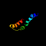
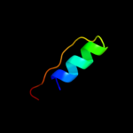
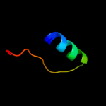
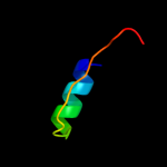
Top model
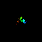
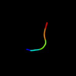
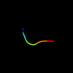
Image coloured by rainbow N → C terminus
Model (left) based on template d1z9ha1
Top template information
Fold:GST C-terminal domain-like
Superfamily:GST C-terminal domain-like
Family:Glutathione S-transferase (GST), C-terminal domain
Confidence and coverage
Confidence: 56.9% Coverage: 34%
50 residues ( 34% of your sequence) have been modelled with 56.9% confidence by the single highest scoring template.
You may wish to submit your sequence to Phyrealarm. This will automatically scan your sequence every week for new potential templates as they appear in the Phyre2 library.
Please note: You must be registered and logged in to use Phyrealarm.
3D viewing
Interactive 3D view in Jmol

Sequence analysis 

Secondary structure and disorder prediction 

   1........10.........20.........30.........40.........50.........60
Sequence  MFSPQSRLRHAVADTFAMVVYCSVVNMCIEVFLSGMSFEQSFYSRLVAIPVNILIAWPYG
Secondary structure 


SS confidence 


Disorder  ???


?


Disorder confidence 


   .........70.........80.........90.........100.........110.........120
Sequence  MYRDLFMRAARKVSPSGWIKNLADILAYVTFQSPVYVAILLVVGADWHQIMAAVSSNIVV
Secondary structure 


SS confidence 


Disorder 


??????


?


Disorder confidence 


   .........130.........140.........
Sequence  SMLMGAVYGYFLDYCRRLFKVSRYQQVKA
Secondary structure 
SS confidence 


Disorder 


?

????
Disorder confidence 


Confidence Key
High(9)                    Low (0)
?Disordered
Alpha helix
Beta strand

Domain analysis 

Hover over an aligned region to see model and summary info

Please note, only up to the top 20 hits are modelled to reduce computer load

RankAligned region

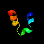
PDB 1z9h chain A domain 1

3D model

Region: 7 - 56
Aligned: 50
Modelled: 50
Confidence: 56.9%
Identity: 14%
Fold: GST C-terminal domain-like
Superfamily: GST C-terminal domain-like
Family: Glutathione S-transferase (GST), C-terminal domain

Phyre2

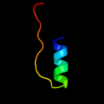
PDB 1nku chain A

3D model

Region: 27 - 45
Aligned: 19
Modelled: 19
Confidence: 40.1%
Identity: 26%
Fold: DNA-glycosylase
Superfamily: DNA-glycosylase
Family: 3-Methyladenine DNA glycosylase I (Tag)

Phyre2

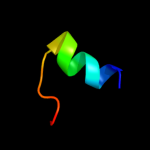
PDB 2jg6 chain A

3D model

Region: 27 - 45
Aligned: 19
Modelled: 19
Confidence: 32.6%
Identity: 16%
PDB header:hydrolase
Chain: A: PDB Molecule:dna-3-methyladenine glycosidase;
PDBTitle: crystal structure of a 3-methyladenine dna glycosylase i2 from staphylococcus aureus

Phyre2

PDB 2xf5 chain C

3D model

Region: 128 - 133
Aligned: 6
Modelled: 6
Confidence: 20.1%
Identity: 100%
PDB header:viral protein
Chain: C: PDB Molecule:gp23.1;
PDBTitle: crystal structure of bacillus subtilis spp1 phage gp23.1, a2 putative chaperone.

Phyre2

PDB 2ntk chain A domain 1

3D model

Region: 25 - 48
Aligned: 24
Modelled: 24
Confidence: 17.6%
Identity: 17%
Fold: Ntn hydrolase-like
Superfamily: Archaeal IMP cyclohydrolase PurO
Family: Archaeal IMP cyclohydrolase PurO

Phyre2

PDB 1wyp chain A

3D model

Region: 129 - 145
Aligned: 16
Modelled: 17
Confidence: 14.5%
Identity: 19%
PDB header:structural protein
Chain: A: PDB Molecule:calponin 1;
PDBTitle: solution structure of the ch domain of human calponin 1

Phyre2

PDB 2csh chain A domain 2

3D model

Region: 61 - 68
Aligned: 8
Modelled: 8
Confidence: 13.3%
Identity: 63%
Fold: beta-beta-alpha zinc fingers
Superfamily: beta-beta-alpha zinc fingers
Family: Classic zinc finger, C2H2

Phyre2

PDB 1ei5 chain A

3D model

Region: 22 - 47
Aligned: 25
Modelled: 26
Confidence: 13.0%
Identity: 16%
PDB header:hydrolase
Chain: A: PDB Molecule:d-aminopeptidase;
PDBTitle: crystal structure of a d-aminopeptidase from ochrobactrum2 anthropi

Phyre2

PDB 3br8 chain A

3D model

Region: 62 - 84
Aligned: 23
Modelled: 23
Confidence: 12.7%
Identity: 35%
PDB header:hydrolase
Chain: A: PDB Molecule:probable acylphosphatase;
PDBTitle: crystal structure of acylphosphatase from bacillus subtilis

Phyre2

PDB 2acy chain A

3D model

Region: 62 - 84
Aligned: 23
Modelled: 23
Confidence: 11.9%
Identity: 22%
Fold: Ferredoxin-like
Superfamily: Acylphosphatase/BLUF domain-like
Family: Acylphosphatase-like

Phyre2

PDB 1e7l chain A domain 2

3D model

Region: 60 - 86
Aligned: 27
Modelled: 27
Confidence: 11.8%
Identity: 30%
Fold: His-Me finger endonucleases
Superfamily: His-Me finger endonucleases
Family: Recombination endonuclease VII, N-terminal domain

Phyre2

PDB 2bje chain A

3D model

Region: 62 - 84
Aligned: 23
Modelled: 23
Confidence: 11.0%
Identity: 30%
PDB header:hydrolase
Chain: A: PDB Molecule:acylphosphatase;
PDBTitle: acylphosphatase from sulfolobus solfataricus. monclinic p212 space group

Phyre2

PDB 2d86 chain A

3D model

Region: 129 - 145
Aligned: 17
Modelled: 17
Confidence: 10.9%
Identity: 24%
PDB header:signaling protein, protein binding
Chain: A: PDB Molecule:vav-3 protein;
PDBTitle: solution structure of the ch domain from human vav-3 protein

Phyre2

PDB 1w2i chain A

3D model

Region: 62 - 84
Aligned: 23
Modelled: 23
Confidence: 10.1%
Identity: 43%
Fold: Ferredoxin-like
Superfamily: Acylphosphatase/BLUF domain-like
Family: Acylphosphatase-like

Phyre2

PDB 1bmx chain A

3D model

Region: 131 - 146
Aligned: 16
Modelled: 16
Confidence: 9.9%
Identity: 44%
PDB header:viral protein
Chain: A: PDB Molecule:human immunodeficiency virus type 1 capsid;
PDBTitle: hiv-1 capsid protein major homology region peptide analog,2 nmr, 8 structures

Phyre2

PDB 3o3v chain B

3D model

Region: 22 - 47
Aligned: 25
Modelled: 26
Confidence: 9.9%
Identity: 28%
PDB header:hydrolase
Chain: B: PDB Molecule:beta-lactamase;
PDBTitle: crystal structure of clbp peptidase domain

Phyre2

PDB 1ulr chain A

3D model

Region: 62 - 84
Aligned: 23
Modelled: 23
Confidence: 9.6%
Identity: 35%
Fold: Ferredoxin-like
Superfamily: Acylphosphatase/BLUF domain-like
Family: Acylphosphatase-like

Phyre2

PDB 1i8y chain A

3D model

Region: 58 - 62
Aligned: 5
Modelled: 5
Confidence: 9.5%
Identity: 60%
PDB header:cytokine
Chain: A: PDB Molecule:granulin-1;
PDBTitle: semi-automatic structure determination of the cg1 3-302 peptide based on aria

Phyre2

PDB 1i8y chain A

3D model

Region: 58 - 62
Aligned: 5
Modelled: 5
Confidence: 9.5%
Identity: 60%
Fold: Knottins (small inhibitors, toxins, lectins)
Superfamily: Granulin repeat
Family: Granulin repeat

Phyre2

PDB 1gxu chain A

3D model

Region: 62 - 84
Aligned: 23
Modelled: 23
Confidence: 8.8%
Identity: 22%
Fold: Ferredoxin-like
Superfamily: Acylphosphatase/BLUF domain-like
Family: Acylphosphatase-like

Phyre2
1

d1z9ha1
2

d1nkua_
3

c2jg6A_
4

c2xf5C_
5

d2ntka1
6

c1wypA_
7

d2csha2
8

c1ei5A_
9

c3br8A_
10

d2acya_
11

d1e7la2
12

c2bjeA_
13

c2d86A_
14

d1w2ia_
15

c1bmxA_
16

c3o3vB_
17

d1ulra_
18

c1i8yA_
19

d1i8ya_
20

d1gxua_
21


22


23


24


25


26


27


28


29


30


31


32


Detailed template information 

#
Template Alignment Coverage3D Model Confidence
% i.d. Template Information
1d1z9ha1


56.9 14 Fold:GST C-terminal domain-like
Superfamily:GST C-terminal domain-like
Family:Glutathione S-transferase (GST), C-terminal domain
2d1nkua_


40.1 26 Fold:DNA-glycosylase
Superfamily:DNA-glycosylase
Family:3-Methyladenine DNA glycosylase I (Tag)
3c2jg6A_


32.6 16 PDB header:hydrolase
Chain: A: PDB Molecule:dna-3-methyladenine glycosidase;
PDBTitle: crystal structure of a 3-methyladenine dna glycosylase i2 from staphylococcus aureus
4c2xf5C_


20.1 100 PDB header:viral protein
Chain: C: PDB Molecule:gp23.1;
PDBTitle: crystal structure of bacillus subtilis spp1 phage gp23.1, a2 putative chaperone.
5d2ntka1


17.6 17 Fold:Ntn hydrolase-like
Superfamily:Archaeal IMP cyclohydrolase PurO
Family:Archaeal IMP cyclohydrolase PurO
6c1wypA_


14.5 19 PDB header:structural protein
Chain: A: PDB Molecule:calponin 1;
PDBTitle: solution structure of the ch domain of human calponin 1
7d2csha2


13.3 63 Fold:beta-beta-alpha zinc fingers
Superfamily:beta-beta-alpha zinc fingers
Family:Classic zinc finger, C2H2
8c1ei5A_


13.0 16 PDB header:hydrolase
Chain: A: PDB Molecule:d-aminopeptidase;
PDBTitle: crystal structure of a d-aminopeptidase from ochrobactrum2 anthropi
9c3br8A_


12.7 35 PDB header:hydrolase
Chain: A: PDB Molecule:probable acylphosphatase;
PDBTitle: crystal structure of acylphosphatase from bacillus subtilis
10d2acya_


11.9 22 Fold:Ferredoxin-like
Superfamily:Acylphosphatase/BLUF domain-like
Family:Acylphosphatase-like
11d1e7la2


11.8 30 Fold:His-Me finger endonucleases
Superfamily:His-Me finger endonucleases
Family:Recombination endonuclease VII, N-terminal domain
12c2bjeA_


11.0 30 PDB header:hydrolase
Chain: A: PDB Molecule:acylphosphatase;
PDBTitle: acylphosphatase from sulfolobus solfataricus. monclinic p212 space group
13c2d86A_


10.9 24 PDB header:signaling protein, protein binding
Chain: A: PDB Molecule:vav-3 protein;
PDBTitle: solution structure of the ch domain from human vav-3 protein
14d1w2ia_


10.1 43 Fold:Ferredoxin-like
Superfamily:Acylphosphatase/BLUF domain-like
Family:Acylphosphatase-like
15c1bmxA_


9.9 44 PDB header:viral protein
Chain: A: PDB Molecule:human immunodeficiency virus type 1 capsid;
PDBTitle: hiv-1 capsid protein major homology region peptide analog,2 nmr, 8 structures
16c3o3vB_


9.9 28 PDB header:hydrolase
Chain: B: PDB Molecule:beta-lactamase;
PDBTitle: crystal structure of clbp peptidase domain
17d1ulra_


9.6 35 Fold:Ferredoxin-like
Superfamily:Acylphosphatase/BLUF domain-like
Family:Acylphosphatase-like
18c1i8yA_


9.5 60 PDB header:cytokine
Chain: A: PDB Molecule:granulin-1;
PDBTitle: semi-automatic structure determination of the cg1 3-302 peptide based on aria
19d1i8ya_


9.5 60 Fold:Knottins (small inhibitors, toxins, lectins)
Superfamily:Granulin repeat
Family:Granulin repeat
20d1gxua_


8.8 22 Fold:Ferredoxin-like
Superfamily:Acylphosphatase/BLUF domain-like
Family:Acylphosphatase-like
21d1urra_


not modelled 8.7 26 Fold:Ferredoxin-like
Superfamily:Acylphosphatase/BLUF domain-like
Family:Acylphosphatase-like
22d1h67a_


not modelled 8.7 25 Fold:CH domain-like
Superfamily:Calponin-homology domain, CH-domain
Family:Calponin-homology domain, CH-domain
23d2hkua2


not modelled 8.6 23 Fold:Tetracyclin repressor-like, C-terminal domain
Superfamily:Tetracyclin repressor-like, C-terminal domain
Family:Tetracyclin repressor-like, C-terminal domain
24c3o59X_


not modelled 8.0 31 PDB header:transferase
Chain: X: PDB Molecule:dna polymerase ii large subunit;
PDBTitle: dna polymerase d large subunit dp2(1-300) from pyrococcus horikoshii
25c3a7kD_


not modelled 7.9 19 PDB header:membrane protein
Chain: D: PDB Molecule:halorhodopsin;
PDBTitle: crystal structure of halorhodopsin from natronomonas2 pharaonis
26c3tg9A_


not modelled 7.7 16 PDB header:penicillin binding protein
Chain: A: PDB Molecule:penicillin-binding protein;
PDBTitle: the crystal structure of penicillin binding protein from bacillus2 halodurans
27d1ujoa_


not modelled 7.2 21 Fold:CH domain-like
Superfamily:Calponin-homology domain, CH-domain
Family:Calponin-homology domain, CH-domain
28d1ui5a2


not modelled 6.0 9 Fold:Tetracyclin repressor-like, C-terminal domain
Superfamily:Tetracyclin repressor-like, C-terminal domain
Family:Tetracyclin repressor-like, C-terminal domain
29c1zbhA_


not modelled 5.9 18 PDB header:hydrolase/rna
Chain: A: PDB Molecule:3'-5' exonuclease eri1;
PDBTitle: 3'-end specific recognition of histone mrna stem-loop by 3'-2 exonuclease
30c1wymA_


not modelled 5.6 19 PDB header:structural protein
Chain: A: PDB Molecule:transgelin-2;
PDBTitle: solution structure of the ch domain of human transgelin-2
31c2wnmA_


not modelled 5.6 38 PDB header:hydrolase
Chain: A: PDB Molecule:gene 2;
PDBTitle: solution structure of gp2
32c3i6xC_


not modelled 5.4 6 PDB header:calmodulin-binding, membrane protein
Chain: C: PDB Molecule:ras gtpase-activating-like protein iqgap1;
PDBTitle: crystal structure of the calponin homology domain of iqgap1

Binding site prediction 

Due to computational demand, binding site predictions are not run for batch jobs

If you want to predict binding sites, please manually submit your model of choice to 3DLigandSite


Phyre is for academic use only

Please cite: Protein structure prediction on the web: a case study using the Phyre server
Kelley LA and Sternberg MJE. Nature Protocols 4, 363 - 371 (2009) [pdf] [Import into BibTeX]
 
If you use the binding site predictions from 3DLigandSite, please also cite:
3DLigandSite: predicting ligand-binding sites using similar structures.
Wass MN, Kelley LA and Sternberg MJ Nucleic Acids Research 38, W469-73 (2010) [PubMed]
 
© Structural Bioinformatics Group
Imperial College London
Lawrence Kelley, Benjamin Jefferys
Disclaimer
Terms and Conditions
Component software
Template detection: HHpred 1.51
Secondary structure prediction: Psi-pred 2.5
Disorder prediction: Disopred 2.4
Transmembrane prediction: Memsat_SVM
Multi-template modelling and ab initio: Poing 1.0