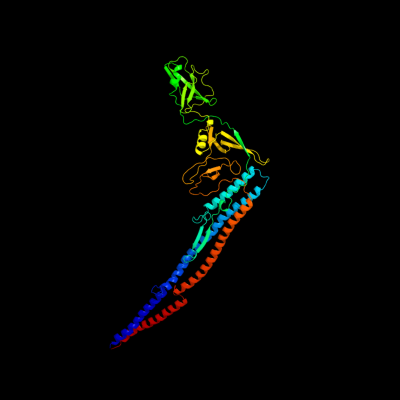
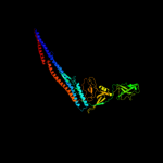
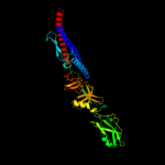
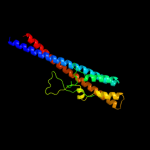
1 d1ucua_
100.0
45
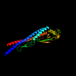
Fold: Phase 1 flagellinSuperfamily: Phase 1 flagellinFamily: Phase 1 flagellin2 c3k8vB_
100.0
39
PDB header: structural proteinChain: B: PDB Molecule: flagellin homolog;PDBTitle: crysatl structure of a bacterial cell-surface flagellin n20c20
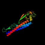
3 d1io1a_
100.0
37
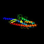
Fold: Phase 1 flagellinSuperfamily: Phase 1 flagellinFamily: Phase 1 flagellin4 c3k8wA_
100.0
36
PDB header: structural proteinChain: A: PDB Molecule: flagellin homolog;PDBTitle: crysatl structure of a bacterial cell-surface flagellin n20c45
5 c2zbiB_
100.0
33
PDB header: structural proteinChain: B: PDB Molecule: flagellin homolog;PDBTitle: crysatl structure of a bacterial cell-surface flagellin
6 c2d4xA_
100.0
17
PDB header: structural proteinChain: A: PDB Molecule: flagellar hook-associated protein 3;PDBTitle: crystal structure of a 26k fragment of hap3 (flgl)
7 c3pwxB_
100.0
18
PDB header: structural proteinChain: B: PDB Molecule: putative flagellar hook-associated protein;PDBTitle: structure of putative flagellar hook-associated protein from vibrio2 parahaemolyticus
8 c1oryB_
99.4
45
PDB header: chaperoneChain: B: PDB Molecule: flagellin;PDBTitle: flagellar export chaperone in complex with its cognate binding partner
9 c3a69A_
90.7
22
PDB header: motor proteinChain: A: PDB Molecule: flagellar hook protein flge;PDBTitle: atomic model of the bacterial flagellar hook based on2 docking an x-ray derived structure and terminal two alpha-3 helices into an 7.1 angstrom resolution cryoem map
10 c3k1hA_
64.5
19
PDB header: chaperoneChain: A: PDB Molecule: putative uncharacterized protein;PDBTitle: crystal structure of hp1076 from h.pylori
11 c3hfeC_
29.8
30
PDB header: transport proteinChain: C: PDB Molecule: potassium voltage-gated channel subfamily kqt member 1;PDBTitle: a trimeric form of the kv7.1 a domain tail
12 d1dt9a3
29.1
16
Fold: N-terminal domain of eukaryotic peptide chain release factor subunit 1, ERF1Superfamily: N-terminal domain of eukaryotic peptide chain release factor subunit 1, ERF1Family: N-terminal domain of eukaryotic peptide chain release factor subunit 1, ERF113 c3bj4B_
28.1
24
PDB header: signaling proteinChain: B: PDB Molecule: potassium voltage-gated channel subfamily kqtPDBTitle: the kcnq1 (kv7.1) c-terminus, a multi-tiered scaffold for2 subunit assembly and protein interaction
14 d256ba_
21.5
13
Fold: Four-helical up-and-down bundleSuperfamily: CytochromesFamily: Cytochrome b56215 c3c6vB_
17.3
16
PDB header: structural genomics, unknown functionChain: B: PDB Molecule: probable tautomerase/dehalogenase au4130;PDBTitle: crystal structure of au4130/apc7354, a probable enzyme from the2 thermophilic fungus aspergillus fumigatus
16 d2jdig1
16.4
19
Fold: Pyruvate kinase C-terminal domain-likeSuperfamily: ATP synthase (F1-ATPase), gamma subunitFamily: ATP synthase (F1-ATPase), gamma subunit17 c3r47J_
15.1
25
PDB header: de novo proteinChain: J: PDB Molecule: coiled coil helix l24h;PDBTitle: crystal structure of a 6-helix coiled coil cc-hex-h24
18 c3r47I_
15.1
25
PDB header: de novo proteinChain: I: PDB Molecule: coiled coil helix l24h;PDBTitle: crystal structure of a 6-helix coiled coil cc-hex-h24
19 c3r47B_
15.1
25
PDB header: de novo proteinChain: B: PDB Molecule: coiled coil helix l24h;PDBTitle: crystal structure of a 6-helix coiled coil cc-hex-h24
20 c2w6jG_
14.3
19
PDB header: hydrolaseChain: G: PDB Molecule: atp synthase subunit gamma, mitochondrial;PDBTitle: low resolution structures of bovine mitochondrial f1-atpase2 during controlled dehydration: hydration state 5.
21 c3r47F_
not modelled
14.1
25
PDB header: de novo proteinChain: F: PDB Molecule: coiled coil helix l24h;PDBTitle: crystal structure of a 6-helix coiled coil cc-hex-h24
22 c3r47C_
not modelled
14.1
25
PDB header: de novo proteinChain: C: PDB Molecule: coiled coil helix l24h;PDBTitle: crystal structure of a 6-helix coiled coil cc-hex-h24
23 c3r47L_
not modelled
14.1
25
PDB header: de novo proteinChain: L: PDB Molecule: coiled coil helix l24h;PDBTitle: crystal structure of a 6-helix coiled coil cc-hex-h24
24 c3r47M_
not modelled
14.1
25
PDB header: de novo proteinChain: M: PDB Molecule: coiled coil helix l24h;PDBTitle: crystal structure of a 6-helix coiled coil cc-hex-h24
25 c3r47H_
not modelled
13.5
25
PDB header: de novo proteinChain: H: PDB Molecule: coiled coil helix l24h;PDBTitle: crystal structure of a 6-helix coiled coil cc-hex-h24
26 c3r47K_
not modelled
13.5
25
PDB header: de novo proteinChain: K: PDB Molecule: coiled coil helix l24h;PDBTitle: crystal structure of a 6-helix coiled coil cc-hex-h24
27 c3r48F_
not modelled
13.5
25
PDB header: de novo proteinChain: F: PDB Molecule: coiled coil helix w22-l24h;PDBTitle: crystal structure of a hetero-hexamer coiled coil
28 c3r47A_
not modelled
13.4
25
PDB header: de novo proteinChain: A: PDB Molecule: coiled coil helix l24h;PDBTitle: crystal structure of a 6-helix coiled coil cc-hex-h24
29 c3r47E_
not modelled
13.4
25
PDB header: de novo proteinChain: E: PDB Molecule: coiled coil helix l24h;PDBTitle: crystal structure of a 6-helix coiled coil cc-hex-h24
30 c3r47G_
not modelled
13.4
25
PDB header: de novo proteinChain: G: PDB Molecule: coiled coil helix l24h;PDBTitle: crystal structure of a 6-helix coiled coil cc-hex-h24
31 c3r46G_
not modelled
13.3
25
PDB header: de novo proteinChain: G: PDB Molecule: coiled coil helix l24d;PDBTitle: crystal structure of a parallel 6-helix coiled coil cc-hex-d24
32 c3r46B_
not modelled
13.3
25
PDB header: de novo proteinChain: B: PDB Molecule: coiled coil helix l24d;PDBTitle: crystal structure of a parallel 6-helix coiled coil cc-hex-d24
33 c3r46A_
not modelled
13.3
25
PDB header: de novo proteinChain: A: PDB Molecule: coiled coil helix l24d;PDBTitle: crystal structure of a parallel 6-helix coiled coil cc-hex-d24
34 c3r46E_
not modelled
13.3
25
PDB header: de novo proteinChain: E: PDB Molecule: coiled coil helix l24d;PDBTitle: crystal structure of a parallel 6-helix coiled coil cc-hex-d24
35 c3r46F_
not modelled
13.3
25
PDB header: de novo proteinChain: F: PDB Molecule: coiled coil helix l24d;PDBTitle: crystal structure of a parallel 6-helix coiled coil cc-hex-d24
36 c3r46C_
not modelled
13.3
25
PDB header: de novo proteinChain: C: PDB Molecule: coiled coil helix l24d;PDBTitle: crystal structure of a parallel 6-helix coiled coil cc-hex-d24
37 c3at7B_
not modelled
13.1
17
PDB header: structural proteinChain: B: PDB Molecule: alginate-binding flagellin;PDBTitle: crystal structure of bacterial cell-surface alginate-binding protein2 algp7
38 c2oszA_
not modelled
12.8
15
PDB header: structural proteinChain: A: PDB Molecule: nucleoporin p58/p45;PDBTitle: structure of nup58/45 suggests flexible nuclear pore diameter by2 intermolecular sliding
39 c3r48A_
not modelled
12.7
25
PDB header: de novo proteinChain: A: PDB Molecule: coiled coil helix w22-l24h;PDBTitle: crystal structure of a hetero-hexamer coiled coil
40 c3r48G_
not modelled
12.7
25
PDB header: de novo proteinChain: G: PDB Molecule: coiled coil helix w22-l24h;PDBTitle: crystal structure of a hetero-hexamer coiled coil
41 c3r3kC_
not modelled
12.7
25
PDB header: de novo proteinChain: C: PDB Molecule: cchex-phi22 helix;PDBTitle: crystal structure of a parallel 6-helix coiled coil
42 c3r3kB_
not modelled
12.7
25
PDB header: de novo proteinChain: B: PDB Molecule: cchex-phi22 helix;PDBTitle: crystal structure of a parallel 6-helix coiled coil
43 c3r3kA_
not modelled
12.7
25
PDB header: de novo proteinChain: A: PDB Molecule: cchex-phi22 helix;PDBTitle: crystal structure of a parallel 6-helix coiled coil
44 c3fksY_
not modelled
11.6
19
PDB header: hydrolaseChain: Y: PDB Molecule: atp synthase subunit gamma, mitochondrial;PDBTitle: yeast f1 atpase in the absence of bound nucleotides
45 c2xokG_
not modelled
10.4
19
PDB header: hydrolaseChain: G: PDB Molecule: atp synthase subunit gamma, mitochondrial;PDBTitle: refined structure of yeast f1c10 atpase complex to 3 a2 resolution
46 c2flzC_
not modelled
9.3
8
PDB header: hydrolaseChain: C: PDB Molecule: cis-3-chloroacrylic acid dehalogenase;PDBTitle: the x-ray structure of cis-3-chloroacrylic acid dehalogenase (cis-2 caad) with a sulfate ion bound in the active site
47 c3e20C_
not modelled
9.1
16
PDB header: translationChain: C: PDB Molecule: eukaryotic peptide chain release factor subunit 1;PDBTitle: crystal structure of s.pombe erf1/erf3 complex
48 d2g0ua1
not modelled
8.4
16
Fold: Long alpha-hairpinSuperfamily: MxiH-likeFamily: MxiH-like49 c3fyqA_
not modelled
8.2
14
PDB header: cell adhesionChain: A: PDB Molecule: cg6831-pa (talin);PDBTitle: structure of drosophila melanogaster talin ibs2 domain2 (residues 1981-2168)
50 c1junB_
not modelled
7.9
19
PDB header: transcription regulationChain: B: PDB Molecule: c-jun homodimer;PDBTitle: nmr study of c-jun homodimer
51 c3oaaO_
not modelled
6.8
17
PDB header: hydrolase/transport proteinChain: O: PDB Molecule: atp synthase gamma chain;PDBTitle: structure of the e.coli f1-atp synthase inhibited by subunit epsilon
52 d1vcsa1
not modelled
6.6
16
Fold: STAT-likeSuperfamily: t-snare proteinsFamily: t-snare proteins53 d2g3qa1
not modelled
6.5
14
Fold: RuvA C-terminal domain-likeSuperfamily: UBA-likeFamily: UBA domain54 c3d5cX_
not modelled
6.0
10
PDB header: ribosomeChain: X: PDB Molecule: peptide chain release factor 1;PDBTitle: structural basis for translation termination on the 70s ribosome. this2 file contains the 30s subunit, release factor 1 (rf1), two trna, and3 mrna molecules of the second 70s ribosome. the entire crystal4 structure contains two 70s ribosomes as described in remark 400.
55 c2d4yA_
not modelled
6.0
22
PDB header: structural proteinChain: A: PDB Molecule: flagellar hook-associated protein 1;PDBTitle: crystal structure of a 49k fragment of hap1 (flgk)
56 c2k6sB_
not modelled
5.8
21
PDB header: protein transportChain: B: PDB Molecule: rab11fip2 protein;PDBTitle: structure of rab11-fip2 c-terminal coiled-coil domain
57 c3ls1A_
not modelled
5.7
12
PDB header: photosynthesisChain: A: PDB Molecule: sll1638 protein;PDBTitle: crystal structure of cyanobacterial psbq from synechocystis2 sp. pcc 6803 complexed with zn2+
58 d1sgga_
not modelled
5.6
14
Fold: SAM domain-likeSuperfamily: SAM/Pointed domainFamily: SAM (sterile alpha motif) domain59 d1hkxa_
not modelled
5.5
16
Fold: Cystatin-likeSuperfamily: NTF2-likeFamily: Association domain of calcium/calmodulin-dependent protein kinase type II alpha subunit, CAMK2A60 c3pzdB_
not modelled
5.4
13
PDB header: motor protein/apoptosisChain: B: PDB Molecule: netrin receptor dcc;PDBTitle: structure of the myosin x myth4-ferm/dcc complex
61 c2v8sV_
not modelled
5.3
8
PDB header: protein transportChain: V: PDB Molecule: vesicle transport through interaction withPDBTitle: vti1b habc domain - epsinr enth domain complex
62 d1b0xa_
not modelled
5.0
8
Fold: SAM domain-likeSuperfamily: SAM/Pointed domainFamily: SAM (sterile alpha motif) domain