| 1 |
|
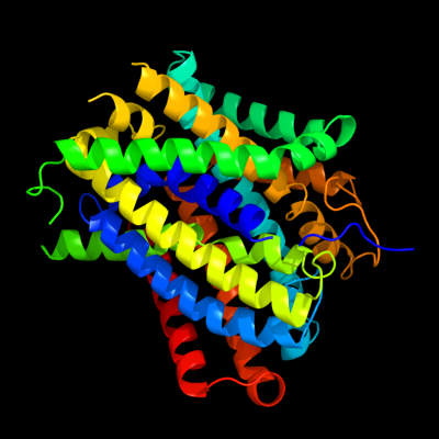
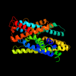
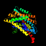
PDB 3gia chain A
Region: 1 - 391
Aligned: 387
Modelled: 387
Confidence: 100.0%
Identity: 12%
PDB header:transport protein
Chain: A: PDB Molecule:uncharacterized protein mj0609;
PDBTitle: crystal structure of apct transporter
Phyre2
| 2 |
|
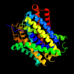
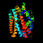
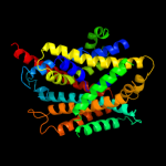
PDB 3lrc chain C
Region: 2 - 389
Aligned: 366
Modelled: 366
Confidence: 100.0%
Identity: 15%
PDB header:transport protein
Chain: C: PDB Molecule:arginine/agmatine antiporter;
PDBTitle: structure of e. coli adic (p1)
Phyre2
| 3 |
|
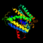
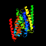
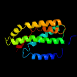
PDB 2jln chain A
Region: 2 - 396
Aligned: 387
Modelled: 387
Confidence: 100.0%
Identity: 11%
PDB header:membrane protein
Chain: A: PDB Molecule:mhp1;
PDBTitle: structure of mhp1, a nucleobase-cation-symport-1 family2 transporter
Phyre2
| 4 |
|
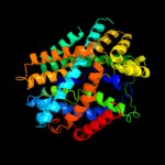
PDB 2xq2 chain A
Region: 10 - 393
Aligned: 368
Modelled: 368
Confidence: 99.0%
Identity: 9%
PDB header:transport protein
Chain: A: PDB Molecule:sodium/glucose cotransporter;
PDBTitle: structure of the k294a mutant of vsglt
Phyre2
| 5 |
|
PDB 3dh4 chain A
Region: 2 - 393
Aligned: 375
Modelled: 392
Confidence: 98.9%
Identity: 10%
PDB header:transport protein
Chain: A: PDB Molecule:sodium/glucose cotransporter;
PDBTitle: crystal structure of sodium/sugar symporter with bound galactose from2 vibrio parahaemolyticus
Phyre2
| 6 |
|
PDB 2a65 chain A domain 1
Region: 3 - 400
Aligned: 395
Modelled: 398
Confidence: 98.4%
Identity: 14%
Fold: SNF-like
Superfamily: SNF-like
Family: SNF-like
Phyre2
| 7 |
|
PDB 2w8a chain C
Region: 4 - 369
Aligned: 366
Modelled: 366
Confidence: 96.4%
Identity: 12%
PDB header:membrane protein
Chain: C: PDB Molecule:glycine betaine transporter betp;
PDBTitle: crystal structure of the sodium-coupled glycine betaine2 symporter betp from corynebacterium glutamicum with bound3 substrate
Phyre2
| 8 |
|
PDB 3hfx chain A
Region: 4 - 365
Aligned: 362
Modelled: 362
Confidence: 84.8%
Identity: 12%
PDB header:transport protein
Chain: A: PDB Molecule:l-carnitine/gamma-butyrobetaine antiporter;
PDBTitle: crystal structure of carnitine transporter
Phyre2
| 9 |
|
PDB 1iwg chain A domain 8
Region: 205 - 365
Aligned: 161
Modelled: 161
Confidence: 6.3%
Identity: 7%
Fold: Multidrug efflux transporter AcrB transmembrane domain
Superfamily: Multidrug efflux transporter AcrB transmembrane domain
Family: Multidrug efflux transporter AcrB transmembrane domain
Phyre2