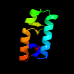
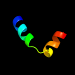
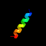
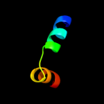
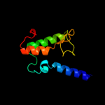
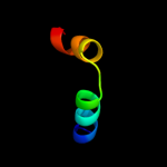
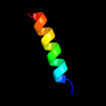
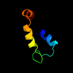
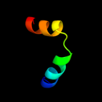
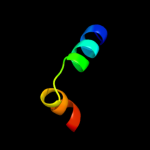
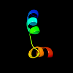
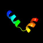
1 d1oeya_
77.0
37
Fold: beta-Grasp (ubiquitin-like)Superfamily: CAD & PB1 domainsFamily: PB1 domain2 d2o3la1
53.0
16
Fold: Left-handed superhelixSuperfamily: BH3980-likeFamily: BH3980-like3 d1gt0d_
50.3
26
Fold: HMG-boxSuperfamily: HMG-boxFamily: HMG-box4 c2wvrB_
49.8
11
PDB header: replicationChain: B: PDB Molecule: geminin;PDBTitle: human cdt1:geminin complex
5 d1wgfa_
47.1
22
Fold: HMG-boxSuperfamily: HMG-boxFamily: HMG-box6 d2hh6a1
46.6
14
Fold: Left-handed superhelixSuperfamily: BH3980-likeFamily: BH3980-like7 c2e6oA_
46.0
26
PDB header: transcription, cell cycleChain: A: PDB Molecule: hmg box-containing protein 1;PDBTitle: solution structure of the hmg box domain from human hmg-box2 transcription factor 1
8 c2zxxA_
44.4
11
PDB header: cell cycle/replicationChain: A: PDB Molecule: geminin;PDBTitle: crystal structure of cdt1/geminin complex
9 c3u2bC_
44.3
17
PDB header: transcription/dnaChain: C: PDB Molecule: transcription factor sox-4;PDBTitle: structure of the sox4 hmg domain bound to dna
10 d2lefa_
42.8
17
Fold: HMG-boxSuperfamily: HMG-boxFamily: HMG-box11 c2eqzA_
42.0
22
PDB header: transcriptionChain: A: PDB Molecule: high mobility group protein b3;PDBTitle: solution structure of the first hmg-box domain from high2 mobility group protein b3
12 c1j3xA_
41.5
22
PDB header: dna binding proteinChain: A: PDB Molecule: high mobility group protein 2;PDBTitle: solution structure of the n-terminal domain of the hmgb2
13 d1j3xa_
41.5
22
Fold: HMG-boxSuperfamily: HMG-boxFamily: HMG-box14 d2gzka2
40.1
26
Fold: HMG-boxSuperfamily: HMG-boxFamily: HMG-box15 c1hryA_
40.1
26
PDB header: dna binding protein/dnaChain: A: PDB Molecule: human sry;PDBTitle: the 3d structure of the human sry-dna complex solved by2 multid-dimensional heteronuclear-edited and-filtered nmr
16 c1hrzA_
40.1
26
PDB header: dna binding protein/dnaChain: A: PDB Molecule: human sry;PDBTitle: the 3d structure of the human sry-dna complex solved by2 multi-dimensional heteronuclear-edited and-filtered nmr
17 c2cs1A_
39.3
22
PDB header: dna binding proteinChain: A: PDB Molecule: pms1 protein homolog 1;PDBTitle: solution structure of the hmg domain of human dna mismatch2 repair protein
18 d1j46a_
39.2
26
Fold: HMG-boxSuperfamily: HMG-boxFamily: HMG-box19 c2d7lA_
38.3
4
PDB header: gene regulation, dna binding proteinChain: A: PDB Molecule: wd repeat and hmg-box dna binding protein 1;PDBTitle: solution structure of the hmg box domain from human wd2 repeat and hmg-box dna binding protein 1
20 c2yulA_
38.3
26
PDB header: transcriptionChain: A: PDB Molecule: transcription factor sox-17;PDBTitle: solution structure of the hmg box of human transcription2 factor sox-17
21 c1wz6A_
not modelled
37.4
22
PDB header: transcriptionChain: A: PDB Molecule: hmg-box transcription factor bbx;PDBTitle: solution structure of the hmg_box domain of murine bobby2 sox homolog
22 c3fghA_
not modelled
35.7
13
PDB header: transcriptionChain: A: PDB Molecule: transcription factor a, mitochondrial;PDBTitle: human mitochondrial transcription factor a box b
23 c2crjA_
not modelled
33.0
26
PDB header: gene regulationChain: A: PDB Molecule: swi/snf-related matrix-associated actin-PDBTitle: solution structure of the hmg domain of mouse hmg domain2 protein hmgx2
24 c1hmfA_
not modelled
31.5
17
PDB header: dna-bindingChain: A: PDB Molecule: high mobility group protein fragment-b;PDBTitle: structure of the hmg box motif in the b-domain of hmg1
25 d1hmfa_
not modelled
31.5
17
Fold: HMG-boxSuperfamily: HMG-boxFamily: HMG-box26 d1jhga_
not modelled
30.5
18
Fold: DNA/RNA-binding 3-helical bundleSuperfamily: TrpR-likeFamily: Trp repressor, TrpR27 d1lwma_
not modelled
30.0
26
Fold: HMG-boxSuperfamily: HMG-boxFamily: HMG-box28 c2co9A_
not modelled
28.5
22
PDB header: transcriptionChain: A: PDB Molecule: thymus high mobility group box protein tox;PDBTitle: solution structure of the hmg_box domain of thymus high2 mobility group box protein tox from mouse
29 c2jvfA_
not modelled
28.2
28
PDB header: de novo proteinChain: A: PDB Molecule: de novo protein m7;PDBTitle: solution structure of m7, a computationally-designed2 artificial protein
30 d1v54m_
not modelled
28.0
18
Fold: Single transmembrane helixSuperfamily: Mitochondrial cytochrome c oxidase subunit VIIIb (aka IX)Family: Mitochondrial cytochrome c oxidase subunit VIIIb (aka IX)31 c1qysA_
not modelled
27.7
13
PDB header: de novo proteinChain: A: PDB Molecule: top7;PDBTitle: crystal structure of top7: a computationally designed2 protein with a novel fold
32 d1i11a_
not modelled
27.2
26
Fold: HMG-boxSuperfamily: HMG-boxFamily: HMG-box33 c1aabA_
not modelled
27.0
22
PDB header: dna-bindingChain: A: PDB Molecule: high mobility group protein;PDBTitle: nmr structure of rat hmg1 hmga fragment
34 d1aaba_
not modelled
27.0
22
Fold: HMG-boxSuperfamily: HMG-boxFamily: HMG-box35 d1k99a_
not modelled
26.0
22
Fold: HMG-boxSuperfamily: HMG-boxFamily: HMG-box36 d1ckta_
not modelled
26.0
22
Fold: HMG-boxSuperfamily: HMG-boxFamily: HMG-box37 d2o4ta1
not modelled
25.9
16
Fold: Left-handed superhelixSuperfamily: BH3980-likeFamily: BH3980-like38 d1hsma_
not modelled
24.2
17
Fold: HMG-boxSuperfamily: HMG-boxFamily: HMG-box39 d1j3da_
not modelled
22.5
17
Fold: HMG-boxSuperfamily: HMG-boxFamily: HMG-box40 c2qguA_
not modelled
22.4
11
PDB header: structural genomics, unknown functionChain: A: PDB Molecule: probable signal peptide protein;PDBTitle: three-dimensional structure of the phospholipid-binding protein from2 ralstonia solanacearum q8xv73_ralsq in complex with a phospholipid at3 the resolution 1.53 a. northeast structural genomics consortium4 target rsr89
41 d1r8ja1
not modelled
22.2
13
Fold: KaiA/RbsU domainSuperfamily: KaiA/RbsU domainFamily: Circadian clock protein KaiA, C-terminal domain42 c3cxjB_
not modelled
21.7
19
PDB header: structural genomics, unknown functionChain: B: PDB Molecule: uncharacterized protein;PDBTitle: crystal structure of an uncharacterized protein from2 methanothermobacter thermautotrophicus
43 c3frwF_
not modelled
20.8
15
PDB header: structural genomics, unknown functionChain: F: PDB Molecule: putative trp repressor protein;PDBTitle: crystal structure of putative trpr protein from ruminococcus obeum
44 d1puza_
not modelled
20.8
16
Fold: YgfY-likeSuperfamily: YgfY-likeFamily: YgfY-like45 c1e1hD_
not modelled
20.4
42
PDB header: hydrolaseChain: D: PDB Molecule: botulinum neurotoxin type a light chain;PDBTitle: crystal structure of recombinant botulinum neurotoxin type2 a light chain, self-inhibiting zn endopeptidase.
46 d1cc5a_
not modelled
20.0
19
Fold: Cytochrome cSuperfamily: Cytochrome cFamily: monodomain cytochrome c47 d1v64a_
not modelled
19.2
13
Fold: HMG-boxSuperfamily: HMG-boxFamily: HMG-box48 d1gl4a1
not modelled
18.7
35
Fold: GFP-likeSuperfamily: GFP-likeFamily: Domain G2 of nidogen-149 d1v63a_
not modelled
18.7
9
Fold: HMG-boxSuperfamily: HMG-boxFamily: HMG-box50 c1x6iB_
not modelled
18.3
13
PDB header: structural genomics, unknown functionChain: B: PDB Molecule: hypothetical protein ygfy;PDBTitle: crystal structure of ygfy from escherichia coli
51 d1sv1a_
not modelled
18.0
13
Fold: KaiA/RbsU domainSuperfamily: KaiA/RbsU domainFamily: Circadian clock protein KaiA, C-terminal domain52 c2awyB_
not modelled
17.7
11
PDB header: oxygen storage/transportChain: B: PDB Molecule: hemerythrin-like domain protein dcrh;PDBTitle: met-dcrh-hr
53 c2y69Z_
not modelled
17.6
19
PDB header: electron transportChain: Z: PDB Molecule: cytochrome c oxidase polypeptide 8h;PDBTitle: bovine heart cytochrome c oxidase re-refined with molecular2 oxygen
54 c2l66B_
not modelled
17.1
60
PDB header: transcription regulatorChain: B: PDB Molecule: transcriptional regulator, abrb family;PDBTitle: the dna-recognition fold of sso7c4 suggests a new member of spovt-abrb2 superfamily from archaea.
55 c1rh1A_
not modelled
16.9
14
PDB header: antibioticChain: A: PDB Molecule: colicin b;PDBTitle: crystal structure of the cytotoxic bacterial protein2 colicin b at 2.5 a resolution
56 c3lisB_
not modelled
16.9
16
PDB header: transcriptionChain: B: PDB Molecule: csp231i c protein;PDBTitle: crystal structure of the restriction-modification controller protein2 c.csp231i (monoclinic form)
57 c2rlwA_
not modelled
16.6
30
PDB header: toxinChain: A: PDB Molecule: plnf;PDBTitle: three-dimensional structure of the two peptides that2 constitute the two-peptide bacteriocin plantaracin ef
58 c1h4uA_
not modelled
16.5
35
PDB header: extracellular matrix proteinChain: A: PDB Molecule: nidogen-1;PDBTitle: domain g2 of mouse nidogen-1
59 d2je8a5
not modelled
16.1
13
Fold: TIM beta/alpha-barrelSuperfamily: (Trans)glycosidasesFamily: beta-glycanases60 d1vp8a_
not modelled
16.1
21
Fold: Pyruvate kinase C-terminal domain-likeSuperfamily: PK C-terminal domain-likeFamily: MTH1675-like61 c3korD_
not modelled
16.0
19
PDB header: transcriptionChain: D: PDB Molecule: possible trp repressor;PDBTitle: crystal structure of a putative trp repressor from staphylococcus2 aureus
62 d2al3a1
not modelled
15.7
14
Fold: beta-Grasp (ubiquitin-like)Superfamily: Ubiquitin-likeFamily: UBX domain63 d2akja1
not modelled
15.5
18
Fold: Ferredoxin-likeSuperfamily: Nitrite/Sulfite reductase N-terminal domain-likeFamily: Duplicated SiR/NiR-like domains 1 and 364 d1kx7a_
not modelled
14.9
11
Fold: Cytochrome cSuperfamily: Cytochrome cFamily: monodomain cytochrome c65 c2lm4A_
not modelled
14.6
26
PDB header: protein bindingChain: A: PDB Molecule: succinate dehydrogenase assembly factor 2, mitochondrial;PDBTitle: solution nmr structure of mitochondrial succinate dehydrogenase2 assembly factor 2 from saccharomyces cerevisiae, northeast structural3 genomics consortium target yt682a
66 c2jr5A_
not modelled
14.4
13
PDB header: structural genomics, unknown functionChain: A: PDB Molecule: upf0350 protein vc_2471;PDBTitle: solution structure of upf0350 protein vc_2471. northeast2 structural genomics target vcr36
67 d1ctda_
not modelled
14.4
38
Fold: EF Hand-likeSuperfamily: EF-handFamily: Calmodulin-like68 d1tlha_
not modelled
13.8
16
Fold: Anti-sigma factor AsiASuperfamily: Anti-sigma factor AsiAFamily: Anti-sigma factor AsiA69 c1r8jB_
not modelled
13.8
13
PDB header: circadian clock proteinChain: B: PDB Molecule: kaia;PDBTitle: crystal structure of circadian clock protein kaia from2 synechococcus elongatus
70 c1ojhK_
not modelled
13.7
17
PDB header: protein bindingChain: K: PDB Molecule: nbla;PDBTitle: crystal structure of nbla from pcc 7120
71 d1m47a_
not modelled
13.7
24
Fold: 4-helical cytokinesSuperfamily: 4-helical cytokinesFamily: Short-chain cytokines72 c2nxbB_
not modelled
13.1
13
PDB header: signaling proteinChain: B: PDB Molecule: bromodomain-containing protein 3;PDBTitle: crystal structure of human bromodomain containing protein 3 (brd3)
73 d1ojha_
not modelled
12.5
17
Fold: NblA-likeSuperfamily: NblA-likeFamily: NblA-like74 c3lpgA_
not modelled
12.3
15
PDB header: hydrolase/hydrolase inhibitorChain: A: PDB Molecule: beta-glucuronidase;PDBTitle: structure of e. coli beta-glucuronidase bound with a novel, potent2 inhibitor 3-(2-fluorophenyl)-1-(2-hydroxyethyl)-1-((6-methyl-2-oxo-1,3 2-dihydroquinolin-3-yl)methyl)urea
75 c2je8B_
not modelled
11.8
15
PDB header: hydrolaseChain: B: PDB Molecule: beta-mannosidase;PDBTitle: structure of a beta-mannosidase from bacteroides2 thetaiotaomicron
76 d1fcdc1
not modelled
11.6
11
Fold: Cytochrome cSuperfamily: Cytochrome cFamily: Two-domain cytochrome c77 c3d0wD_
not modelled
11.6
26
PDB header: structural genomics, unknown functionChain: D: PDB Molecule: yflh protein;PDBTitle: crystal structure of yflh protein from bacillus subtilis.2 northeast structural genomics consortium target sr326
78 c3bzjA_
not modelled
11.6
22
PDB header: hydrolaseChain: A: PDB Molecule: uv endonuclease;PDBTitle: uvde k229l
79 d1pgja1
not modelled
11.3
16
Fold: 6-phosphogluconate dehydrogenase C-terminal domain-likeSuperfamily: 6-phosphogluconate dehydrogenase C-terminal domain-likeFamily: Hydroxyisobutyrate and 6-phosphogluconate dehydrogenase domain80 c2zxyA_
not modelled
11.2
22
PDB header: oxygen binding, transport proteinChain: A: PDB Molecule: cytochrome c552;PDBTitle: crystal structure of cytochrome c555 from aquifex aeolicus
81 d1ql3a_
not modelled
11.2
28
Fold: Cytochrome cSuperfamily: Cytochrome cFamily: monodomain cytochrome c82 d1rh1a2
not modelled
11.1
16
Fold: Toxins' membrane translocation domainsSuperfamily: ColicinFamily: Colicin83 c1zb7A_
not modelled
10.8
50
PDB header: toxinChain: A: PDB Molecule: neurotoxin;PDBTitle: crystal structure of botulinum neurotoxin type g light chain
84 d1dvha_
not modelled
10.7
22
Fold: Cytochrome cSuperfamily: Cytochrome cFamily: monodomain cytochrome c85 d1peqa1
not modelled
10.5
7
Fold: R1 subunit of ribonucleotide reductase, N-terminal domainSuperfamily: R1 subunit of ribonucleotide reductase, N-terminal domainFamily: R1 subunit of ribonucleotide reductase, N-terminal domain86 c2r7tA_
not modelled
10.5
24
PDB header: transferase/rnaChain: A: PDB Molecule: rna-dependent rna polymerase;PDBTitle: crystal structure of rotavirus sa11 vp1/rna (ugugaacc)2 complex
87 d1c53a_
not modelled
10.5
17
Fold: Cytochrome cSuperfamily: Cytochrome cFamily: monodomain cytochrome c88 c3ogrA_
not modelled
10.5
23
PDB header: hydrolaseChain: A: PDB Molecule: beta-galactosidase;PDBTitle: complex structure of beta-galactosidase from trichoderma reesei with2 galactose
89 d1v2za_
not modelled
10.4
12
Fold: KaiA/RbsU domainSuperfamily: KaiA/RbsU domainFamily: Circadian clock protein KaiA, C-terminal domain90 d1txla_
not modelled
10.1
20
Fold: LipocalinsSuperfamily: LipocalinsFamily: Hypothetical protein YodA91 c1txlA_
not modelled
10.1
20
PDB header: structural genomics, unknown functionChain: A: PDB Molecule: metal-binding protein yoda;PDBTitle: crystal structure of metal-binding protein yoda from e.2 coli, pfam duf149
92 d1zj8a1
not modelled
10.1
18
Fold: Ferredoxin-likeSuperfamily: Nitrite/Sulfite reductase N-terminal domain-likeFamily: Duplicated SiR/NiR-like domains 1 and 393 c2zonG_
not modelled
9.9
13
PDB header: oxidoreductase/electron transportChain: G: PDB Molecule: cytochrome c551;PDBTitle: crystal structure of electron transfer complex of nitrite2 reductase with cytochrome c
94 d1h9xa1
not modelled
9.8
28
Fold: Cytochrome cSuperfamily: Cytochrome cFamily: N-terminal (heme c) domain of cytochrome cd1-nitrite reductase95 c3ivpD_
not modelled
9.7
9
PDB header: dna binding proteinChain: D: PDB Molecule: putative transposon-related dna-binding protein;PDBTitle: the structure of a possible transposon-related dna-binding protein2 from clostridium difficile 630.
96 c3op9A_
not modelled
9.7
18
PDB header: transcription regulatorChain: A: PDB Molecule: pli0006 protein;PDBTitle: crystal structure of transcriptional regulator from listeria innocua
97 c2da4A_
not modelled
9.6
10
PDB header: structural genomics, unknown functionChain: A: PDB Molecule: hypothetical protein dkfzp686k21156;PDBTitle: solution structure of the homeobox domain of the2 hypothetical protein, dkfzp686k21156
98 c2xhlA_
not modelled
9.6
50
PDB header: hydrolaseChain: A: PDB Molecule: botulinum neurotoxin b light chain;PDBTitle: structure of a functional derivative of clostridium2 botulinum neurotoxin type b
99 c2xrhA_
not modelled
9.4
17
PDB header: unknown functionChain: A: PDB Molecule: protein hp0721;PDBTitle: crystal structure of the truncated form of hp0721


