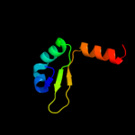
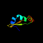
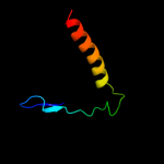
1 d2a6qa1
100.0
100
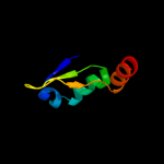
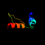
Fold: YefM-likeSuperfamily: YefM-likeFamily: YefM-like2 c3g5oA_
99.9
27
PDB header: toxin/antitoxinChain: A: PDB Molecule: uncharacterized protein rv2865;PDBTitle: the crystal structure of the toxin-antitoxin complex relbe2 (rv2865-2 2866) from mycobacterium tuberculosis
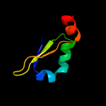
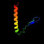
3 d2a6qb1
99.8
100
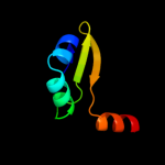
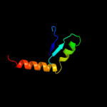
Fold: YefM-likeSuperfamily: YefM-likeFamily: YefM-like4 c3hs2H_
99.6
29
PDB header: antitoxinChain: H: PDB Molecule: prevent host death protein;PDBTitle: crystal structure of phd truncated to residue 57 in an orthorhombic2 space group
5 c3hryA_
99.5
27
PDB header: antitoxinChain: A: PDB Molecule: prevent host death protein;PDBTitle: crystal structure of phd in a trigonal space group and partially2 disordered
6 c2odkD_
99.2
18
PDB header: structural genomics, unknown functionChain: D: PDB Molecule: hypothetical protein;PDBTitle: putative prevent-host-death protein from nitrosomonas europaea
7 d2odka1
99.1
18
Fold: YefM-likeSuperfamily: YefM-likeFamily: YefM-like8 c3k6qB_
95.4
27
PDB header: ligand binding proteinChain: B: PDB Molecule: putative ligand binding protein;PDBTitle: crystal structure of an antitoxin part of a putative toxin/antitoxin2 system (swol_0700) from syntrophomonas wolfei subsp. wolfei at 1.80 a3 resolution
9 d1grja1
35.3
21
Fold: Long alpha-hairpinSuperfamily: GreA transcript cleavage protein, N-terminal domainFamily: GreA transcript cleavage protein, N-terminal domain10 c2iyfA_
30.5
30
PDB header: transferaseChain: A: PDB Molecule: oleandomycin glycosyltransferase;PDBTitle: the crystal structure of macrolide glycosyltransferases: a2 blueprint for antibiotic engineering
11 d1n0ea_
27.2
14
Fold: Double-split beta-barrelSuperfamily: AbrB/MazE/MraZ-likeFamily: Hypothetical protein MraZ12 d1ez4a2
27.0
16
Fold: LDH C-terminal domain-likeSuperfamily: LDH C-terminal domain-likeFamily: Lactate & malate dehydrogenases, C-terminal domain13 c1n0fF_
21.8
14
PDB header: biosynthetic proteinChain: F: PDB Molecule: protein mraz;PDBTitle: crystal structure of a cell division and cell wall2 biosynthesis protein upf0040 from mycoplasma pneumoniae:3 indication of a novel fold with a possible new conserved4 sequence motif
14 d1pzga2
20.0
23
Fold: LDH C-terminal domain-likeSuperfamily: LDH C-terminal domain-likeFamily: Lactate & malate dehydrogenases, C-terminal domain15 c2p6pB_
18.5
18
PDB header: transferaseChain: B: PDB Molecule: glycosyl transferase;PDBTitle: x-ray crystal structure of c-c bond-forming dtdp-d-olivose-transferase2 urdgt2
16 c2iyaB_
16.8
15
PDB header: transferaseChain: B: PDB Molecule: oleandomycin glycosyltransferase;PDBTitle: the crystal structure of macrolide glycosyltransferases: a2 blueprint for antibiotic engineering
17 d1y0na_
16.6
19
Fold: YehU-likeSuperfamily: YehU-likeFamily: YehU-like18 c3othB_
15.2
15
PDB header: transferase/antibioticChain: B: PDB Molecule: calg1;PDBTitle: crystal structure of calg1, calicheamicin glycostyltransferase, tdp2 and calicheamicin alpha3i bound form
19 c3ia7A_
15.1
16
PDB header: transferaseChain: A: PDB Molecule: calg4;PDBTitle: crystal structure of calg4, the calicheamicin glycosyltransferase
20 c2qzsA_
14.3
12
PDB header: transferaseChain: A: PDB Molecule: glycogen synthase;PDBTitle: crystal structure of wild-type e.coli gs in complex with adp2 and glucose(wtgsb)
21 d2iw1a1
not modelled
12.7
15
Fold: UDP-Glycosyltransferase/glycogen phosphorylaseSuperfamily: UDP-Glycosyltransferase/glycogen phosphorylaseFamily: Glycosyl transferases group 122 c3dzcA_
not modelled
12.6
23
PDB header: isomeraseChain: A: PDB Molecule: udp-n-acetylglucosamine 2-epimerase;PDBTitle: 2.35 angstrom resolution structure of wecb (vc0917), a udp-n-2 acetylglucosamine 2-epimerase from vibrio cholerae.
23 d1f6da_
not modelled
11.9
21
Fold: UDP-Glycosyltransferase/glycogen phosphorylaseSuperfamily: UDP-Glycosyltransferase/glycogen phosphorylaseFamily: UDP-N-acetylglucosamine 2-epimerase24 d1g8ma1
not modelled
11.9
8
Fold: Methylglyoxal synthase-likeSuperfamily: Methylglyoxal synthase-likeFamily: Inosicase25 c2p4vA_
not modelled
11.8
18
PDB header: transcriptionChain: A: PDB Molecule: transcription elongation factor greb;PDBTitle: crystal structure of the transcript cleavage factor, greb2 at 2.6a resolution
26 c2jjmH_
not modelled
11.7
20
PDB header: transferaseChain: H: PDB Molecule: glycosyl transferase, group 1 family protein;PDBTitle: crystal structure of a family gt4 glycosyltransferase from2 bacillus anthracis orf ba1558.
27 c1grjA_
not modelled
11.6
21
PDB header: transcription regulationChain: A: PDB Molecule: grea protein;PDBTitle: grea transcript cleavage factor from escherichia coli
28 d2vgna2
not modelled
11.6
20
Fold: Ribonuclease H-like motifSuperfamily: Translational machinery componentsFamily: ERF1/Dom34 middle domain-like29 c3iaaB_
not modelled
11.3
15
PDB header: transferaseChain: B: PDB Molecule: calg2;PDBTitle: crystal structure of calg2, calicheamicin glycosyltransferase, tdp2 bound form
30 c1ez4B_
not modelled
10.6
16
PDB header: oxidoreductaseChain: B: PDB Molecule: lactate dehydrogenase;PDBTitle: crystal structure of non-allosteric l-lactate dehydrogenase2 from lactobacillus pentosus at 2.3 angstrom resolution
31 c3s29C_
not modelled
10.2
14
PDB header: transferaseChain: C: PDB Molecule: sucrose synthase 1;PDBTitle: the crystal structure of sucrose synthase-1 from arabidopsis thaliana2 and its functional implications.
32 c2r60A_
not modelled
9.0
17
PDB header: transferaseChain: A: PDB Molecule: glycosyl transferase, group 1;PDBTitle: structure of apo sucrose phosphate synthase (sps) of2 halothermothrix orenii
33 c3zvkG_
not modelled
9.0
3
PDB header: antitoxin/toxin/dnaChain: G: PDB Molecule: antitoxin of toxin-antitoxin system vapb;PDBTitle: crystal structure of vapbc2 from rickettsia felis bound to2 a dna fragment from their promoter
34 d1a5za2
not modelled
8.5
21
Fold: LDH C-terminal domain-likeSuperfamily: LDH C-terminal domain-likeFamily: Lactate & malate dehydrogenases, C-terminal domain35 c3ot5D_
not modelled
8.3
16
PDB header: isomeraseChain: D: PDB Molecule: udp-n-acetylglucosamine 2-epimerase;PDBTitle: 2.2 angstrom resolution crystal structure of putative udp-n-2 acetylglucosamine 2-epimerase from listeria monocytogenes
36 d1o6ca_
not modelled
7.7
18
Fold: UDP-Glycosyltransferase/glycogen phosphorylaseSuperfamily: UDP-Glycosyltransferase/glycogen phosphorylaseFamily: UDP-N-acetylglucosamine 2-epimerase37 d1mlda2
not modelled
7.6
19
Fold: LDH C-terminal domain-likeSuperfamily: LDH C-terminal domain-likeFamily: Lactate & malate dehydrogenases, C-terminal domain38 d2cmda2
not modelled
7.5
19
Fold: LDH C-terminal domain-likeSuperfamily: LDH C-terminal domain-likeFamily: Lactate & malate dehydrogenases, C-terminal domain39 c2fy2A_
not modelled
6.7
16
PDB header: transferaseChain: A: PDB Molecule: choline o-acetyltransferase;PDBTitle: structures of ligand bound human choline acetyltransferase2 provide insight into regulation of acetylcholine synthesis
40 c1zrxA_
not modelled
6.7
17
PDB header: antimicrobial protein, antibioticChain: A: PDB Molecule: stomoxyn;PDBTitle: solution structure of stomoxyn in h20/tfe 50%
41 c1smkD_
not modelled
6.5
19
PDB header: oxidoreductaseChain: D: PDB Molecule: malate dehydrogenase, glyoxysomal;PDBTitle: mature and translocatable forms of glyoxysomal malate2 dehydrogenase have different activities and stabilities3 but similar crystal structures
42 c1sevA_
not modelled
6.5
19
PDB header: oxidoreductaseChain: A: PDB Molecule: malate dehydrogenase, glyoxysomal precursor;PDBTitle: mature and translocatable forms of glyoxysomal malate2 dehydrogenase have different activities and stabilities3 but similar crystal structures
43 d1uxja2
not modelled
6.4
23
Fold: LDH C-terminal domain-likeSuperfamily: LDH C-terminal domain-likeFamily: Lactate & malate dehydrogenases, C-terminal domain44 d1ghca_
not modelled
5.8
14
Fold: DNA/RNA-binding 3-helical bundleSuperfamily: "Winged helix" DNA-binding domainFamily: Linker histone H1/H545 c1vjqB_
not modelled
5.7
14
PDB header: structural genomics, de novo proteinChain: B: PDB Molecule: designed protein;PDBTitle: designed protein based on backbone conformation of2 procarboxypeptidase-a (1aye) with sidechains chosen for maximal3 predicted stability.
46 d1gv0a2
not modelled
5.5
18
Fold: LDH C-terminal domain-likeSuperfamily: LDH C-terminal domain-likeFamily: Lactate & malate dehydrogenases, C-terminal domain47 c2zqeA_
not modelled
5.3
14
PDB header: dna binding proteinChain: A: PDB Molecule: muts2 protein;PDBTitle: crystal structure of the smr domain of thermus thermophilus muts2
48 d1pkxa1
not modelled
5.1
13
Fold: Methylglyoxal synthase-likeSuperfamily: Methylglyoxal synthase-likeFamily: Inosicase