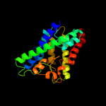
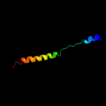
1 c3klzE_
100.0
23
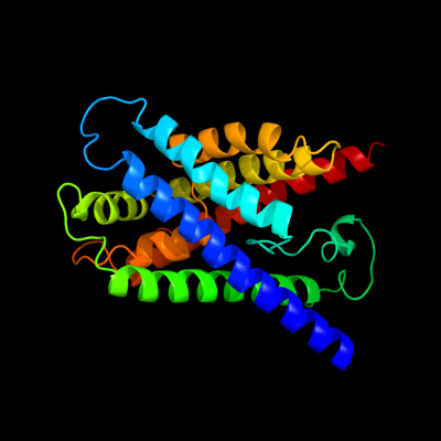
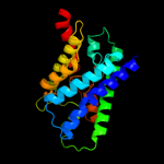
PDB header: membrane proteinChain: E: PDB Molecule: putative formate transporter 1;PDBTitle: pentameric formate channel with formate bound
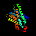
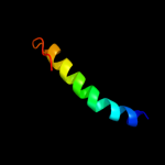
2 c3kcvG_
100.0
18
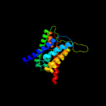
PDB header: transport proteinChain: G: PDB Molecule: probable formate transporter 1;PDBTitle: structure of formate channel
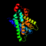
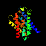
3 c3llqB_
85.2
9
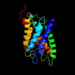
PDB header: membrane proteinChain: B: PDB Molecule: aquaporin z 2;PDBTitle: aquaporin structure from plant pathogen agrobacterium tumerfaciens
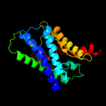
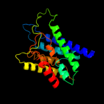
4 c3c02A_
83.5
14
PDB header: membrane proteinChain: A: PDB Molecule: aquaglyceroporin;PDBTitle: x-ray structure of the aquaglyceroporin from plasmodium falciparum
5 c1ymgA_
74.1
10
PDB header: membrane proteinChain: A: PDB Molecule: lens fiber major intrinsic protein;PDBTitle: the channel architecture of aquaporin o at 2.2 angstrom resolution
6 d1ymga1
74.1
10
Fold: Aquaporin-likeSuperfamily: Aquaporin-likeFamily: Aquaporin-like7 c1ldaA_
74.0
11
PDB header: transport proteinChain: A: PDB Molecule: glycerol uptake facilitator protein;PDBTitle: crystal structure of the e. coli glycerol facilitator (glpf) without2 substrate glycerol
8 d1fx8a_
74.0
11
Fold: Aquaporin-likeSuperfamily: Aquaporin-likeFamily: Aquaporin-like9 c2kncA_
58.8
12
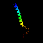
PDB header: cell adhesionChain: A: PDB Molecule: integrin alpha-iib;PDBTitle: platelet integrin alfaiib-beta3 transmembrane-cytoplasmic2 heterocomplex
10 c2w2eA_
58.8
13
PDB header: membrane proteinChain: A: PDB Molecule: aquaporin;PDBTitle: 1.15 angstrom crystal structure of p.pastoris aquaporin,2 aqy1, in a closed conformation at ph 3.5
11 d1rc2a_
45.2
7
Fold: Aquaporin-likeSuperfamily: Aquaporin-likeFamily: Aquaporin-like12 c2kncB_
45.0
24
PDB header: cell adhesionChain: B: PDB Molecule: integrin beta-3;PDBTitle: platelet integrin alfaiib-beta3 transmembrane-cytoplasmic2 heterocomplex
13 c3qnqD_
38.7
16
PDB header: membrane protein, transport proteinChain: D: PDB Molecule: pts system, cellobiose-specific iic component;PDBTitle: crystal structure of the transporter chbc, the iic component from the2 n,n'-diacetylchitobiose-specific phosphotransferase system
14 c2b5fD_
37.4
12
PDB header: transport protein,membrane proteinChain: D: PDB Molecule: aquaporin;PDBTitle: crystal structure of the spinach aquaporin sopip2;1 in an2 open conformation to 3.9 resolution
15 d1c17m_
24.6
39
Fold: F1F0 ATP synthase subunit ASuperfamily: F1F0 ATP synthase subunit AFamily: F1F0 ATP synthase subunit A16 c3iyzA_
19.1
12
PDB header: transport proteinChain: A: PDB Molecule: aquaporin-4;PDBTitle: structure of aquaporin-4 s180d mutant at 10.0 a resolution from2 electron micrograph
17 c3d9sB_
12.1
11
PDB header: membrane proteinChain: B: PDB Molecule: aquaporin-5;PDBTitle: human aquaporin 5 (aqp5) - high resolution x-ray structure
18 c3s0xB_
11.8
14
PDB header: hydrolaseChain: B: PDB Molecule: peptidase a24b, flak domain protein;PDBTitle: the crystal structure of gxgd membrane protease flak
19 c2jy0A_
10.5
33
PDB header: membrane protein, viral proteinChain: A: PDB Molecule: protease ns2-3;PDBTitle: solution nmr structure of hcv ns2 protein, membrane segment2 (1-27)
20 c2h3oA_
9.6
19
PDB header: membrane proteinChain: A: PDB Molecule: merf;PDBTitle: structure of merft, a membrane protein with two trans-2 membrane helices
21 d2c1wa1
not modelled
7.4
33
Fold: EndoU-likeSuperfamily: EndoU-likeFamily: Eukaryotic EndoU ribonuclease22 c2q7cC_
not modelled
7.2
29
PDB header: viral proteinChain: C: PDB Molecule: fusion protein between yeast variant gcn4 andPDBTitle: crystal structure of iqn17
23 c2ywxA_
not modelled
6.9
13
PDB header: lyaseChain: A: PDB Molecule: phosphoribosylaminoimidazole carboxylase catalytic subunit;PDBTitle: crystal structure of phosphoribosylaminoimidazole carboxylase2 catalytic subunit from methanocaldococcus jannaschii
24 c2voyG_
not modelled
6.2
39
PDB header: hydrolaseChain: G: PDB Molecule: sarcoplasmic/endoplasmic reticulum calciumPDBTitle: cryoem model of copa, the copper transporting atpase from2 archaeoglobus fulgidus
25 d1wpna_
not modelled
6.0
19
Fold: DHH phosphoesterasesSuperfamily: DHH phosphoesterasesFamily: Manganese-dependent inorganic pyrophosphatase (family II)26 d2hawa1
not modelled
6.0
19
Fold: DHH phosphoesterasesSuperfamily: DHH phosphoesterasesFamily: Manganese-dependent inorganic pyrophosphatase (family II)27 d1k20a_
not modelled
5.7
9
Fold: DHH phosphoesterasesSuperfamily: DHH phosphoesterasesFamily: Manganese-dependent inorganic pyrophosphatase (family II)28 d1qkla_
not modelled
5.6
19
Fold: RPB6/omega subunit-likeSuperfamily: RPB6/omega subunit-likeFamily: RPB629 d2foka2
not modelled
5.5
40
Fold: DNA/RNA-binding 3-helical bundleSuperfamily: "Winged helix" DNA-binding domainFamily: Restriction endonuclease FokI, N-terminal (recognition) domain30 d1i74a_
not modelled
5.4
9
Fold: DHH phosphoesterasesSuperfamily: DHH phosphoesterasesFamily: Manganese-dependent inorganic pyrophosphatase (family II)31 c1q90L_
not modelled
5.0
11
PDB header: photosynthesisChain: L: PDB Molecule: cytochrome b6f complex subunit petl;PDBTitle: structure of the cytochrome b6f (plastohydroquinone : plastocyanin2 oxidoreductase) from chlamydomonas reinhardtii