Phyre Home Retrieve Phyre Job Id
Emaill.a.kelley@imperial.ac.uk
DescriptionP39317
DateThu Jan 5 11:59:19 GMT 2012
Unique Job ID68f6712da43f894a

Summary 

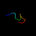
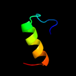
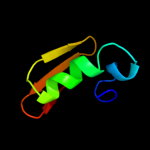
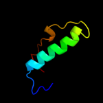
Top model
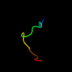
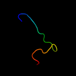
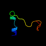
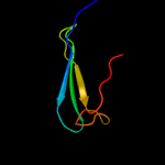
Image coloured by rainbow N → C terminus
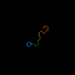
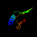
Model (left) based on template d2enga_
Top template information
Fold:Double psi beta-barrel
Superfamily:Barwin-like endoglucanases
Family:Eng V-like
Confidence and coverage
Confidence: 44.9% Coverage: 5%
17 residues ( 5% of your sequence) have been modelled with 44.9% confidence by the single highest scoring template.
You may wish to submit your sequence to Phyrealarm. This will automatically scan your sequence every week for new potential templates as they appear in the Phyre2 library.
Please note: You must be registered and logged in to use Phyrealarm.
3D viewing
Interactive 3D view in Jmol

Sequence analysis 

Secondary structure and disorder prediction 

   1........10.........20.........30.........40.........50.........60
Sequence  MLPRIRHNNFIGAVELFVKSSYTKTHSNNFFNNIHHAFKKKDWISNYDSLLTLREFFRCA
Secondary structure 


SS confidence 


Disorder  ???????


?


Disorder confidence 


   .........70.........80.........90.........100.........110.........120
Sequence  TQIDKSGYQVLSSKNETVHAMDKFLISFSLKDNGAEYTMTLRGSGFEYEEIPITINEYNS
Secondary structure 


SS confidence 


Disorder 


?


Disorder confidence 


   .........130.........140.........150.........160.........170.........180
Sequence  FMDFKNREFPLEQNRRLYAWDILQKKQSDIPKRIKGYIHQAIGDVSLGYALLEDIVSKLK
Secondary structure 


SS confidence 


Disorder 


???????


?
Disorder confidence 


   .........190.........200.........210.........220.........230.........240
Sequence  RGKFELQIPGGGIKECDGWYIYEKIIDDNFAIVIESLGFALKIYGGDERFRNGSSVVLED
Secondary structure 


SS confidence 


Disorder  ??


???
?


Disorder confidence 


   .........250.........260.........270.........280.........290.........300
Sequence  EDYSLIYNFLVNAGCQQVELAEQVDAIVSANLAADSDITKEKICEKYKSTIEAFKKEQLA
Secondary structure 


SS confidence 


Disorder 


?


??
???????


?


Disorder confidence 


   .........310..
Sequence  LPVLVRRKNSET
Secondary structure 


SS confidence 


Disorder 


???????
Disorder confidence 


Confidence Key
High(9)                    Low (0)
?Disordered
Alpha helix
Beta strand

Domain analysis 

Hover over an aligned region to see model and summary info

Please note, only up to the top 20 hits are modelled to reduce computer load

RankAligned region

PDB 2eng chain A

3D model

Region: 182 - 198
Aligned: 17
Modelled: 17
Confidence: 44.9%
Identity: 53%
Fold: Double psi beta-barrel
Superfamily: Barwin-like endoglucanases
Family: Eng V-like

Phyre2

PDB 1l8f chain A

3D model

Region: 179 - 198
Aligned: 20
Modelled: 20
Confidence: 42.4%
Identity: 50%
Fold: Double psi beta-barrel
Superfamily: Barwin-like endoglucanases
Family: Eng V-like

Phyre2

PDB 1miu chain A domain 3

3D model

Region: 196 - 242
Aligned: 47
Modelled: 47
Confidence: 37.0%
Identity: 23%
Fold: OB-fold
Superfamily: Nucleic acid-binding proteins
Family: Single strand DNA-binding domain, SSB

Phyre2

PDB 1iyj chain B domain 3

3D model

Region: 189 - 242
Aligned: 54
Modelled: 54
Confidence: 36.2%
Identity: 22%
Fold: OB-fold
Superfamily: Nucleic acid-binding proteins
Family: Single strand DNA-binding domain, SSB

Phyre2

PDB 1j6x chain A

3D model

Region: 170 - 200
Aligned: 23
Modelled: 29
Confidence: 34.2%
Identity: 35%
Fold: LuxS/MPP-like metallohydrolase
Superfamily: LuxS/MPP-like metallohydrolase
Family: Autoinducer-2 production protein LuxS

Phyre2

PDB 1vje chain A

3D model

Region: 170 - 200
Aligned: 24
Modelled: 31
Confidence: 29.7%
Identity: 42%
Fold: LuxS/MPP-like metallohydrolase
Superfamily: LuxS/MPP-like metallohydrolase
Family: Autoinducer-2 production protein LuxS

Phyre2

PDB 1i5p chain A domain 1

3D model

Region: 88 - 116
Aligned: 29
Modelled: 29
Confidence: 25.6%
Identity: 38%
Fold: Galactose-binding domain-like
Superfamily: Galactose-binding domain-like
Family: delta-Endotoxin, C-terminal domain

Phyre2

PDB 1j98 chain A

3D model

Region: 187 - 200
Aligned: 13
Modelled: 14
Confidence: 24.0%
Identity: 38%
Fold: LuxS/MPP-like metallohydrolase
Superfamily: LuxS/MPP-like metallohydrolase
Family: Autoinducer-2 production protein LuxS

Phyre2

PDB 1j6w chain A

3D model

Region: 187 - 200
Aligned: 13
Modelled: 14
Confidence: 22.6%
Identity: 46%
Fold: LuxS/MPP-like metallohydrolase
Superfamily: LuxS/MPP-like metallohydrolase
Family: Autoinducer-2 production protein LuxS

Phyre2

PDB 2qqp chain D

3D model

Region: 143 - 165
Aligned: 23
Modelled: 23
Confidence: 22.4%
Identity: 48%
PDB header:virus
Chain: D: PDB Molecule:small capsid protein;
PDBTitle: crystal structure of authentic providence virus

Phyre2

PDB 1wy5 chain A domain 2

3D model

Region: 158 - 201
Aligned: 41
Modelled: 44
Confidence: 21.3%
Identity: 24%
Fold: MesJ substrate recognition domain-like
Superfamily: MesJ substrate recognition domain-like
Family: MesJ substrate recognition domain-like

Phyre2

PDB 1cfr chain A

3D model

Region: 158 - 202
Aligned: 44
Modelled: 45
Confidence: 16.8%
Identity: 23%
Fold: Restriction endonuclease-like
Superfamily: Restriction endonuclease-like
Family: Cfr10I/Bse634I

Phyre2

PDB 3na2 chain C

3D model

Region: 192 - 245
Aligned: 54
Modelled: 54
Confidence: 14.0%
Identity: 30%
PDB header:structural genomics, unknown function
Chain: C: PDB Molecule:uncharacterized protein;
PDBTitle: crystal structure of protein of unknown function from mine drainage2 metagenome leptospirillum rubarum

Phyre2

PDB 1tbf chain A

3D model

Region: 126 - 189
Aligned: 64
Modelled: 64
Confidence: 13.4%
Identity: 27%
Fold: HD-domain/PDEase-like
Superfamily: HD-domain/PDEase-like
Family: PDEase

Phyre2

PDB 2fvu chain A

3D model

Region: 193 - 241
Aligned: 37
Modelled: 49
Confidence: 13.2%
Identity: 41%
PDB header:transcription
Chain: A: PDB Molecule:regulatory protein sir3;
PDBTitle: structure of the yeast sir3 bah domain

Phyre2

PDB 1xoz chain A

3D model

Region: 126 - 189
Aligned: 64
Modelled: 64
Confidence: 13.0%
Identity: 27%
PDB header:hydrolase
Chain: A: PDB Molecule:cgmp-specific 3',5'-cyclic phosphodiesterase;
PDBTitle: catalytic domain of human phosphodiesterase 5a in complex2 with tadalafil

Phyre2

PDB 1qmj chain A

3D model

Region: 94 - 140
Aligned: 41
Modelled: 47
Confidence: 12.4%
Identity: 27%
Fold: Concanavalin A-like lectins/glucanases
Superfamily: Concanavalin A-like lectins/glucanases
Family: Galectin (animal S-lectin)

Phyre2

PDB 2cos chain A

3D model

Region: 250 - 257
Aligned: 8
Modelled: 8
Confidence: 11.7%
Identity: 88%
PDB header:transferase
Chain: A: PDB Molecule:serine/threonine protein kinase lats2;
PDBTitle: solution structure of rsgi ruh-038, a uba domain from mouse2 lats2 (large tumor suppressor homolog 2)

Phyre2

PDB 2vy8 chain A

3D model

Region: 137 - 156
Aligned: 20
Modelled: 20
Confidence: 11.1%
Identity: 40%
PDB header:transcription
Chain: A: PDB Molecule:polymerase basic protein 2;
PDBTitle: the 627-domain from influenza a virus polymerase pb22 subunit with glu-627

Phyre2

PDB 1z0k chain B domain 1

3D model

Region: 154 - 161
Aligned: 8
Modelled: 8
Confidence: 9.9%
Identity: 50%
Fold: Long alpha-hairpin
Superfamily: Rabenosyn-5 Rab-binding domain-like
Family: Rabenosyn-5 Rab-binding domain-like

Phyre2
1

d2enga_
2

d1l8fa_
3

d1miua3
4

d1iyjb3
5

d1j6xa_
6

d1vjea_
7

d1i5pa1
8

d1j98a_
9

d1j6wa_
10

c2qqpD_
11

d1wy5a2
12

d1cfra_
13

c3na2C_
14

d1tbfa_
15

c2fvuA_
16

c1xozA_
17

d1qmja_
18

c2cosA_
19

c2vy8A_
20

d1z0kb1
21


22


23


24


25


26


27


28


29


30


31


32


33


34


35


36


37


38


39


40


41


42


43


44


45


46


47


Detailed template information 

#
Template Alignment Coverage3D Model Confidence
% i.d. Template Information
1d2enga_


44.9 53 Fold:Double psi beta-barrel
Superfamily:Barwin-like endoglucanases
Family:Eng V-like
2d1l8fa_


42.4 50 Fold:Double psi beta-barrel
Superfamily:Barwin-like endoglucanases
Family:Eng V-like
3d1miua3


37.0 23 Fold:OB-fold
Superfamily:Nucleic acid-binding proteins
Family:Single strand DNA-binding domain, SSB
4d1iyjb3


36.2 22 Fold:OB-fold
Superfamily:Nucleic acid-binding proteins
Family:Single strand DNA-binding domain, SSB
5d1j6xa_


34.2 35 Fold:LuxS/MPP-like metallohydrolase
Superfamily:LuxS/MPP-like metallohydrolase
Family:Autoinducer-2 production protein LuxS
6d1vjea_


29.7 42 Fold:LuxS/MPP-like metallohydrolase
Superfamily:LuxS/MPP-like metallohydrolase
Family:Autoinducer-2 production protein LuxS
7d1i5pa1


25.6 38 Fold:Galactose-binding domain-like
Superfamily:Galactose-binding domain-like
Family:delta-Endotoxin, C-terminal domain
8d1j98a_


24.0 38 Fold:LuxS/MPP-like metallohydrolase
Superfamily:LuxS/MPP-like metallohydrolase
Family:Autoinducer-2 production protein LuxS
9d1j6wa_


22.6 46 Fold:LuxS/MPP-like metallohydrolase
Superfamily:LuxS/MPP-like metallohydrolase
Family:Autoinducer-2 production protein LuxS
10c2qqpD_


22.4 48 PDB header:virus
Chain: D: PDB Molecule:small capsid protein;
PDBTitle: crystal structure of authentic providence virus
11d1wy5a2


21.3 24 Fold:MesJ substrate recognition domain-like
Superfamily:MesJ substrate recognition domain-like
Family:MesJ substrate recognition domain-like
12d1cfra_


16.8 23 Fold:Restriction endonuclease-like
Superfamily:Restriction endonuclease-like
Family:Cfr10I/Bse634I
13c3na2C_


14.0 30 PDB header:structural genomics, unknown function
Chain: C: PDB Molecule:uncharacterized protein;
PDBTitle: crystal structure of protein of unknown function from mine drainage2 metagenome leptospirillum rubarum
14d1tbfa_


13.4 27 Fold:HD-domain/PDEase-like
Superfamily:HD-domain/PDEase-like
Family:PDEase
15c2fvuA_


13.2 41 PDB header:transcription
Chain: A: PDB Molecule:regulatory protein sir3;
PDBTitle: structure of the yeast sir3 bah domain
16c1xozA_


13.0 27 PDB header:hydrolase
Chain: A: PDB Molecule:cgmp-specific 3',5'-cyclic phosphodiesterase;
PDBTitle: catalytic domain of human phosphodiesterase 5a in complex2 with tadalafil
17d1qmja_


12.4 27 Fold:Concanavalin A-like lectins/glucanases
Superfamily:Concanavalin A-like lectins/glucanases
Family:Galectin (animal S-lectin)
18c2cosA_


11.7 88 PDB header:transferase
Chain: A: PDB Molecule:serine/threonine protein kinase lats2;
PDBTitle: solution structure of rsgi ruh-038, a uba domain from mouse2 lats2 (large tumor suppressor homolog 2)
19c2vy8A_


11.1 40 PDB header:transcription
Chain: A: PDB Molecule:polymerase basic protein 2;
PDBTitle: the 627-domain from influenza a virus polymerase pb22 subunit with glu-627
20d1z0kb1


9.9 50 Fold:Long alpha-hairpin
Superfamily:Rabenosyn-5 Rab-binding domain-like
Family:Rabenosyn-5 Rab-binding domain-like
21c1mjeA_


not modelled 9.8 23 PDB header:gene regulation/antitumor protein/dna
Chain: A: PDB Molecule:breast cancer 2;
PDBTitle: structure of a brca2-dss1-ssdna complex
22d1ddba_


not modelled 9.6 30 Fold:Toxins' membrane translocation domains
Superfamily:Bcl-2 inhibitors of programmed cell death
Family:Bcl-2 inhibitors of programmed cell death
23d1yzma1


not modelled 9.5 50 Fold:Long alpha-hairpin
Superfamily:Rabenosyn-5 Rab-binding domain-like
Family:Rabenosyn-5 Rab-binding domain-like
24c3cw4A_


not modelled 9.2 40 PDB header:transferase
Chain: A: PDB Molecule:polymerase basic protein 2;
PDBTitle: large c-terminal domain of influenza a virus rna-dependent polymerase2 pb2
25c1x50A_


not modelled 9.0 16 PDB header:sugar binding protein
Chain: A: PDB Molecule:galectin-4;
PDBTitle: solution structure of the c-terminal gal-bind lectin domain2 from human galectin-4
26c2q2kA_


not modelled 9.0 58 PDB header:dna binding protein/dna
Chain: A: PDB Molecule:hypothetical protein;
PDBTitle: structure of nucleic-acid binding protein
27c3c8lB_


not modelled 8.8 29 PDB header:unknown function
Chain: B: PDB Molecule:ftsz-like protein of unknown function;
PDBTitle: crystal structure of a ftsz-like protein of unknown function2 (npun_r1471) from nostoc punctiforme pcc 73102 at 1.22 a resolution
28c2wg6L_


not modelled 8.4 24 PDB header:transcription,hydrolase
Chain: L: PDB Molecule:general control protein gcn4,
PDBTitle: proteasome-activating nucleotidase (pan) n-domain (57-134)2 from archaeoglobus fulgidus fused to gcn4, p61a mutant
29d1hlca_


not modelled 8.4 21 Fold:Concanavalin A-like lectins/glucanases
Superfamily:Concanavalin A-like lectins/glucanases
Family:Galectin (animal S-lectin)
30c3duiB_


not modelled 7.5 20 PDB header:sugar binding protein
Chain: B: PDB Molecule:beta-galactoside-binding lectin;
PDBTitle: crystal structure of the oxidized cg-1b: an adhesion/growth-2 regulatory lectin from chicken
31d2cosa1


not modelled 7.4 88 Fold:RuvA C-terminal domain-like
Superfamily:UBA-like
Family:UBA domain
32d1ok7a2


not modelled 7.2 17 Fold:DNA clamp
Superfamily:DNA clamp
Family:DNA polymerase III, beta subunit
33c3h6nA_


not modelled 6.7 21 PDB header:signaling protein
Chain: A: PDB Molecule:plexin-d1;
PDBTitle: crystal structure of the ubiquitin-like domain of plexin d1
34c3bjcA_


not modelled 6.5 23 PDB header:hydrolase
Chain: A: PDB Molecule:cgmp-specific 3',5'-cyclic phosphodiesterase;
PDBTitle: crystal structure of the pde5a catalytic domain in complex2 with a novel inhibitor
35c1iyjB_


not modelled 6.5 22 PDB header:gene regulation/antitumor protein
Chain: B: PDB Molecule:breast cancer susceptibility;
PDBTitle: structure of a brca2-dss1 complex
36c3najA_


not modelled 6.3 18 PDB header:sugar binding protein
Chain: A: PDB Molecule:galectin-8;
PDBTitle: crystal structure of a protease-resistant mutant form of human2 galectin-8
37c3zwfA_


not modelled 6.3 36 PDB header:hydrolase
Chain: A: PDB Molecule:zinc phosphodiesterase elac protein 1;
PDBTitle: crystal structure of human trnase z, short form (elac1).
38c3bg4D_


not modelled 6.2 30 PDB header:hydrolase/hydrolase inhibitor
Chain: D: PDB Molecule:guamerin;
PDBTitle: the crystal structure of guamerin in complex with2 chymotrypsin and the development of an elastase-specific3 inhibitor
39d1f3va_


not modelled 6.0 44 Fold:Ferredoxin-like
Superfamily:TRADD, N-terminal domain
Family:TRADD, N-terminal domain
40c2l6pA_


not modelled 6.0 19 PDB header:structure genomics, unknown function
Chain: A: PDB Molecule:phac1, phac2 and phad genes;
PDBTitle: nmr solution structure of the protein np_253742.1
41c4a0kB_


not modelled 5.9 83 PDB header:ligase/dna-binding protein/dna
Chain: B: PDB Molecule:e3 ubiquitin-protein ligase rbx1;
PDBTitle: structure of ddb1-ddb2-cul4a-rbx1 bound to a 12 bp abasic2 site containing dna-duplex
42c3phfX_


not modelled 5.8 23 PDB header:viral protein
Chain: X: PDB Molecule:envelope glycoprotein l;
PDBTitle: crystal structure of the epstein-barr virus gh and gl complex
43c1gl9B_


not modelled 5.7 22 PDB header:topoisomerase
Chain: B: PDB Molecule:reverse gyrase;
PDBTitle: archaeoglobus fulgidus reverse gyrase complexed with adpnp
44c2jphA_


not modelled 5.7 21 PDB header:signaling protein, protein binding
Chain: A: PDB Molecule:plexin-b1;
PDBTitle: nmr solution structure of the rho gtpase binding domain of2 human plexin-b1
45c1xtcC_


not modelled 5.6 47 PDB header:toxin
Chain: C: PDB Molecule:cholera toxin;
PDBTitle: cholera toxin
46c3h43F_


not modelled 5.6 19 PDB header:hydrolase
Chain: F: PDB Molecule:proteasome-activating nucleotidase;
PDBTitle: n-terminal domain of the proteasome-activating nucleotidase2 of methanocaldococcus jannaschii
47c2ounA_


not modelled 5.2 24 PDB header:hydrolase
Chain: A: PDB Molecule:camp and camp-inhibited cgmp 3',5'-cyclic
PDBTitle: crystal structure of pde10a2 in complex with amp

Binding site prediction 

Due to computational demand, binding site predictions are not run for batch jobs

If you want to predict binding sites, please manually submit your model of choice to 3DLigandSite


Phyre is for academic use only

Please cite: Protein structure prediction on the web: a case study using the Phyre server
Kelley LA and Sternberg MJE. Nature Protocols 4, 363 - 371 (2009) [pdf] [Import into BibTeX]
 
If you use the binding site predictions from 3DLigandSite, please also cite:
3DLigandSite: predicting ligand-binding sites using similar structures.
Wass MN, Kelley LA and Sternberg MJ Nucleic Acids Research 38, W469-73 (2010) [PubMed]
 
© Structural Bioinformatics Group
Imperial College London
Lawrence Kelley, Benjamin Jefferys
Disclaimer
Terms and Conditions
Component software
Template detection: HHpred 1.51
Secondary structure prediction: Psi-pred 2.5
Disorder prediction: Disopred 2.4
Transmembrane prediction: Memsat_SVM
Multi-template modelling and ab initio: Poing 1.0