1 c2k4jA_
48.2
16
PDB header: transcriptionChain: A: PDB Molecule: putative transcriptional regulator;PDBTitle: arsr dna binding domain
2 d1opca_
35.8
14
Fold: DNA/RNA-binding 3-helical bundleSuperfamily: C-terminal effector domain of the bipartite response regulatorsFamily: PhoB-like3 c2v1yA_
33.6
24
PDB header: transferaseChain: A: PDB Molecule: phosphatidylinositol-4,5-bisphosphate 3-kinase catalyticPDBTitle: structure of a phosphoinositide 3-kinase alpha adaptor-2 binding domain (abd) in a complex with the ish2 domain3 from p85 alpha
4 d1gxqa_
31.1
14
Fold: DNA/RNA-binding 3-helical bundleSuperfamily: C-terminal effector domain of the bipartite response regulatorsFamily: PhoB-like5 c3q9vB_
31.1
25
PDB header: dna binding proteinChain: B: PDB Molecule: dna-binding response regulator;PDBTitle: crystal structure of rra c-terminal domain(123-221) from deinococcus2 radiodurans
6 c2hqnA_
29.5
19
PDB header: signaling proteinChain: A: PDB Molecule: putative transcriptional regulator;PDBTitle: structure of a atypical orphan response regulator protein revealed a2 new phosphorylation-independent regulatory mechanism
7 c2hwvA_
27.1
11
PDB header: transcriptionChain: A: PDB Molecule: dna-binding response regulator vicr;PDBTitle: crystal structure of an essential response regulator dna binding2 domain, vicrc in enterococcus faecalis, a member of the yycf3 subfamily.
8 c2kq0A_
25.2
36
PDB header: ligaseChain: A: PDB Molecule: e3 ubiquitin-protein ligase nedd4;PDBTitle: human nedd4 3rd ww domain complex with ebola zaire virus matrix2 protein vp40 derived peptide ilptappeymea
9 c2vxzA_
14.9
7
PDB header: viral proteinChain: A: PDB Molecule: pyrsv_gp04;PDBTitle: crystal structure of hypothetical protein pyrsv_gp04 from2 pyrobaculum spherical virus
10 c3q9sA_
13.2
23
PDB header: dna binding proteinChain: A: PDB Molecule: dna-binding response regulator;PDBTitle: crystal structure of rra(1-215) from deinococcus radiodurans
11 c2j7uA_
12.7
28
PDB header: viral proteinChain: A: PDB Molecule: rna dependent rna polymerase;PDBTitle: dengue virus ns5 rna dependent rna polymerase domain
12 d2f21a1
12.7
40
Fold: WW domain-likeSuperfamily: WW domainFamily: WW domain13 c2kykA_
11.4
38
PDB header: ligaseChain: A: PDB Molecule: e3 ubiquitin-protein ligase itchy homolog;PDBTitle: the sandwich region between two lmp2a py motif regulates the2 interaction between aip4ww2domain and py motif
14 c2zxjB_
10.4
12
PDB header: transcriptionChain: B: PDB Molecule: transcriptional regulatory protein walr;PDBTitle: crystal structure of yycf dna-binding domain from staphylococcus2 aureus
15 c2lazA_
10.1
40
PDB header: signaling protein/transcriptionChain: A: PDB Molecule: e3 ubiquitin-protein ligase smurf1;PDBTitle: structure of the first ww domain of human smurf1 in complex with a2 mono-phosphorylated human smad1 derived peptide
16 c1wmvA_
9.9
50
PDB header: oxidoreductase, apoptosisChain: A: PDB Molecule: ww domain containing oxidoreductase;PDBTitle: solution structure of the second ww domain of wwox
17 c3ml6D_
9.8
44
PDB header: protein transportChain: D: PDB Molecule: chimeric complex between protein dishevlled2 homolog dvl-2PDBTitle: a complex between dishevlled2 and clathrin adaptor ap-2
18 c2jzyA_
9.7
14
PDB header: transcriptionChain: A: PDB Molecule: transcriptional regulatory protein pcor;PDBTitle: solution structure of c-terminal effector domain of2 putative two-component-system response regulator involved3 in copper resistance from klebsiella pneumoniae
19 c2zajA_
9.7
40
PDB header: protein bindingChain: A: PDB Molecule: membrane-associated guanylate kinase, ww and pdzPDBTitle: solution structure of the short-isoform of the second ww2 domain from the human membrane-associated guanylate kinase,3 ww and pdz domain-containing protein 1 (magi-1)
20 c1k6nH_
9.6
37
PDB header: photosynthesisChain: H: PDB Molecule: photosynthetic reaction center h subunit;PDBTitle: e(l212)a,d(l213)a double mutant structure of photosynthetic reaction2 center from rhodobacter sphaeroides
21 c2lb0A_
not modelled
9.0
40
PDB header: signaling protein/transcriptionChain: A: PDB Molecule: e3 ubiquitin-protein ligase smurf1;PDBTitle: structure of the first ww domain of human smurf1 in complex with a di-2 phosphorylated human smad1 derived peptide
22 c2pmuD_
not modelled
8.9
18
PDB header: transcription regulationChain: D: PDB Molecule: response regulator phop;PDBTitle: crystal structure of the dna-binding domain of phop
23 d1tk7a2
not modelled
8.8
50
Fold: WW domain-likeSuperfamily: WW domainFamily: WW domain24 c2dmvA_
not modelled
8.8
40
PDB header: ligaseChain: A: PDB Molecule: itchy homolog e3 ubiquitin protein ligase;PDBTitle: solution structure of the second ww domain of itchy homolog2 e3 ubiquitin protein ligase (itch)
25 c3l81A_
not modelled
8.7
44
PDB header: transport proteinChain: A: PDB Molecule: ap-4 complex subunit mu-1;PDBTitle: crystal structure of adaptor protein complex 4 (ap-4) mu4 subunit c-2 terminal domain, in complex with a sorting peptide from the amyloid3 precursor protein (app)
26 d1wfxa_
not modelled
8.4
18
Fold: ADP-ribosylationSuperfamily: ADP-ribosylationFamily: Tpt1/KptA27 c2ysbA_
not modelled
8.3
50
PDB header: protein bindingChain: A: PDB Molecule: salvador homolog 1 protein;PDBTitle: solution structure of the first ww domain from the mouse2 salvador homolog 1 protein (sav1)
28 c1bw8A_
not modelled
8.3
44
PDB header: peptide binding proteinChain: A: PDB Molecule: protein (mu2 adaptin subunit);PDBTitle: mu2 adaptin subunit (ap50) of ap2 adaptor (second domain),2 complexed with egfr internalization peptide fyralm
29 c2lawA_
not modelled
8.0
50
PDB header: signaling protein/transcriptionChain: A: PDB Molecule: yorkie homolog;PDBTitle: structure of the second ww domain from human yap in complex with a2 human smad1 derived peptide
30 c1eysH_
not modelled
7.9
41
PDB header: electron transportChain: H: PDB Molecule: photosynthetic reaction center;PDBTitle: crystal structure of photosynthetic reaction center from a2 thermophilic bacterium, thermochromatium tepidum
31 d1i8gb_
not modelled
7.8
40
Fold: WW domain-likeSuperfamily: WW domainFamily: WW domain32 c2jmfA_
not modelled
7.7
50
PDB header: ligase/signaling proteinChain: A: PDB Molecule: e3 ubiquitin-protein ligase suppressor of deltex;PDBTitle: solution structure of the su(dx) ww4- notch py peptide2 complex
33 d2pr9a1
not modelled
7.6
44
Fold: Common fold of diphtheria toxin/transcription factors/cytochrome fSuperfamily: Second domain of Mu2 adaptin subunit (ap50) of ap2 adaptorFamily: Second domain of Mu2 adaptin subunit (ap50) of ap2 adaptor34 c1ymzA_
not modelled
7.5
50
PDB header: unknown functionChain: A: PDB Molecule: cc45;PDBTitle: cc45, an artificial ww domain designed using statistical2 coupling analysis
35 c1wr7A_
not modelled
7.3
50
PDB header: ligaseChain: A: PDB Molecule: nedd4-2;PDBTitle: solution structure of the third ww domain of nedd4-2
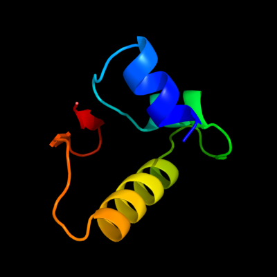
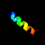
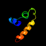
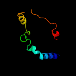
36 c2l4jA_
not modelled
7.2
60
PDB header: transcriptionChain: A: PDB Molecule: yes-associated protein 2 (yap2);PDBTitle: yap ww2
37 c2yshA_
not modelled
7.2
40
PDB header: protein bindingChain: A: PDB Molecule: growth-arrest-specific protein 7;PDBTitle: solution structure of the ww domain from the human growth-2 arrest-specific protein 7, gas-7
38 d1ys7a1
not modelled
7.2
16
Fold: DNA/RNA-binding 3-helical bundleSuperfamily: C-terminal effector domain of the bipartite response regulatorsFamily: PhoB-like39 c2gwwB_
not modelled
7.1
50
PDB header: cell adhesion, structural proteinChain: B: PDB Molecule: ipaa;PDBTitle: human vinculin (head domain, vh1, residues 1-258) in2 complex with shigella's ipaa vinculin binding site3 (residues 602-633)
40 c3ll6B_
not modelled
7.1
30
PDB header: transferaseChain: B: PDB Molecule: cyclin g-associated kinase;PDBTitle: crystal structure of the human cyclin g associated kinase (gak)
41 d1nmva1
not modelled
7.0
40
Fold: WW domain-likeSuperfamily: WW domainFamily: WW domain42 d2jmfa1
not modelled
6.8
50
Fold: WW domain-likeSuperfamily: WW domainFamily: WW domain43 d1f8ab1
not modelled
6.7
40
Fold: WW domain-likeSuperfamily: WW domainFamily: WW domain44 c1wr4A_
not modelled
6.7
40
PDB header: ligaseChain: A: PDB Molecule: ubiquitin-protein ligase nedd4-2;PDBTitle: solution structure of the second ww domain of nedd4-2
45 c3ogfA_
not modelled
6.6
43
PDB header: de novo proteinChain: A: PDB Molecule: de novo designed dimeric trefoil-fold sub-domain whichPDBTitle: crystal structure of difoil-4p homo-trimer: de novo designed dimeric2 trefoil-fold sub-domain which forms homo-trimer assembly
46 c1e0mA_
not modelled
6.5
50
PDB header: de novo proteinChain: A: PDB Molecule: wwprototype;PDBTitle: prototype ww domain
47 c2ez5W_
not modelled
6.5
30
PDB header: signalling protein,ligaseChain: W: PDB Molecule: e3 ubiquitin-protein ligase nedd4;PDBTitle: solution structure of the dnedd4 ww3* domain- comm lpsy2 peptide complex
48 c1w63P_
not modelled
6.3
56
PDB header: endocytosisChain: P: PDB Molecule: adaptor-related protein complex 1, mu 1 subunit;PDBTitle: ap1 clathrin adaptor core
49 d1tk7a1
not modelled
6.2
50
Fold: WW domain-likeSuperfamily: WW domainFamily: WW domain50 c1yiuA_
not modelled
6.2
40
PDB header: ligaseChain: A: PDB Molecule: itchy e3 ubiquitin protein ligase;PDBTitle: itch e3 ubiquitin ligase ww3 domain
51 c2oqrA_
not modelled
6.2
8
PDB header: transcription,signaling proteinChain: A: PDB Molecule: sensory transduction protein regx3;PDBTitle: the structure of the response regulator regx3 from mycobacterium2 tuberculosis
52 d1iqoa_
not modelled
6.0
21
Fold: Hypothetical protein MTH1880Superfamily: Hypothetical protein MTH1880Family: Hypothetical protein MTH188053 c2dwvB_
not modelled
5.9
50
PDB header: protein bindingChain: B: PDB Molecule: salvador homolog 1 protein;PDBTitle: solution structure of the second ww domain from mouse2 salvador homolog 1 protein (mww45)
54 d1k9ra_
not modelled
5.9
50
Fold: WW domain-likeSuperfamily: WW domainFamily: WW domain55 c2ysgA_
not modelled
5.7
60
PDB header: protein bindingChain: A: PDB Molecule: syntaxin-binding protein 4;PDBTitle: solution structure of the ww domain from the human syntaxin-2 binding protein 4
56 d1flca2
not modelled
5.7
57
Fold: Flavodoxin-likeSuperfamily: SGNH hydrolaseFamily: Esterase domain of haemagglutinin-esterase-fusion glycoprotein HEF157 c2ysdA_
not modelled
5.7
33
PDB header: protein bindingChain: A: PDB Molecule: membrane-associated guanylate kinase, ww and pdzPDBTitle: solution structure of the first ww domain from the human2 membrane-associated guanylate kinase, ww and pdz domain-3 containing protein 1. magi-1
58 c2ysfA_
not modelled
5.4
50
PDB header: protein bindingChain: A: PDB Molecule: e3 ubiquitin-protein ligase itchy homolog;PDBTitle: solution structure of the fourth ww domain from the human2 e3 ubiquitin-protein ligase itchy homolog, itch
59 c3ol0C_
not modelled
5.3
46
PDB header: de novo proteinChain: C: PDB Molecule: de novo designed monomer trefoil-fold sub-domain whichPDBTitle: crystal structure of monofoil-4p homo-trimer: de novo designed monomer2 trefoil-fold sub-domain which forms homo-trimer assembly
60 d2csba2
not modelled
5.3
36
Fold: SAM domain-likeSuperfamily: RuvA domain 2-likeFamily: Topoisomerase V repeat domain61 c2jkrM_
not modelled
5.2
44
PDB header: endocytosisChain: M: PDB Molecule: ap-2 complex subunit mu-1;PDBTitle: ap2 clathrin adaptor core with dileucine peptide rm(2 phosphos)qikrllse
62 c2ftcK_
not modelled
5.2
0
PDB header: ribosomeChain: K: PDB Molecule: 39s ribosomal protein l19, mitochondrial;PDBTitle: structural model for the large subunit of the mammalian mitochondrial2 ribosome