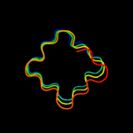
1 c3cxbA_
96.6
23
PDB header: signaling proteinChain: A: PDB Molecule: protein sifa;PDBTitle: crystal structure of sifa and skip
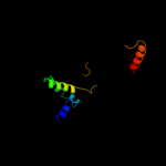
2 c3n90A_
47.0
18
PDB header: unknown functionChain: A: PDB Molecule: thylakoid lumenal 15 kda protein 1, chloroplastic;PDBTitle: the 1.7 angstrom resolution crystal structure of at2g44920, a2 pentapeptide repeat protein from arabidopsis thaliana thylakoid3 lumen.
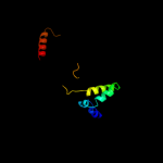
3 d2j8ia1
43.0
20
Fold: Single-stranded right-handed beta-helixSuperfamily: Pentapeptide repeat-likeFamily: Pentapeptide repeats4 c1v0dA_
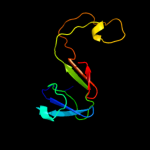
32.0
22
PDB header: hydrolaseChain: A: PDB Molecule: dna fragmentation factor 40 kda subunit;PDBTitle: crystal structure of caspase-activated dnase (cad)
5 d1v0da_
32.0
22
Fold: His-Me finger endonucleasesSuperfamily: His-Me finger endonucleasesFamily: Caspase-activated DNase, CAD (DffB, DFF40)6 d2fvka1
27.0
14
Fold: Composite domain of metallo-dependent hydrolasesSuperfamily: Composite domain of metallo-dependent hydrolasesFamily: Hydantoinase (dihydropyrimidinase)7 d2j8ka1
25.0
29
Fold: Single-stranded right-handed beta-helixSuperfamily: Pentapeptide repeat-likeFamily: Pentapeptide repeats8 c2j8iB_
18.1
20
PDB header: toxinChain: B: PDB Molecule: np275;PDBTitle: structure of np275, a pentapeptide repeat protein from2 nostoc punctiforme
9 c2xtwB_
15.9
14
PDB header: cell cycleChain: B: PDB Molecule: qnrb1;PDBTitle: structure of qnrb1 (full length), a plasmid-mediated2 fluoroquinolone resistance protein
10 c2j8kA_
15.0
29
PDB header: toxinChain: A: PDB Molecule: np275-np276;PDBTitle: structure of the fusion of np275 and np276, pentapeptide2 repeat proteins from nostoc punctiforme
11 c3du1X_
14.1
29
PDB header: structural proteinChain: X: PDB Molecule: all3740 protein;PDBTitle: the 2.0 angstrom resolution crystal structure of hetl, a pentapeptide2 repeat protein involved in heterocyst differentiation regulation from3 the cyanobacterium nostoc sp. strain pcc 7120
12 c2jx3A_
14.1
24
PDB header: dna binding proteinChain: A: PDB Molecule: protein dek;PDBTitle: nmr solution structure of the n-terminal domain of dek
13 c1soxB_
13.8
13
PDB header: oxidoreductaseChain: B: PDB Molecule: sulfite oxidase;PDBTitle: sulfite oxidase from chicken liver
14 d1zh5a1
13.2
14
Fold: DNA/RNA-binding 3-helical bundleSuperfamily: "Winged helix" DNA-binding domainFamily: La domain15 c1i8nA_
13.1
48
PDB header: toxinChain: A: PDB Molecule: anti-platelet protein;PDBTitle: crystal structure of leech anti-platelet protein
16 d1i8na_
13.1
48
Fold: Hairpin loop containing domain-likeSuperfamily: Hairpin loop containing domain-likeFamily: Anti-platelet protein17 c2o6wA_
12.9
18
PDB header: unknown functionChain: A: PDB Molecule: repeat five residue (rfr) protein orPDBTitle: crystal structure of a pentapeptide repeat protein (rfr23)2 from the cyanobacterium cyanothece 51142
18 d2f3la1
11.8
24
Fold: Single-stranded right-handed beta-helixSuperfamily: Pentapeptide repeat-likeFamily: Pentapeptide repeats19 d1tuaa2
11.6
10
Fold: Eukaryotic type KH-domain (KH-domain type I)Superfamily: Eukaryotic type KH-domain (KH-domain type I)Family: Eukaryotic type KH-domain (KH-domain type I)20 c1nh1A_
11.4
27
PDB header: avirulence proteinChain: A: PDB Molecule: avirulence b protein;PDBTitle: crystal structure of the type iii effector avrb from2 pseudomonas syringae.
21 d1nh1a_
not modelled
11.4
27
Fold: Antivirulence factorSuperfamily: Antivirulence factorFamily: Antivirulence factor22 c3ia0c_
not modelled
11.3
26
PDB header: structural proteinChain: C: PDB Molecule: ethanolamine utilization protein euts;PDBTitle: ethanolamine utilization microcompartment shell subunit,2 euts-g39v mutant
23 c2g0yA_
not modelled
11.1
18
PDB header: unknown functionChain: A: PDB Molecule: pentapeptide repeat protein;PDBTitle: crystal structure of a lumenal pentapeptide repeat protein from2 cyanothece sp 51142 at 2.3 angstrom resolution. tetragonal crystal3 form
24 c3lo0A_
not modelled
10.9
28
PDB header: hydrolaseChain: A: PDB Molecule: inorganic pyrophosphatase;PDBTitle: crystal structure of inorganic pyrophosphatase from2 ehrlichia chaffeensis
25 c2wknE_
not modelled
10.7
31
PDB header: hydrolaseChain: E: PDB Molecule: formamidase;PDBTitle: gamma lactamase from delftia acidovorans
26 c2wvmA_
not modelled
9.6
53
PDB header: transferaseChain: A: PDB Molecule: mannosyl-3-phosphoglycerate synthase;PDBTitle: h309a mutant of mannosyl-3-phosphoglycerate synthase from2 thermus thermophilus hb27 in complex with3 gdp-alpha-d-mannose and mg(ii)
27 c2xt4B_
not modelled
9.4
14
PDB header: cell cycleChain: B: PDB Molecule: mcbg-like protein;PDBTitle: structure of the pentapeptide repeat protein albg, a2 resistance factor for the topoisomerase poison albicidin.
28 c3ifuA_
not modelled
9.1
26
PDB header: transcriptionChain: A: PDB Molecule: non-structural protein;PDBTitle: the crystal structure of porcine reproductive and2 respiratory syndrome virus (prrsv) leader protease nsp1
29 d1yj5a1
not modelled
9.0
20
Fold: HAD-likeSuperfamily: HAD-likeFamily: phosphatase domain of polynucleotide kinase30 c2xt4A_
not modelled
8.7
11
PDB header: cell cycleChain: A: PDB Molecule: mcbg-like protein;PDBTitle: structure of the pentapeptide repeat protein albg, a2 resistance factor for the topoisomerase poison albicidin.
31 c3mjjD_
not modelled
8.5
26
PDB header: hydrolaseChain: D: PDB Molecule: predicted acetamidase/formamidase;PDBTitle: crystal structure analysis of a recombinant predicted2 acetamidase/formamidase from the thermophile thermoanaerobacter3 tengcongensis
32 d1isia_
not modelled
8.4
21
Fold: Flavodoxin-likeSuperfamily: N-(deoxy)ribosyltransferase-likeFamily: ADP ribosyl cyclase-like33 d1j2za_
not modelled
8.4
15
Fold: Single-stranded left-handed beta-helixSuperfamily: Trimeric LpxA-like enzymesFamily: UDP N-acetylglucosamine acyltransferase34 c2w7zB_
not modelled
8.3
21
PDB header: inhibitorChain: B: PDB Molecule: pentapeptide repeat family protein;PDBTitle: structure of the pentapeptide repeat protein efsqnr, a dna2 gyrase inhibitor. free amines modified by cyclic3 pentylation with glutaraldehyde.
35 c3t2dA_
not modelled
8.0
41
PDB header: lyase, hydrolaseChain: A: PDB Molecule: fructose-1,6-bisphosphate aldolase/phosphatase;PDBTitle: fructose-1,6-bisphosphate aldolase/phosphatase from thermoproteus2 neutrophilus, fbp-bound form
36 c1jrjA_
not modelled
8.0
26
PDB header: hormone/growth factorChain: A: PDB Molecule: exendin-4;PDBTitle: solution structure of exendin-4 in 30-vol% trifluoroethanol
37 c3b9eA_
not modelled
7.9
26
PDB header: hydrolaseChain: A: PDB Molecule: chitinase a;PDBTitle: crystal structure of inactive mutant e315m chitinase a from2 vibrio harveyi
38 c3b9tD_
not modelled
7.9
40
PDB header: hydrolaseChain: D: PDB Molecule: twin-arginine translocation pathway signal protein;PDBTitle: crystal structure of predicted acetamidase/formamidase (yp_546212.1)2 from methylobacillus flagellatus kt at 1.58 a resolution
39 c2zu8A_
not modelled
7.6
47
PDB header: transferaseChain: A: PDB Molecule: mannosyl-3-phosphoglycerate synthase;PDBTitle: crystal structure of mannosyl-3-phosphoglycerate synthase2 from pyrococcus horikoshii
40 c3pssB_
not modelled
7.6
17
PDB header: cell cycleChain: B: PDB Molecule: qnr;PDBTitle: crystal structure of ahqnr, the qnr protein from aeromonas hydrophila2 (p21 crystal form)
41 c2eg9B_
not modelled
7.1
26
PDB header: hydrolaseChain: B: PDB Molecule: adp-ribosyl cyclase 1;PDBTitle: crystal structure of the truncated extracellular domain of2 mouse cd38
42 d2cqka1
not modelled
7.1
15
Fold: DNA/RNA-binding 3-helical bundleSuperfamily: "Winged helix" DNA-binding domainFamily: La domain43 d1xnta_
not modelled
6.9
27
Fold: Galactose-binding domain-likeSuperfamily: Galactose-binding domain-likeFamily: N-terminal domain of xrcc144 c1xnaA_
not modelled
6.9
27
PDB header: dna binding proteinChain: A: PDB Molecule: protein (dna-repair protein xrcc1);PDBTitle: nmr solution structure of the single-strand break repair2 protein xrcc1-n-terminal domain
45 d1sura_
not modelled
6.8
16
Fold: Adenine nucleotide alpha hydrolase-likeSuperfamily: Adenine nucleotide alpha hydrolases-likeFamily: PAPS reductase-like46 c3n3fB_
not modelled
6.7
14
PDB header: protein bindingChain: B: PDB Molecule: collagen alpha-1(xv) chain;PDBTitle: crystal structure of the human collagen xv trimerization domain: a2 potent trimerizing unit common to multiplexin collagens
47 c2xz2A_
not modelled
6.6
26
PDB header: rna-binding proteinChain: A: PDB Molecule: cstf-50, isoform b;PDBTitle: crystal structure of cstf-50 homodimerization domain
48 c2vbeA_
not modelled
6.6
39
PDB header: viral proteinChain: A: PDB Molecule: tailspike-protein;PDBTitle: tailspike protein of bacteriophage sf6
49 c3cgiD_
not modelled
6.4
50
PDB header: unknown functionChain: D: PDB Molecule: propanediol utilization protein pduu;PDBTitle: crystal structure of the pduu shell protein from the pdu2 microcompartment
50 d1hs5a_
not modelled
6.4
34
Fold: p53 tetramerization domainSuperfamily: p53 tetramerization domainFamily: p53 tetramerization domain51 c2goyC_
not modelled
6.4
22
PDB header: oxidoreductaseChain: C: PDB Molecule: adenosine phosphosulfate reductase;PDBTitle: crystal structure of assimilatory adenosine 5'-2 phosphosulfate reductase with bound aps
52 c1d0rA_
not modelled
6.3
26
PDB header: hormone/growth factorChain: A: PDB Molecule: glucagon-like peptide-1-(7-36)-amide;PDBTitle: solution structure of glucagon-like peptide-1-(7-36)-amide2 in trifluoroethanol/water
53 c2bhlB_
not modelled
6.3
43
PDB header: oxidoreductase (choh(d)-nadp)Chain: B: PDB Molecule: glucose-6-phosphate 1-dehydrogenase;PDBTitle: x-ray structure of human glucose-6-phosphate dehydrogenase2 (deletion variant) complexed with glucose-6-phosphate
54 c1yj5B_
not modelled
6.3
20
PDB header: transferaseChain: B: PDB Molecule: 5' polynucleotide kinase-3' phosphatase catalytic domain;PDBTitle: molecular architecture of mammalian polynucleotide kinase, a dna2 repair enzyme
55 d2a1ja1
not modelled
6.2
17
Fold: SAM domain-likeSuperfamily: RuvA domain 2-likeFamily: Hef domain-like56 c3nb2B_
not modelled
6.1
25
PDB header: ligaseChain: B: PDB Molecule: secreted effector protein;PDBTitle: crystal structure of e. coli o157:h7 effector protein nlel
57 c2j9wB_
not modelled
6.1
19
PDB header: protein transportChain: B: PDB Molecule: vps28-prov protein;PDBTitle: structural insight into the escrt-i-ii link and its role in2 mvb trafficking
58 d1khia2
not modelled
6.1
36
Fold: OB-foldSuperfamily: Nucleic acid-binding proteinsFamily: Cold shock DNA-binding domain-like59 d1whra_
not modelled
6.0
14
Fold: IF3-likeSuperfamily: R3H domainFamily: R3H domain60 d1wvfa1
not modelled
5.9
31
Fold: Ferredoxin-likeSuperfamily: FAD-linked oxidases, C-terminal domainFamily: Vanillyl-alcohol oxidase-like61 d2f4la1
not modelled
5.8
24
Fold: CUB-likeSuperfamily: Acetamidase/Formamidase-likeFamily: Acetamidase/Formamidase-like62 c3hugJ_
not modelled
5.8
30
PDB header: transcription/membrane proteinChain: J: PDB Molecule: probable conserved membrane protein;PDBTitle: crystal structure of mycobacterium tuberculosis anti-sigma factor rsla2 in complex with -35 promoter binding domain of sigl
63 d1edqa2
not modelled
5.8
25
Fold: TIM beta/alpha-barrelSuperfamily: (Trans)glycosidasesFamily: Type II chitinase64 d2bm5a1
not modelled
5.7
4
Fold: Single-stranded right-handed beta-helixSuperfamily: Pentapeptide repeat-likeFamily: Pentapeptide repeats65 d1mjea2
not modelled
5.7
28
Fold: BRCA2 tower domainSuperfamily: BRCA2 tower domainFamily: BRCA2 tower domain66 c2c1tA_
not modelled
5.6
17
PDB header: protein transport/membrane proteinChain: A: PDB Molecule: importin alpha subunit;PDBTitle: structure of the kap60p:nup2 complex
67 c1qkiE_
not modelled
5.6
43
PDB header: oxidoreductaseChain: E: PDB Molecule: glucose-6-phosphate 1-dehydrogenase;PDBTitle: x-ray structure of human glucose 6-phosphate dehydrogenase2 (variant canton r459l) complexed with structural nadp+
68 d1r4pb_
not modelled
5.5
39
Fold: OB-foldSuperfamily: Bacterial enterotoxinsFamily: Bacterial AB5 toxins, B-subunits69 d1l8na1
not modelled
5.5
45
Fold: TIM beta/alpha-barrelSuperfamily: (Trans)glycosidasesFamily: alpha-D-glucuronidase/Hyaluronidase catalytic domain70 d2ef1a1
not modelled
5.5
23
Fold: Flavodoxin-likeSuperfamily: N-(deoxy)ribosyltransferase-likeFamily: ADP ribosyl cyclase-like71 d1c4qa_
not modelled
5.3
33
Fold: OB-foldSuperfamily: Bacterial enterotoxinsFamily: Bacterial AB5 toxins, B-subunits72 d1zs4a1
not modelled
5.3
23
Fold: lambda repressor-like DNA-binding domainsSuperfamily: lambda repressor-like DNA-binding domainsFamily: Bacteriophage CII protein73 c2o8vA_
not modelled
5.2
21
PDB header: oxidoreductaseChain: A: PDB Molecule: phosphoadenosine phosphosulfate reductase;PDBTitle: paps reductase in a covalent complex with thioredoxin c35a