Phyre Home Retrieve Phyre Job Id
Emaill.a.kelley@imperial.ac.uk
DescriptionQ9JMS7
DateThu Jan 5 12:38:03 GMT 2012
Unique Job ID55c067a56976a548

Summary 

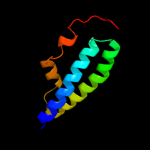
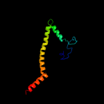
Top model
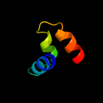
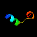
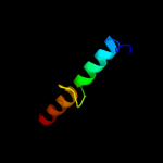
Image coloured by rainbow N → C terminus
Model (left) based on template c3fseB_
Top template information
PDB header:hydrolase
Chain: B: PDB Molecule:two-domain protein containing dj-1/thij/pfpi-like and
PDBTitle: crystal structure of a two-domain protein containing dj-1/thij/pfpi-2 like and ferritin-like domains (ava_4496) from anabaena variabilis3 atcc 29413 at 1.90 a resolution
Confidence and coverage
Confidence: 63.8% Coverage: 34%
78 residues ( 34% of your sequence) have been modelled with 63.8% confidence by the single highest scoring template.
You may wish to submit your sequence to Phyrealarm. This will automatically scan your sequence every week for new potential templates as they appear in the Phyre2 library.
Please note: You must be registered and logged in to use Phyrealarm.
3D viewing
Interactive 3D view in Jmol

Sequence analysis 

Secondary structure and disorder prediction 

   1........10.........20.........30.........40.........50.........60
Sequence  MMYRVNHIMRTINEMSSYTPHMKVNRIAERLSKVQKISFCISVISFFLLAIITLTYGPFN
Secondary structure 


SS confidence 


Disorder  ????


??????


Disorder confidence 


   .........70.........80.........90.........100.........110.........120
Sequence  TKSNLSFISALSLYFINVIMGVTYLSVPVINTIKYIYNFKGEVVNELIYDIDSDEQHIEA
Secondary structure 


SS confidence 


Disorder 

??


Disorder confidence 


   .........130.........140.........150.........160.........170.........180
Sequence  LLPYSLEELTYVSNCIQVRIPKIKSKCFLWGGGKTAIISILCLSYSAICIVNGGSIDGIF
Secondary structure 


SS confidence 


Disorder 


?


?
Disorder confidence 


   .........190.........200.........210.........220.........230.
Sequence  VGETGDKIIVAIMFFILYTSLMNMFFKQKLLYLQNLKMIIDMTIKIKRNFT
Secondary structure 


SS confidence 


Disorder 


?????
Disorder confidence 


Confidence Key
High(9)                    Low (0)
?Disordered
Alpha helix
Beta strand

Domain analysis 

Hover over an aligned region to see model and summary info

Please note, only up to the top 20 hits are modelled to reduce computer load

RankAligned region

PDB 3fse chain B

3D model

Region: 99 - 177
Aligned: 78
Modelled: 79
Confidence: 63.8%
Identity: 23%
PDB header:hydrolase
Chain: B: PDB Molecule:two-domain protein containing dj-1/thij/pfpi-like and
PDBTitle: crystal structure of a two-domain protein containing dj-1/thij/pfpi-2 like and ferritin-like domains (ava_4496) from anabaena variabilis3 atcc 29413 at 1.90 a resolution

Phyre2

PDB 3ipd chain B

3D model

Region: 5 - 59
Aligned: 55
Modelled: 55
Confidence: 30.4%
Identity: 18%
PDB header:exocytosis
Chain: B: PDB Molecule:syntaxin-1a;
PDBTitle: helical extension of the neuronal snare complex into the2 membrane, spacegroup i 21 21 21

Phyre2

PDB 2oc5 chain A domain 1

3D model

Region: 119 - 147
Aligned: 29
Modelled: 29
Confidence: 25.6%
Identity: 10%
Fold: Ferritin-like
Superfamily: Ferritin-like
Family: PMT1231-like

Phyre2

PDB 3lt7 chain D

3D model

Region: 6 - 36
Aligned: 29
Modelled: 31
Confidence: 20.6%
Identity: 34%
PDB header:cell adhesion
Chain: D: PDB Molecule:adhesin yada;
PDBTitle: a transition from strong right-handed to canonical left-handed2 supercoiling in a conserved coiled coil segment of trimeric3 autotransporter adhesins - the m3 mutant structure

Phyre2

PDB 2w8a chain C

3D model

Region: 8 - 133
Aligned: 92
Modelled: 101
Confidence: 19.7%
Identity: 14%
PDB header:membrane protein
Chain: C: PDB Molecule:glycine betaine transporter betp;
PDBTitle: crystal structure of the sodium-coupled glycine betaine2 symporter betp from corynebacterium glutamicum with bound3 substrate

Phyre2

PDB 1v8g chain A domain 1

3D model

Region: 90 - 108
Aligned: 19
Modelled: 19
Confidence: 18.0%
Identity: 16%
Fold: Methionine synthase domain-like
Superfamily: Nucleoside phosphorylase/phosphoribosyltransferase N-terminal domain
Family: Nucleoside phosphorylase/phosphoribosyltransferase N-terminal domain

Phyre2

PDB 1o17 chain A domain 1

3D model

Region: 90 - 108
Aligned: 19
Modelled: 19
Confidence: 17.6%
Identity: 32%
Fold: Methionine synthase domain-like
Superfamily: Nucleoside phosphorylase/phosphoribosyltransferase N-terminal domain
Family: Nucleoside phosphorylase/phosphoribosyltransferase N-terminal domain

Phyre2

PDB 1uou chain A domain 1

3D model

Region: 90 - 108
Aligned: 19
Modelled: 19
Confidence: 16.6%
Identity: 16%
Fold: Methionine synthase domain-like
Superfamily: Nucleoside phosphorylase/phosphoribosyltransferase N-terminal domain
Family: Nucleoside phosphorylase/phosphoribosyltransferase N-terminal domain

Phyre2

PDB 1khd chain A domain 1

3D model

Region: 90 - 107
Aligned: 18
Modelled: 18
Confidence: 14.1%
Identity: 17%
Fold: Methionine synthase domain-like
Superfamily: Nucleoside phosphorylase/phosphoribosyltransferase N-terminal domain
Family: Nucleoside phosphorylase/phosphoribosyltransferase N-terminal domain

Phyre2

PDB 3izc chain K

3D model

Region: 5 - 41
Aligned: 37
Modelled: 37
Confidence: 14.0%
Identity: 27%
PDB header:ribosome
Chain: K: PDB Molecule:60s ribosomal protein rpl16 (l13p);
PDBTitle: localization of the large subunit ribosomal proteins into a 6.1 a2 cryo-em map of saccharomyces cerevisiae translating 80s ribosome

Phyre2

PDB 2y0f chain D

3D model

Region: 130 - 187
Aligned: 49
Modelled: 58
Confidence: 13.9%
Identity: 27%
PDB header:oxidoreductase
Chain: D: PDB Molecule:4-hydroxy-3-methylbut-2-en-1-yl diphosphate synthase;
PDBTitle: structure of gcpe (ispg) from thermus thermophilus hb27

Phyre2

PDB 2d0o chain A domain 3

3D model

Region: 167 - 186
Aligned: 20
Modelled: 20
Confidence: 13.0%
Identity: 35%
Fold: Ribonuclease H-like motif
Superfamily: Actin-like ATPase domain
Family: ATPase domain of dehydratase reactivase alpha subunit

Phyre2

PDB 1brw chain A domain 1

3D model

Region: 90 - 108
Aligned: 19
Modelled: 19
Confidence: 12.8%
Identity: 16%
Fold: Methionine synthase domain-like
Superfamily: Nucleoside phosphorylase/phosphoribosyltransferase N-terminal domain
Family: Nucleoside phosphorylase/phosphoribosyltransferase N-terminal domain

Phyre2

PDB 2tpt chain A domain 1

3D model

Region: 90 - 108
Aligned: 19
Modelled: 19
Confidence: 11.8%
Identity: 16%
Fold: Methionine synthase domain-like
Superfamily: Nucleoside phosphorylase/phosphoribosyltransferase N-terminal domain
Family: Nucleoside phosphorylase/phosphoribosyltransferase N-terminal domain

Phyre2

PDB 2qam chain 3 domain 1

3D model

Region: 112 - 125
Aligned: 14
Modelled: 14
Confidence: 11.7%
Identity: 36%
Fold: L35p-like
Superfamily: L35p-like
Family: Ribosomal protein L35p

Phyre2

PDB 1mug chain A

3D model

Region: 125 - 156
Aligned: 32
Modelled: 32
Confidence: 11.6%
Identity: 16%
Fold: Uracil-DNA glycosylase-like
Superfamily: Uracil-DNA glycosylase-like
Family: Mug-like

Phyre2

PDB 1rzs chain A

3D model

Region: 144 - 168
Aligned: 25
Modelled: 25
Confidence: 10.5%
Identity: 24%
Fold: lambda repressor-like DNA-binding domains
Superfamily: lambda repressor-like DNA-binding domains
Family: Phage repressors

Phyre2

PDB 2voy chain G

3D model

Region: 14 - 51
Aligned: 32
Modelled: 38
Confidence: 10.3%
Identity: 28%
PDB header:hydrolase
Chain: G: PDB Molecule:sarcoplasmic/endoplasmic reticulum calcium
PDBTitle: cryoem model of copa, the copper transporting atpase from2 archaeoglobus fulgidus

Phyre2

PDB 2q9q chain F

3D model

Region: 99 - 145
Aligned: 47
Modelled: 47
Confidence: 10.1%
Identity: 17%
PDB header:replication
Chain: F: PDB Molecule:gins complex subunit 4;
PDBTitle: the crystal structure of full length human gins complex

Phyre2

PDB 1nbw chain A domain 3

3D model

Region: 167 - 185
Aligned: 19
Modelled: 19
Confidence: 9.8%
Identity: 37%
Fold: Ribonuclease H-like motif
Superfamily: Actin-like ATPase domain
Family: ATPase domain of dehydratase reactivase alpha subunit

Phyre2
1

c3fseB_
2

c3ipdB_
3

d2oc5a1
4

c3lt7D_
5

c2w8aC_
6

d1v8ga1
7

d1o17a1
8

d1uoua1
9

d1khda1
10

c3izck_
11

c2y0fD_
12

d2d0oa3
13

d1brwa1
14

d2tpta1
15

d2qam31
16

d1muga_
17

d1rzsa_
18

c2voyG_
19

c2q9qF_
20

d1nbwa3
21


22


23


24


25


26


27


28


29


30


31


32


33


34


35


36


37


38


39


40


41


42


43


44


45


Detailed template information 

#
Template Alignment Coverage3D Model Confidence
% i.d. Template Information
1c3fseB_


63.8 23 PDB header:hydrolase
Chain: B: PDB Molecule:two-domain protein containing dj-1/thij/pfpi-like and
PDBTitle: crystal structure of a two-domain protein containing dj-1/thij/pfpi-2 like and ferritin-like domains (ava_4496) from anabaena variabilis3 atcc 29413 at 1.90 a resolution
2c3ipdB_


30.4 18 PDB header:exocytosis
Chain: B: PDB Molecule:syntaxin-1a;
PDBTitle: helical extension of the neuronal snare complex into the2 membrane, spacegroup i 21 21 21
3d2oc5a1


25.6 10 Fold:Ferritin-like
Superfamily:Ferritin-like
Family:PMT1231-like
4c3lt7D_


20.6 34 PDB header:cell adhesion
Chain: D: PDB Molecule:adhesin yada;
PDBTitle: a transition from strong right-handed to canonical left-handed2 supercoiling in a conserved coiled coil segment of trimeric3 autotransporter adhesins - the m3 mutant structure
5c2w8aC_


19.7 14 PDB header:membrane protein
Chain: C: PDB Molecule:glycine betaine transporter betp;
PDBTitle: crystal structure of the sodium-coupled glycine betaine2 symporter betp from corynebacterium glutamicum with bound3 substrate
6d1v8ga1


18.0 16 Fold:Methionine synthase domain-like
Superfamily:Nucleoside phosphorylase/phosphoribosyltransferase N-terminal domain
Family:Nucleoside phosphorylase/phosphoribosyltransferase N-terminal domain
7d1o17a1


17.6 32 Fold:Methionine synthase domain-like
Superfamily:Nucleoside phosphorylase/phosphoribosyltransferase N-terminal domain
Family:Nucleoside phosphorylase/phosphoribosyltransferase N-terminal domain
8d1uoua1


16.6 16 Fold:Methionine synthase domain-like
Superfamily:Nucleoside phosphorylase/phosphoribosyltransferase N-terminal domain
Family:Nucleoside phosphorylase/phosphoribosyltransferase N-terminal domain
9d1khda1


14.1 17 Fold:Methionine synthase domain-like
Superfamily:Nucleoside phosphorylase/phosphoribosyltransferase N-terminal domain
Family:Nucleoside phosphorylase/phosphoribosyltransferase N-terminal domain
10c3izck_


14.0 27 PDB header:ribosome
Chain: K: PDB Molecule:60s ribosomal protein rpl16 (l13p);
PDBTitle: localization of the large subunit ribosomal proteins into a 6.1 a2 cryo-em map of saccharomyces cerevisiae translating 80s ribosome
11c2y0fD_


13.9 27 PDB header:oxidoreductase
Chain: D: PDB Molecule:4-hydroxy-3-methylbut-2-en-1-yl diphosphate synthase;
PDBTitle: structure of gcpe (ispg) from thermus thermophilus hb27
12d2d0oa3


13.0 35 Fold:Ribonuclease H-like motif
Superfamily:Actin-like ATPase domain
Family:ATPase domain of dehydratase reactivase alpha subunit
13d1brwa1


12.8 16 Fold:Methionine synthase domain-like
Superfamily:Nucleoside phosphorylase/phosphoribosyltransferase N-terminal domain
Family:Nucleoside phosphorylase/phosphoribosyltransferase N-terminal domain
14d2tpta1


11.8 16 Fold:Methionine synthase domain-like
Superfamily:Nucleoside phosphorylase/phosphoribosyltransferase N-terminal domain
Family:Nucleoside phosphorylase/phosphoribosyltransferase N-terminal domain
15d2qam31


11.7 36 Fold:L35p-like
Superfamily:L35p-like
Family:Ribosomal protein L35p
16d1muga_


11.6 16 Fold:Uracil-DNA glycosylase-like
Superfamily:Uracil-DNA glycosylase-like
Family:Mug-like
17d1rzsa_


10.5 24 Fold:lambda repressor-like DNA-binding domains
Superfamily:lambda repressor-like DNA-binding domains
Family:Phage repressors
18c2voyG_


10.3 28 PDB header:hydrolase
Chain: G: PDB Molecule:sarcoplasmic/endoplasmic reticulum calcium
PDBTitle: cryoem model of copa, the copper transporting atpase from2 archaeoglobus fulgidus
19c2q9qF_


10.1 17 PDB header:replication
Chain: F: PDB Molecule:gins complex subunit 4;
PDBTitle: the crystal structure of full length human gins complex
20d1nbwa3


9.8 37 Fold:Ribonuclease H-like motif
Superfamily:Actin-like ATPase domain
Family:ATPase domain of dehydratase reactivase alpha subunit
21c3kdpG_


not modelled 9.3 33 PDB header:hydrolase
Chain: G: PDB Molecule:na+/k+ atpase gamma subunit transcript variant a;
PDBTitle: crystal structure of the sodium-potassium pump
22c3kdpH_


not modelled 9.3 33 PDB header:hydrolase
Chain: H: PDB Molecule:na+/k+ atpase gamma subunit transcript variant a;
PDBTitle: crystal structure of the sodium-potassium pump
23c4a19Q_


not modelled 8.4 13 PDB header:ribosome
Chain: Q: PDB Molecule:60s ribosomal protein l36;
PDBTitle: t.thermophila 60s ribosomal subunit in complex with2 initiation factor 6. this file contains 26s rrna and3 proteins of molecule 2.
24c3mk7F_


not modelled 8.4 15 PDB header:oxidoreductase
Chain: F: PDB Molecule:cytochrome c oxidase, cbb3-type, subunit p;
PDBTitle: the structure of cbb3 cytochrome oxidase
25d1nb9a_


not modelled 8.3 20 Fold:Reductase/isomerase/elongation factor common domain
Superfamily:Riboflavin kinase-like
Family:ATP-dependent riboflavin kinase-like
26c3iz5k_


not modelled 7.7 22 PDB header:ribosome
Chain: K: PDB Molecule:60s ribosomal protein l13a (l13p);
PDBTitle: localization of the large subunit ribosomal proteins into a 5.5 a2 cryo-em map of triticum aestivum translating 80s ribosome
27d2elca1


not modelled 7.5 17 Fold:Methionine synthase domain-like
Superfamily:Nucleoside phosphorylase/phosphoribosyltransferase N-terminal domain
Family:Nucleoside phosphorylase/phosphoribosyltransferase N-terminal domain
28d1t3ua_


not modelled 7.5 15 Fold:Cell division protein ZapA-like
Superfamily:Cell division protein ZapA-like
Family:Cell division protein ZapA-like
29c2f8sA_


not modelled 7.3 15 PDB header:rna binding protein/rna
Chain: A: PDB Molecule:argonaute protein;
PDBTitle: crystal structure of aa-ago with externally-bound sirna
30d2guka1


not modelled 7.0 25 Fold:PG1857-like
Superfamily:PG1857-like
Family:PG1857-like
31c2zdjA_


not modelled 6.7 18 PDB header:structural genomics, unknown function
Chain: A: PDB Molecule:hypothetical protein ttma177;
PDBTitle: crystal structure of ttma177, a hypothetical protein from2 thermus thermophilus phage tma
32c1nbwA_


not modelled 6.6 35 PDB header:hydrolase
Chain: A: PDB Molecule:glycerol dehydratase reactivase alpha subunit;
PDBTitle: glycerol dehydratase reactivase
33c1khdD_


not modelled 6.6 17 PDB header:transferase
Chain: D: PDB Molecule:anthranilate phosphoribosyltransferase;
PDBTitle: crystal structure analysis of the anthranilate2 phosphoribosyltransferase from erwinia carotovora at 1.93 resolution (current name, pectobacterium carotovorum)
34c3p45F_


not modelled 6.5 14 PDB header:hydrolase
Chain: F: PDB Molecule:caspase-6;
PDBTitle: crystal structure of apo-caspase-6 at physiological ph
35d1xmec1


not modelled 6.3 24 Fold:Single transmembrane helix
Superfamily:Bacterial ba3 type cytochrome c oxidase subunit IIa
Family:Bacterial ba3 type cytochrome c oxidase subunit IIa
36d1tzyc_


not modelled 6.3 15 Fold:Histone-fold
Superfamily:Histone-fold
Family:Nucleosome core histones
37c3iuoA_


not modelled 6.2 21 PDB header:hydrolase
Chain: A: PDB Molecule:atp-dependent dna helicase recq;
PDBTitle: the crystal structure of the c-terminal domain of the atp-dependent2 dna helicase recq from porphyromonas gingivalis to 1.6a
38c3mmyF_


not modelled 6.2 31 PDB header:nuclear protein
Chain: F: PDB Molecule:nuclear pore complex protein nup98;
PDBTitle: structural and functional analysis of the interaction between the2 nucleoporin nup98 and the mrna export factor rae1
39d1f3va_


not modelled 6.1 30 Fold:Ferredoxin-like
Superfamily:TRADD, N-terminal domain
Family:TRADD, N-terminal domain
40c1unvA_


not modelled 6.0 30 PDB header:four helix bundle
Chain: A: PDB Molecule:general control protein gcn4;
PDBTitle: structure based engineering of internal molecular surfaces2 of four helix bundles
41c1unvB_


not modelled 5.9 29 PDB header:four helix bundle
Chain: B: PDB Molecule:general control protein gcn4;
PDBTitle: structure based engineering of internal molecular surfaces2 of four helix bundles
42c2ke4A_


not modelled 5.7 15 PDB header:membrane protein
Chain: A: PDB Molecule:cdc42-interacting protein 4;
PDBTitle: the nmr structure of the tc10 and cdc42 interacting domain2 of cip4
43c3p56B_


not modelled 5.3 17 PDB header:hydrolase/replication
Chain: B: PDB Molecule:ribonuclease h2 subunit b;
PDBTitle: the structure of the human rnase h2 complex defines key interaction2 interfaces relevant to enzyme function and human disease
44c1v8gB_


not modelled 5.3 16 PDB header:transferase
Chain: B: PDB Molecule:anthranilate phosphoribosyltransferase;
PDBTitle: crystal structure of anthranilate phosphoribosyltransferase2 (trpd) from thermus thermophilus hb8
45d2v7qi1


not modelled 5.2 57 Fold:Non-globular all-alpha subunits of globular proteins
Superfamily:Epsilon subunit of mitochondrial F1F0-ATP synthase
Family:Epsilon subunit of mitochondrial F1F0-ATP synthase

Binding site prediction 

Due to computational demand, binding site predictions are not run for batch jobs

If you want to predict binding sites, please manually submit your model of choice to 3DLigandSite


Phyre is for academic use only

Please cite: Protein structure prediction on the web: a case study using the Phyre server
Kelley LA and Sternberg MJE. Nature Protocols 4, 363 - 371 (2009) [pdf] [Import into BibTeX]
 
If you use the binding site predictions from 3DLigandSite, please also cite:
3DLigandSite: predicting ligand-binding sites using similar structures.
Wass MN, Kelley LA and Sternberg MJ Nucleic Acids Research 38, W469-73 (2010) [PubMed]
 
© Structural Bioinformatics Group
Imperial College London
Lawrence Kelley, Benjamin Jefferys
Disclaimer
Terms and Conditions
Component software
Template detection: HHpred 1.51
Secondary structure prediction: Psi-pred 2.5
Disorder prediction: Disopred 2.4
Transmembrane prediction: Memsat_SVM
Multi-template modelling and ab initio: Poing 1.0