Phyre Home Retrieve Phyre Job Id
Emaill.a.kelley@imperial.ac.uk
DescriptionP0ADQ5
DateThu Jan 5 11:21:33 GMT 2012
Unique Job ID534b0a197c67fba8

Summary 

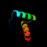
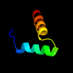
Top model
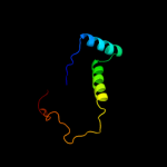
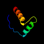
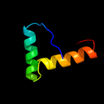
Image coloured by rainbow N → C terminus
Model (left) based on template c2k5jB_
Top template information
PDB header:structural genomics, unknown function
Chain: B: PDB Molecule:uncharacterized protein yiif;
PDBTitle: solution structure of protein yiif from shigella flexneri2 serotype 5b (strain 8401) . northeast structural genomics3 consortium target sft1
Confidence and coverage
Confidence:100.0% Coverage:100%
70 residues (100% of your sequence) have been modelled with 100.0% confidence by the single highest scoring template.
3D viewing
Interactive 3D view in Jmol

Sequence analysis 

Secondary structure and disorder prediction 

   1........10.........20.........30.........40.........50.........60
Sequence  MNTVFLHLSEEAIKRLNKLRGWRKVSRSAILREAVEQYLERQQFPVRKAKGGRQKGEVVG
Secondary structure 


SS confidence 


Disorder  ?????


?


???????????????????
Disorder confidence 


   .........70
Sequence  VDDQCKEHKE
Secondary structure 
SS confidence 


Disorder  ?


????
Disorder confidence 


Confidence Key
High(9)                    Low (0)
?Disordered
Alpha helix
Beta strand

Domain analysis 

Hover over an aligned region to see model and summary info

Please note, only up to the top 20 hits are modelled to reduce computer load

RankAligned region

PDB 2k5j chain B

3D model

Region: 1 - 70
Aligned: 70
Modelled: 70
Confidence: 100.0%
Identity: 41%
PDB header:structural genomics, unknown function
Chain: B: PDB Molecule:uncharacterized protein yiif;
PDBTitle: solution structure of protein yiif from shigella flexneri2 serotype 5b (strain 8401) . northeast structural genomics3 consortium target sft1

Phyre2

PDB 1q5v chain B

3D model

Region: 1 - 49
Aligned: 49
Modelled: 49
Confidence: 97.9%
Identity: 20%
PDB header:transcription
Chain: B: PDB Molecule:nickel responsive regulator;
PDBTitle: apo-nikr

Phyre2

PDB 2bj3 chain D

3D model

Region: 1 - 52
Aligned: 52
Modelled: 52
Confidence: 97.9%
Identity: 12%
PDB header:transcription
Chain: D: PDB Molecule:nickel responsive regulator;
PDBTitle: nikr-apo

Phyre2

PDB 2bj7 chain A domain 1

3D model

Region: 1 - 45
Aligned: 45
Modelled: 45
Confidence: 97.9%
Identity: 9%
Fold: Ribbon-helix-helix
Superfamily: Ribbon-helix-helix
Family: CopG-like

Phyre2

PDB 2ca9 chain B

3D model

Region: 1 - 42
Aligned: 42
Modelled: 42
Confidence: 97.7%
Identity: 17%
PDB header:transcriptional regulation
Chain: B: PDB Molecule:putative nickel-responsive regulator;
PDBTitle: apo-nikr from helicobacter pylori in closed trans-2 conformation

Phyre2

PDB 2hza chain A domain 1

3D model

Region: 3 - 42
Aligned: 40
Modelled: 40
Confidence: 97.1%
Identity: 23%
Fold: Ribbon-helix-helix
Superfamily: Ribbon-helix-helix
Family: CopG-like

Phyre2

PDB 2hza chain B domain 1

3D model

Region: 3 - 42
Aligned: 40
Modelled: 40
Confidence: 96.9%
Identity: 23%
Fold: Ribbon-helix-helix
Superfamily: Ribbon-helix-helix
Family: CopG-like

Phyre2

PDB 2rbf chain B

3D model

Region: 2 - 41
Aligned: 40
Modelled: 40
Confidence: 94.9%
Identity: 18%
PDB header:oxidoreductase/dna
Chain: B: PDB Molecule:bifunctional protein puta;
PDBTitle: structure of the ribbon-helix-helix domain of escherichia coli puta2 (puta52) complexed with operator dna (o2)

Phyre2

PDB 2cpg chain A

3D model

Region: 3 - 42
Aligned: 40
Modelled: 40
Confidence: 93.9%
Identity: 30%
Fold: Ribbon-helix-helix
Superfamily: Ribbon-helix-helix
Family: CopG-like

Phyre2

PDB 2an7 chain A

3D model

Region: 1 - 45
Aligned: 37
Modelled: 45
Confidence: 91.6%
Identity: 19%
PDB header:dna binding protein
Chain: A: PDB Molecule:protein pard;
PDBTitle: solution structure of the bacterial antidote pard

Phyre2

PDB 2k9i chain B

3D model

Region: 4 - 47
Aligned: 44
Modelled: 44
Confidence: 89.0%
Identity: 18%
PDB header:dna binding protein
Chain: B: PDB Molecule:plasmid prn1, complete sequence;
PDBTitle: nmr structure of plasmid copy control protein orf56 from2 sulfolobus islandicus

Phyre2

PDB 3h87 chain D

3D model

Region: 7 - 69
Aligned: 63
Modelled: 63
Confidence: 88.8%
Identity: 19%
PDB header:toxin/antitoxin
Chain: D: PDB Molecule:putative uncharacterized protein;
PDBTitle: rv0301 rv0300 toxin antitoxin complex from mycobacterium tuberculosis

Phyre2

PDB 2ay0 chain A domain 1

3D model

Region: 2 - 41
Aligned: 40
Modelled: 40
Confidence: 79.1%
Identity: 18%
Fold: Ribbon-helix-helix
Superfamily: Ribbon-helix-helix
Family: PutA pre-N-terminal region-like

Phyre2

PDB 2bsq chain E domain 1

3D model

Region: 7 - 41
Aligned: 35
Modelled: 35
Confidence: 52.1%
Identity: 17%
Fold: Ribbon-helix-helix
Superfamily: Ribbon-helix-helix
Family: Trafficking protein A-like

Phyre2

PDB 2k29 chain A

3D model

Region: 1 - 45
Aligned: 45
Modelled: 45
Confidence: 49.6%
Identity: 22%
PDB header:transcription
Chain: A: PDB Molecule:antitoxin relb;
PDBTitle: structure of the dbd domain of e. coli antitoxin relb

Phyre2

PDB 1p94 chain A

3D model

Region: 3 - 42
Aligned: 40
Modelled: 40
Confidence: 43.2%
Identity: 18%
Fold: Ribbon-helix-helix
Superfamily: Ribbon-helix-helix
Family: CopG-like

Phyre2

PDB 2q2k chain A

3D model

Region: 14 - 40
Aligned: 27
Modelled: 27
Confidence: 25.3%
Identity: 26%
PDB header:dna binding protein/dna
Chain: A: PDB Molecule:hypothetical protein;
PDBTitle: structure of nucleic-acid binding protein

Phyre2

PDB 1fcd chain C domain 1

3D model

Region: 1 - 46
Aligned: 27
Modelled: 27
Confidence: 22.4%
Identity: 19%
Fold: Cytochrome c
Superfamily: Cytochrome c
Family: Two-domain cytochrome c

Phyre2

PDB 1yxe chain A

3D model

Region: 44 - 56
Aligned: 13
Modelled: 13
Confidence: 21.2%
Identity: 23%
PDB header:immune system
Chain: A: PDB Molecule:apical membrane antigen 1;
PDBTitle: structure and inter-domain interactions of domain ii from the blood2 stage malarial protein, apical membrane antigen 1

Phyre2

PDB 2kel chain B

3D model

Region: 6 - 41
Aligned: 36
Modelled: 36
Confidence: 20.4%
Identity: 17%
PDB header:transcription repressor
Chain: B: PDB Molecule:uncharacterized protein 56b;
PDBTitle: structure of the transcription regulator svtr from the2 hyperthermophilic archaeal virus sirv1

Phyre2
1

c2k5jB_
2

c1q5vB_
3

c2bj3D_
4

d2bj7a1
5

c2ca9B_
6

d2hzaa1
7

d2hzab1
8

c2rbfB_
9

d2cpga_
10

c2an7A_
11

c2k9iB_
12

c3h87D_
13

d2ay0a1
14

d2bsqe1
15

c2k29A_
16

d1p94a_
17

c2q2kA_
18

d1fcdc1
19

c1yxeA_
20

c2kelB_
21


22


23


24


25


26


27


28


29


30


31


32


33


34


35


36


37


38


39


40


41


42


43


44


45


46


47


48


49


50


51


52


53


54


55


56


57


58


59


60


61


Detailed template information 

#
Template Alignment Coverage3D Model Confidence
% i.d. Template Information
1c2k5jB_


100.0 41 PDB header:structural genomics, unknown function
Chain: B: PDB Molecule:uncharacterized protein yiif;
PDBTitle: solution structure of protein yiif from shigella flexneri2 serotype 5b (strain 8401) . northeast structural genomics3 consortium target sft1
2c1q5vB_


97.9 20 PDB header:transcription
Chain: B: PDB Molecule:nickel responsive regulator;
PDBTitle: apo-nikr
3c2bj3D_


97.9 12 PDB header:transcription
Chain: D: PDB Molecule:nickel responsive regulator;
PDBTitle: nikr-apo
4d2bj7a1


97.9 9 Fold:Ribbon-helix-helix
Superfamily:Ribbon-helix-helix
Family:CopG-like
5c2ca9B_


97.7 17 PDB header:transcriptional regulation
Chain: B: PDB Molecule:putative nickel-responsive regulator;
PDBTitle: apo-nikr from helicobacter pylori in closed trans-2 conformation
6d2hzaa1


97.1 23 Fold:Ribbon-helix-helix
Superfamily:Ribbon-helix-helix
Family:CopG-like
7d2hzab1


96.9 23 Fold:Ribbon-helix-helix
Superfamily:Ribbon-helix-helix
Family:CopG-like
8c2rbfB_


94.9 18 PDB header:oxidoreductase/dna
Chain: B: PDB Molecule:bifunctional protein puta;
PDBTitle: structure of the ribbon-helix-helix domain of escherichia coli puta2 (puta52) complexed with operator dna (o2)
9d2cpga_


93.9 30 Fold:Ribbon-helix-helix
Superfamily:Ribbon-helix-helix
Family:CopG-like
10c2an7A_


91.6 19 PDB header:dna binding protein
Chain: A: PDB Molecule:protein pard;
PDBTitle: solution structure of the bacterial antidote pard
11c2k9iB_


89.0 18 PDB header:dna binding protein
Chain: B: PDB Molecule:plasmid prn1, complete sequence;
PDBTitle: nmr structure of plasmid copy control protein orf56 from2 sulfolobus islandicus
12c3h87D_


88.8 19 PDB header:toxin/antitoxin
Chain: D: PDB Molecule:putative uncharacterized protein;
PDBTitle: rv0301 rv0300 toxin antitoxin complex from mycobacterium tuberculosis
13d2ay0a1


79.1 18 Fold:Ribbon-helix-helix
Superfamily:Ribbon-helix-helix
Family:PutA pre-N-terminal region-like
14d2bsqe1


52.1 17 Fold:Ribbon-helix-helix
Superfamily:Ribbon-helix-helix
Family:Trafficking protein A-like
15c2k29A_


49.6 22 PDB header:transcription
Chain: A: PDB Molecule:antitoxin relb;
PDBTitle: structure of the dbd domain of e. coli antitoxin relb
16d1p94a_


43.2 18 Fold:Ribbon-helix-helix
Superfamily:Ribbon-helix-helix
Family:CopG-like
17c2q2kA_


25.3 26 PDB header:dna binding protein/dna
Chain: A: PDB Molecule:hypothetical protein;
PDBTitle: structure of nucleic-acid binding protein
18d1fcdc1


22.4 19 Fold:Cytochrome c
Superfamily:Cytochrome c
Family:Two-domain cytochrome c
19c1yxeA_


21.2 23 PDB header:immune system
Chain: A: PDB Molecule:apical membrane antigen 1;
PDBTitle: structure and inter-domain interactions of domain ii from the blood2 stage malarial protein, apical membrane antigen 1
20c2kelB_


20.4 17 PDB header:transcription repressor
Chain: B: PDB Molecule:uncharacterized protein 56b;
PDBTitle: structure of the transcription regulator svtr from the2 hyperthermophilic archaeal virus sirv1
21c3kxeD_


not modelled 20.3 13 PDB header:protein binding
Chain: D: PDB Molecule:antitoxin protein pard-1;
PDBTitle: a conserved mode of protein recognition and binding in a2 pard-pare toxin-antitoxin complex
22d1mnta_


not modelled 18.6 15 Fold:Ribbon-helix-helix
Superfamily:Ribbon-helix-helix
Family:Arc/Mnt-like phage repressors
23c2dbiA_


not modelled 18.5 17 PDB header:structural genomics, unknown function
Chain: A: PDB Molecule:hypothetical protein ybiu;
PDBTitle: crystal structure of a hypothetical protein jw0805 from2 escherichia coli
24c3mayE_


not modelled 16.1 7 PDB header:heme-binding protein
Chain: E: PDB Molecule:possible exported protein;
PDBTitle: crystal structure of a secreted mycobacterium tuberculosis heme-2 binding protein
25c2qhoF_


not modelled 15.2 30 PDB header:protein binding/ligase
Chain: F: PDB Molecule:e3 ubiquitin-protein ligase edd1;
PDBTitle: crystal structure of the uba domain from edd ubiquitin2 ligase in complex with ubiquitin
26c3o39A_


not modelled 13.7 16 PDB header:chaperone
Chain: A: PDB Molecule:periplasmic protein related to spheroblast formation;
PDBTitle: crystal structure of spy
27d1i27a_


not modelled 13.0 31 Fold:DNA/RNA-binding 3-helical bundle
Superfamily:"Winged helix" DNA-binding domain
Family:C-terminal domain of the rap74 subunit of TFIIF
28c1ny9A_


not modelled 11.0 22 PDB header:transcription
Chain: A: PDB Molecule:transcriptional activator tipa-s;
PDBTitle: antibiotic binding domain of a tipa-class multidrug2 resistance transcriptional regulator
29d1ny9a_


not modelled 11.0 22 Fold:Antibiotic binding domain of TipA-like multidrug resistance regulators
Superfamily:Antibiotic binding domain of TipA-like multidrug resistance regulators
Family:Antibiotic binding domain of TipA-like multidrug resistance regulators
30d1l5aa2


not modelled 10.2 20 Fold:CoA-dependent acyltransferases
Superfamily:CoA-dependent acyltransferases
Family:NRPS condensation domain (amide synthase)
31c3qzcA_


not modelled 9.8 12 PDB header:signaling protein
Chain: A: PDB Molecule:periplasmic protein cpxp;
PDBTitle: structure of the periplasmic stress response protein cpxp
32c2rqpA_


not modelled 9.7 15 PDB header:gene regulation
Chain: A: PDB Molecule:heterochromatin protein 1-binding protein 3;
PDBTitle: the solution structure of heterochromatin protein 1-binding2 protein 74 histone h1 like domain
33d1b3ta_


not modelled 9.3 14 Fold:Ferredoxin-like
Superfamily:Viral DNA-binding domain
Family:Viral DNA-binding domain
34d1vhib_


not modelled 9.2 14 Fold:Ferredoxin-like
Superfamily:Viral DNA-binding domain
Family:Viral DNA-binding domain
35c3oeoD_


not modelled 8.7 13 PDB header:signaling protein
Chain: D: PDB Molecule:spheroplast protein y;
PDBTitle: the crystal structure e. coli spy
36d1fcdc2


not modelled 8.6 4 Fold:Cytochrome c
Superfamily:Cytochrome c
Family:Two-domain cytochrome c
37c2hwyB_


not modelled 8.3 14 PDB header:rna binding protein
Chain: B: PDB Molecule:protein smg5;
PDBTitle: structure of pin domain of human smg5.
38c1kcfB_


not modelled 8.3 22 PDB header:hydrolase
Chain: B: PDB Molecule:hypothetical 30.2 kd protein c25g10.02 in
PDBTitle: crystal structure of the yeast mitochondrial holliday2 junction resolvase, ydc2
39c2fu2A_


not modelled 8.0 18 PDB header:structural genomics, unknown function
Chain: A: PDB Molecule:hypothetical protein spy2152;
PDBTitle: crystal structure of protein spy2152 from streptococcus pyogenes
40c1sfeA_


not modelled 7.9 12 PDB header:dna-binding protein
Chain: A: PDB Molecule:ada o6-methylguanine-dna methyltransferase;
PDBTitle: ada o6-methylguanine-dna methyltransferase from escherichia coli
41c1x93B_


not modelled 7.7 21 PDB header:transcription
Chain: B: PDB Molecule:hypothetical protein hp0222;
PDBTitle: nmr structure of helicobacter pylori hp0222
42d1x93a1


not modelled 7.7 21 Fold:Ribbon-helix-helix
Superfamily:Ribbon-helix-helix
Family:CopG-like
43d2phcb1


not modelled 7.0 6 Fold:Cyclophilin-like
Superfamily:Cyclophilin-like
Family:PH0987 C-terminal domain-like
44d2outa2


not modelled 7.0 21 Fold:GINS/PriA/YqbF domain
Superfamily:PriA/YqbF domain
Family:YqbF N-terminal domain-like
45c3g5oC_


not modelled 6.8 23 PDB header:toxin/antitoxin
Chain: C: PDB Molecule:uncharacterized protein rv2866;
PDBTitle: the crystal structure of the toxin-antitoxin complex relbe2 (rv2865-2 2866) from mycobacterium tuberculosis
46c3epvB_


not modelled 6.7 9 PDB header:metal binding protein
Chain: B: PDB Molecule:nickel and cobalt resistance protein cnrr;
PDBTitle: x-ray structure of the metal-sensor cnrx in both the apo- and copper-2 bound forms
47d1uxca_


not modelled 6.3 24 Fold:lambda repressor-like DNA-binding domains
Superfamily:lambda repressor-like DNA-binding domains
Family:GalR/LacI-like bacterial regulator
48c2e4jA_


not modelled 6.2 12 PDB header:isomerase
Chain: A: PDB Molecule:prostaglandin-h2 d-isomerase;
PDBTitle: solution structure of mouse lipocalin-type prostaglandin d2 synthase
49d1yqaa1


not modelled 6.1 19 Fold:DNA/RNA-binding 3-helical bundle
Superfamily:"Winged helix" DNA-binding domain
Family:Linker histone H1/H5
50c3by0B_


not modelled 6.0 12 PDB header:ligand binding protein
Chain: B: PDB Molecule:neutrophil gelatinase-associated lipocalin;
PDBTitle: crystal structure of siderocalin (ngal, lipocalin 2) w79a-r81a2 complexed with ferric enterobactin
51d1znda1


not modelled 6.0 24 Fold:Lipocalins
Superfamily:Lipocalins
Family:Retinol binding protein-like
52c2o3fC_


not modelled 6.0 24 PDB header:transcription
Chain: C: PDB Molecule:putative hth-type transcriptional regulator ybbh;
PDBTitle: structural genomics, the crystal structure of the n-2 terminal domain of the putative transcriptional regulator3 ybbh from bacillus subtilis subsp. subtilis str. 168.
53d2o3fa1


not modelled 6.0 24 Fold:DNA/RNA-binding 3-helical bundle
Superfamily:Homeodomain-like
Family:RpiR-like
54d2csga1


not modelled 5.8 13 Fold:Double-stranded beta-helix
Superfamily:Clavaminate synthase-like
Family:YbiU-like
55c3itfA_


not modelled 5.8 19 PDB header:signaling protein
Chain: A: PDB Molecule:periplasmic adaptor protein cpxp;
PDBTitle: structural basis for the inhibitory function of the cpxp adaptor2 protein
56c2jobA_


not modelled 5.6 11 PDB header:lipid binding protein
Chain: A: PDB Molecule:antilipopolysaccharide factor;
PDBTitle: solution structure of an antilipopolysaccharide factor from2 shrimp and its possible lipid a binding site
57c2zqeA_


not modelled 5.6 33 PDB header:dna binding protein
Chain: A: PDB Molecule:muts2 protein;
PDBTitle: crystal structure of the smr domain of thermus thermophilus muts2
58d1uhma_


not modelled 5.5 29 Fold:DNA/RNA-binding 3-helical bundle
Superfamily:"Winged helix" DNA-binding domain
Family:Linker histone H1/H5
59c2zp2B_


not modelled 5.4 12 PDB header:transferase inhibitor
Chain: B: PDB Molecule:kinase a inhibitor;
PDBTitle: c-terminal domain of kipi from bacillus subtilis
60d2je6b2


not modelled 5.3 13 Fold:Ribonuclease PH domain 2-like
Superfamily:Ribonuclease PH domain 2-like
Family:Ribonuclease PH domain 2-like
61c2lcvA_


not modelled 5.3 24 PDB header:transcription regulator
Chain: A: PDB Molecule:hth-type transcriptional repressor cytr;
PDBTitle: structure of the cytidine repressor dna-binding domain; an alternate2 calculation

Binding site prediction 

Due to computational demand, binding site predictions are not run for batch jobs

If you want to predict binding sites, please manually submit your model of choice to 3DLigandSite


Phyre is for academic use only

Please cite: Protein structure prediction on the web: a case study using the Phyre server
Kelley LA and Sternberg MJE. Nature Protocols 4, 363 - 371 (2009) [pdf] [Import into BibTeX]
 
If you use the binding site predictions from 3DLigandSite, please also cite:
3DLigandSite: predicting ligand-binding sites using similar structures.
Wass MN, Kelley LA and Sternberg MJ Nucleic Acids Research 38, W469-73 (2010) [PubMed]
 
© Structural Bioinformatics Group
Imperial College London
Lawrence Kelley, Benjamin Jefferys
Disclaimer
Terms and Conditions
Component software
Template detection: HHpred 1.51
Secondary structure prediction: Psi-pred 2.5
Disorder prediction: Disopred 2.4
Transmembrane prediction: Memsat_SVM
Multi-template modelling and ab initio: Poing 1.0