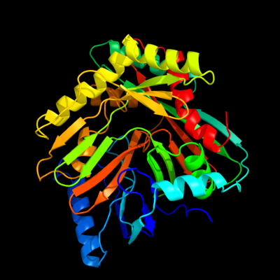
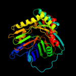
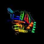
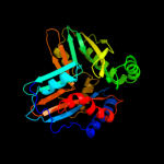
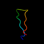
| 1 | c3hwoB_
|
|
 |
100.0 |
100 |
PDB header:isomerase
Chain: B: PDB Molecule:isochorismate synthase entc;
PDBTitle: crystal structure of escherichia coli enterobactin-specific2 isochorismate synthase entc in complex with isochorismate
|
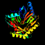
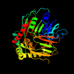
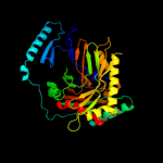
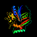
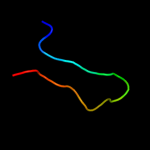
| 2 | c3os6A_
|
|
 |
100.0 |
38 |
PDB header:isomerase
Chain: A: PDB Molecule:isochorismate synthase dhbc;
PDBTitle: crystal structure of putative 2,3-dihydroxybenzoate-specific2 isochorismate synthase, dhbc from bacillus anthracis.
|
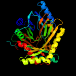
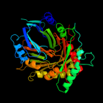
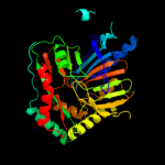
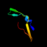
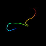
| 3 | d3bzna1
|
|
 |
100.0 |
27 |
Fold:ADC synthase
Superfamily:ADC synthase
Family:ADC synthase |
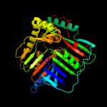
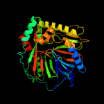
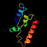
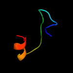
| 4 | d1qdla_
|
|
 |
100.0 |
25 |
Fold:ADC synthase
Superfamily:ADC synthase
Family:ADC synthase |
| 5 | d2fn0a1
|
|
 |
100.0 |
22 |
Fold:ADC synthase
Superfamily:ADC synthase
Family:ADC synthase |
| 6 | c3gseA_
|
|
 |
100.0 |
24 |
PDB header:isomerase
Chain: A: PDB Molecule:menaquinone-specific isochorismate synthase;
PDBTitle: crystal structure of menaquinone-specific isochorismate synthase from2 yersinia pestis co92
|
| 7 | d2g5fa1
|
|
 |
100.0 |
21 |
Fold:ADC synthase
Superfamily:ADC synthase
Family:ADC synthase |
| 8 | d1i1qa_
|
|
 |
100.0 |
22 |
Fold:ADC synthase
Superfamily:ADC synthase
Family:ADC synthase |
| 9 | d1i7qa_
|
|
 |
100.0 |
22 |
Fold:ADC synthase
Superfamily:ADC synthase
Family:ADC synthase |
| 10 | c3h9mA_
|
|
 |
100.0 |
17 |
PDB header:lyase
Chain: A: PDB Molecule:p-aminobenzoate synthetase, component i;
PDBTitle: crystal structure of para-aminobenzoate synthetase,2 component i from cytophaga hutchinsonii
|
| 11 | c2i6yA_
|
|
 |
100.0 |
22 |
PDB header:lyase
Chain: A: PDB Molecule:anthranilate synthase component i, putative;
PDBTitle: structure and mechanism of mycobacterium tuberculosis salicylate2 synthase, mbti
|
| 12 | c3r74B_
|
|
 |
100.0 |
21 |
PDB header:lyase, biosynthetic protein
Chain: B: PDB Molecule:anthranilate/para-aminobenzoate synthases component i;
PDBTitle: crystal structure of 2-amino-2-desoxyisochorismate synthase (adic)2 synthase phze from burkholderia lata 383
|
| 13 | d1k0ga_
|
|
 |
100.0 |
22 |
Fold:ADC synthase
Superfamily:ADC synthase
Family:ADC synthase |
| 14 | c3nqkA_
|
|
 |
41.9 |
16 |
PDB header:structural genomics, unknown function
Chain: A: PDB Molecule:uncharacterized protein;
PDBTitle: crystal structure of a structural genomics, unknown function2 (bacova_03322) from bacteroides ovatus at 2.61 a resolution
|
| 15 | d2ffca1
|
|
 |
23.0 |
18 |
Fold:TIM beta/alpha-barrel
Superfamily:Ribulose-phoshate binding barrel
Family:Decarboxylase |
| 16 | d1hxra_
|
|
 |
19.4 |
22 |
Fold:Mss4-like
Superfamily:Mss4-like
Family:RabGEF Mss4 |
| 17 | d2fu5a1
|
|
 |
19.1 |
22 |
Fold:Mss4-like
Superfamily:Mss4-like
Family:RabGEF Mss4 |
| 18 | d1qusa_
|
|
 |
15.2 |
35 |
Fold:Lysozyme-like
Superfamily:Lysozyme-like
Family:Bacterial muramidase, catalytic domain |
| 19 | c2km1A_
|
|
 |
13.8 |
26 |
PDB header:protein binding
Chain: A: PDB Molecule:protein dre2;
PDBTitle: solution structure of the n-terminal domain of the yeast protein dre2
|
| 20 | d1a6ca2
|
|
 |
12.4 |
11 |
Fold:Nucleoplasmin-like/VP (viral coat and capsid proteins)
Superfamily:Positive stranded ssRNA viruses
Family:Comoviridae-like VP |
| 21 | d1cvra1 |
|
not modelled |
9.6 |
11 |
Fold:Immunoglobulin-like beta-sandwich
Superfamily:E set domains
Family:Gingipain R (RgpB), C-terminal domain |
| 22 | c2z1tA_ |
|
not modelled |
9.0 |
21 |
PDB header:lyase
Chain: A: PDB Molecule:hydrogenase expression/formation protein hype;
PDBTitle: crystal structure of hydrogenase maturation protein hype
|
| 23 | c3fiuD_ |
|
not modelled |
8.7 |
12 |
PDB header:ligase
Chain: D: PDB Molecule:nh(3)-dependent nad(+) synthetase;
PDBTitle: structure of nmn synthetase from francisella tularensis
|
| 24 | c3fuyC_ |
|
not modelled |
8.6 |
30 |
PDB header:structural genomics, unknown function
Chain: C: PDB Molecule:putative integron gene cassette protein;
PDBTitle: structure from the mobile metagenome of cole harbour salt2 marsh: integron cassette protein hfx_cass1
|
| 25 | c1ey2A_ |
|
not modelled |
8.5 |
28 |
PDB header:oxidoreductase
Chain: A: PDB Molecule:homogentisate 1,2-dioxygenase;
PDBTitle: human homogentisate dioxygenase with fe(ii)
|
| 26 | d1eyba_ |
|
not modelled |
8.5 |
28 |
Fold:Double-stranded beta-helix
Superfamily:RmlC-like cupins
Family:Homogentisate dioxygenase |
| 27 | d1d5ta2 |
|
not modelled |
8.1 |
21 |
Fold:FAD-linked reductases, C-terminal domain
Superfamily:FAD-linked reductases, C-terminal domain
Family:GDI-like |
| 28 | c1tzsA_ |
|
not modelled |
7.1 |
13 |
PDB header:hydrolase
Chain: A: PDB Molecule:cathepsin e;
PDBTitle: crystal structure of an activation intermediate of2 cathepsin e
|
| 29 | d2af7a1 |
|
not modelled |
7.0 |
17 |
Fold:AhpD-like
Superfamily:AhpD-like
Family:CMD-like |
| 30 | d3cmsa_ |
|
not modelled |
6.3 |
15 |
Fold:Acid proteases
Superfamily:Acid proteases
Family:Pepsin-like |
| 31 | c3c66B_ |
|
not modelled |
6.0 |
19 |
PDB header:transferase
Chain: B: PDB Molecule:poly(a) polymerase;
PDBTitle: yeast poly(a) polymerase in complex with fip1 residues 80-105
|
| 32 | d2c1wa1 |
|
not modelled |
6.0 |
25 |
Fold:EndoU-like
Superfamily:EndoU-like
Family:Eukaryotic EndoU ribonuclease |
| 33 | c3rrrB_ |
|
not modelled |
6.0 |
29 |
PDB header:viral protein
Chain: B: PDB Molecule:fusion glycoprotein f0;
PDBTitle: structure of the rsv f protein in the post-fusion conformation
|
| 34 | d1rd5a_ |
|
not modelled |
6.0 |
33 |
Fold:TIM beta/alpha-barrel
Superfamily:Ribulose-phoshate binding barrel
Family:Tryptophan biosynthesis enzymes |
| 35 | c1wxnA_ |
|
not modelled |
5.8 |
31 |
PDB header:toxin
Chain: A: PDB Molecule:toxin apetx2;
PDBTitle: solution structure of apetx2, a specific peptide inhibitor2 of asic3 proton-gated channels
|
| 36 | c3rpdB_ |
|
not modelled |
5.5 |
31 |
PDB header:transferase
Chain: B: PDB Molecule:methionine synthase (b12-independent);
PDBTitle: the structure of a b12-independent methionine synthase from shewanella2 sp. w3-18-1 in complex with selenomethionine.
|