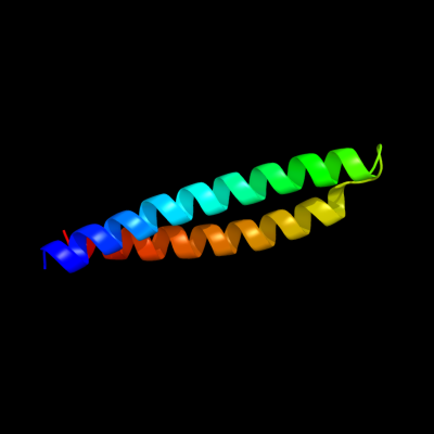
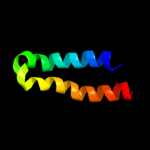
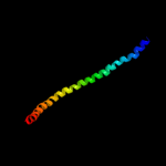
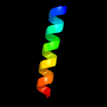
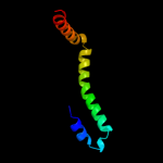
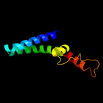
1 c1nfoA_
91.4
15
PDB header: lipid transportChain: A: PDB Molecule: apolipoprotein e2;PDBTitle: apolipoprotein e2 (apoe2, d154a mutation)
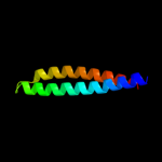
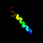
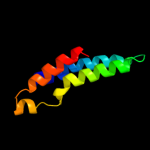
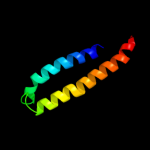
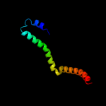
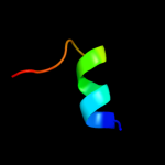
2 d1gs9a_
88.3
16
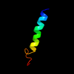
Fold: Four-helical up-and-down bundleSuperfamily: ApolipoproteinFamily: Apolipoprotein3 c2x43S_
80.0
8
PDB header: membrane proteinChain: S: PDB Molecule: sherp;PDBTitle: structural basis of molecular recognition by sherp at membrane2 surfaces
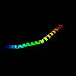
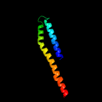
4 c3kdpG_
72.3
31
PDB header: hydrolaseChain: G: PDB Molecule: na+/k+ atpase gamma subunit transcript variant a;PDBTitle: crystal structure of the sodium-potassium pump
5 c3kdpH_
72.3
31
PDB header: hydrolaseChain: H: PDB Molecule: na+/k+ atpase gamma subunit transcript variant a;PDBTitle: crystal structure of the sodium-potassium pump
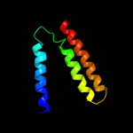
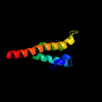
6 c3r2pA_
60.9
13
PDB header: lipid transportChain: A: PDB Molecule: apolipoprotein a-i;PDBTitle: 2.2 angstrom crystal structure of c terminal truncated human2 apolipoprotein a-i reveals the assembly of hdl by dimerization.
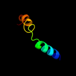
7 c1av1B_
58.8
12
PDB header: lipid transportChain: B: PDB Molecule: apolipoprotein a-i;PDBTitle: crystal structure of human apolipoprotein a-i
8 d1eq1a_
41.8
19
Fold: Apolipophorin-IIISuperfamily: Apolipophorin-IIIFamily: Apolipophorin-III9 c2a01B_
39.4
21
PDB header: lipid transportChain: B: PDB Molecule: apolipoprotein a-i;PDBTitle: crystal structure of lipid-free human apolipoprotein a-i
10 c3gvmA_
35.3
7
PDB header: viral proteinChain: A: PDB Molecule: putative uncharacterized protein sag1039;PDBTitle: structure of the homodimeric wxg-100 family protein from streptococcus2 agalactiae
11 c2jwaA_
31.4
38
PDB header: transferaseChain: A: PDB Molecule: receptor tyrosine-protein kinase erbb-2;PDBTitle: erbb2 transmembrane segment dimer spatial structure
12 d1wa8b1
25.6
11
Fold: Ferritin-likeSuperfamily: EsxAB dimer-likeFamily: ESAT-6 like13 d1ryka_
24.7
5
Fold: SAM domain-likeSuperfamily: Hypothetical protein YjbJFamily: Hypothetical protein YjbJ14 c2vs0B_
23.6
11
PDB header: cell invasionChain: B: PDB Molecule: virulence factor esxa;PDBTitle: structural analysis of homodimeric staphylococcal aureus2 virulence factor esxa
15 d1i6la_
23.4
14
Fold: Adenine nucleotide alpha hydrolase-likeSuperfamily: Nucleotidylyl transferaseFamily: Class I aminoacyl-tRNA synthetases (RS), catalytic domain16 c3prhB_
22.7
18
PDB header: ligaseChain: B: PDB Molecule: tryptophanyl-trna synthetase;PDBTitle: tryptophanyl-trna synthetase val144pro mutant from b. subtilis
17 d2ooca1
21.5
14
Fold: Four-helical up-and-down bundleSuperfamily: Histidine-containing phosphotransfer domain, HPT domainFamily: SphA-like18 c1p58F_
21.4
15
PDB header: virusChain: F: PDB Molecule: envelope protein m;PDBTitle: complex organization of dengue virus membrane proteins as revealed by2 9.5 angstrom cryo-em reconstruction
19 c3e0sA_
21.4
23
PDB header: structural genomics, unknown functionChain: A: PDB Molecule: uncharacterized protein;PDBTitle: crystal structure of an uncharacterized protein from2 chlorobium tepidum
20 c3lw5K_
20.1
44
PDB header: photosynthesisChain: K: PDB Molecule: photosystem i reaction center subunit x psak;PDBTitle: improved model of plant photosystem i
21 c2l5bA_
not modelled
19.4
29
PDB header: apoptosisChain: A: PDB Molecule: activator of apoptosis harakiri;PDBTitle: solution structure of the transmembrane domain of bcl-2 member2 harakiri in micelles
22 d2gtsa1
not modelled
17.1
16
Fold: Ferritin-likeSuperfamily: HP0062-likeFamily: HP0062-like23 d1wa8a1
not modelled
16.7
12
Fold: Ferritin-likeSuperfamily: EsxAB dimer-likeFamily: ESAT-6 like24 d1nekd_
not modelled
16.5
7
Fold: Heme-binding four-helical bundleSuperfamily: Fumarate reductase respiratory complex transmembrane subunitsFamily: Succinate dehydrogenase/Fumarate reductase transmembrane subunits (SdhC/FrdC and SdhD/FrdD)25 c3onjA_
not modelled
12.0
9
PDB header: protein transportChain: A: PDB Molecule: t-snare vti1;PDBTitle: crystal structure of yeast vti1p_habc domain
26 d1vcsa1
not modelled
11.8
11
Fold: STAT-likeSuperfamily: t-snare proteinsFamily: t-snare proteins27 d1s35a1
not modelled
11.7
12
Fold: Spectrin repeat-likeSuperfamily: Spectrin repeatFamily: Spectrin repeat28 d1c99a_
not modelled
10.0
28
Fold: Transmembrane helix hairpinSuperfamily: F1F0 ATP synthase subunit CFamily: F1F0 ATP synthase subunit C29 c2kncB_
not modelled
9.7
5
PDB header: cell adhesionChain: B: PDB Molecule: integrin beta-3;PDBTitle: platelet integrin alfaiib-beta3 transmembrane-cytoplasmic2 heterocomplex
30 d1eiya1
not modelled
9.5
23
Fold: Long alpha-hairpinSuperfamily: tRNA-binding armFamily: Phenylalanyl-tRNA synthetase (PheRS)31 d1k75a_
not modelled
9.3
10
Fold: ALDH-likeSuperfamily: ALDH-likeFamily: L-histidinol dehydrogenase HisD32 d1f16a_
not modelled
8.9
21
Fold: Toxins' membrane translocation domainsSuperfamily: Bcl-2 inhibitors of programmed cell deathFamily: Bcl-2 inhibitors of programmed cell death33 c2g36A_
not modelled
8.3
16
PDB header: ligaseChain: A: PDB Molecule: tryptophanyl-trna synthetase;PDBTitle: crystal structure of tryptophanyl-trna synthetase (ec 6.1.1.2)2 (tryptophan-trna ligase)(trprs) (tm0492) from thermotoga maritima at3 2.50 a resolution
34 c3py7A_
not modelled
7.9
14
PDB header: viral proteinChain: A: PDB Molecule: maltose-binding periplasmic protein,paxillin ld1,protein e6PDBTitle: crystal structure of full-length bovine papillomavirus oncoprotein e62 in complex with ld1 motif of paxillin at 2.3a resolution
35 c1n54A_
not modelled
7.8
19
PDB header: rna binding proteinChain: A: PDB Molecule: 80 kda nuclear cap binding protein;PDBTitle: cap binding complex m7gpppg free
36 c2kbvA_
not modelled
7.8
30
PDB header: membrane proteinChain: A: PDB Molecule: sodium/hydrogen exchanger 1;PDBTitle: structural and functional analysis of tm xi of the nhe12 isoform of the na+/h+ exchanger
37 c2j5dA_
not modelled
7.6
16
PDB header: membrane proteinChain: A: PDB Molecule: bcl2/adenovirus e1b 19 kda protein-interactingPDBTitle: nmr structure of bnip3 transmembrane domain in lipid2 bicelles
38 d1q2ha_
not modelled
7.6
20
Fold: Dimerisation interlockSuperfamily: Phenylalanine zipperFamily: Adapter protein APS, dimerisation domain39 c2v8sV_
not modelled
7.2
15
PDB header: protein transportChain: V: PDB Molecule: vesicle transport through interaction withPDBTitle: vti1b habc domain - epsinr enth domain complex
40 c2ww9B_
not modelled
7.1
21
PDB header: ribosomeChain: B: PDB Molecule: protein transport protein sss1;PDBTitle: cryo-em structure of the active yeast ssh1 complex bound to the2 yeast 80s ribosome
41 c1yi8C_
not modelled
7.1
13
PDB header: ligaseChain: C: PDB Molecule: tryptophanyl-trna synthetase;PDBTitle: crystal structure of tryptophanyl trrna synthetase ii from deinococcus2 radiodurans in complex with l-trp
42 c1q90L_
not modelled
7.1
27
PDB header: photosynthesisChain: L: PDB Molecule: cytochrome b6f complex subunit petl;PDBTitle: structure of the cytochrome b6f (plastohydroquinone : plastocyanin2 oxidoreductase) from chlamydomonas reinhardtii
43 d1q90l_
not modelled
7.1
27
Fold: Single transmembrane helixSuperfamily: PetL subunit of the cytochrome b6f complexFamily: PetL subunit of the cytochrome b6f complex44 c1ywwA_
not modelled
6.9
6
PDB header: structural genomics, unknown functionChain: A: PDB Molecule: hypothetical protein pa4738;PDBTitle: nmr structure of p. aeruginosa protein pa4738: northeast2 structural genomics consortium target pap2
45 c1xq8A_
not modelled
6.8
18
PDB header: lipid binding proteinChain: A: PDB Molecule: alpha-synuclein;PDBTitle: human micelle-bound alpha-synuclein
46 c3hd7A_
not modelled
6.7
14
PDB header: exocytosisChain: A: PDB Molecule: vesicle-associated membrane protein 2;PDBTitle: helical extension of the neuronal snare complex into the membrane,2 spacegroup c 1 2 1
47 c2p2uA_
not modelled
6.6
10
PDB header: dna binding proteinChain: A: PDB Molecule: host-nuclease inhibitor protein gam, putative;PDBTitle: crystal structure of putative host-nuclease inhibitor2 protein gam from desulfovibrio vulgaris
48 d1l2pa_
not modelled
6.5
13
Fold: Single transmembrane helixSuperfamily: F1F0 ATP synthase subunit B, membrane domainFamily: F1F0 ATP synthase subunit B, membrane domain49 c3m5wB_
not modelled
6.3
17
PDB header: ligaseChain: B: PDB Molecule: tryptophanyl-trna synthetase;PDBTitle: crystal structure of tryptophanyl-trna synthetase from2 campylobacter jejuni
50 c3b9yA_
not modelled
6.3
21
PDB header: transport proteinChain: A: PDB Molecule: ammonium transporter family rh-like protein;PDBTitle: crystal structure of the nitrosomonas europaea rh protein
51 d1szia_
not modelled
6.1
22
Fold: Four-helical up-and-down bundleSuperfamily: Mannose-6-phosphate receptor binding protein 1 (Tip47), C-terminal domainFamily: Mannose-6-phosphate receptor binding protein 1 (Tip47), C-terminal domain52 d1tqga_
not modelled
6.0
10
Fold: Four-helical up-and-down bundleSuperfamily: Histidine-containing phosphotransfer domain, HPT domainFamily: Chemotaxis protein CheA P1 domain53 c3kb8A_
not modelled
5.9
14
PDB header: transferaseChain: A: PDB Molecule: hypoxanthine phosphoribosyltransferase;PDBTitle: 2.09 angstrom resolution structure of a hypoxanthine-guanine2 phosphoribosyltransferase (hpt-1) from bacillus anthracis str. 'ames3 ancestor' in complex with gmp
54 d2o97b1
not modelled
5.6
16
Fold: IHF-like DNA-binding proteinsSuperfamily: IHF-like DNA-binding proteinsFamily: Prokaryotic DNA-bending protein55 c2ka2A_
not modelled
5.6
16
PDB header: membrane proteinChain: A: PDB Molecule: bcl2/adenovirus e1b 19 kda protein-interactingPDBTitle: solution nmr structure of bnip3 transmembrane peptide dimer2 in detergent micelles with his173-ser172 intermonomer3 hydrogen bond restraints
56 c2ka1B_
not modelled
5.6
16
PDB header: membrane proteinChain: B: PDB Molecule: bcl2/adenovirus e1b 19 kda protein-interactingPDBTitle: solution nmr structure of bnip3 transmembrane peptide dimer2 in detergent micelles
57 c2ks1B_
not modelled
5.5
14
PDB header: transferaseChain: B: PDB Molecule: epidermal growth factor receptor;PDBTitle: heterodimeric association of transmembrane domains of erbb1 and erbb22 receptors enabling kinase activation
58 c2k1lB_
not modelled
5.5
31
PDB header: signaling proteinChain: B: PDB Molecule: ephrin type-a receptor 1;PDBTitle: nmr structures of dimeric transmembrane domain of the2 receptor tyrosine kinase epha1 in lipid bicelles at ph 6.3
59 c2k1lA_
not modelled
5.5
31
PDB header: signaling proteinChain: A: PDB Molecule: ephrin type-a receptor 1;PDBTitle: nmr structures of dimeric transmembrane domain of the2 receptor tyrosine kinase epha1 in lipid bicelles at ph 6.3
60 c2k1kA_
not modelled
5.5
31
PDB header: signaling proteinChain: A: PDB Molecule: ephrin type-a receptor 1;PDBTitle: nmr structures of dimeric transmembrane domain of the2 receptor tyrosine kinase epha1 in lipid bicelles at ph 4.3
61 c2k1kB_
not modelled
5.5
31
PDB header: signaling proteinChain: B: PDB Molecule: ephrin type-a receptor 1;PDBTitle: nmr structures of dimeric transmembrane domain of the2 receptor tyrosine kinase epha1 in lipid bicelles at ph 4.3
62 d1dqna_
not modelled
5.5
19
Fold: PRTase-likeSuperfamily: PRTase-likeFamily: Phosphoribosyltransferases (PRTases)63 d1g9sa_
not modelled
5.5
14
Fold: PRTase-likeSuperfamily: PRTase-likeFamily: Phosphoribosyltransferases (PRTases)64 c2el7A_
not modelled
5.3
25
PDB header: ligaseChain: A: PDB Molecule: tryptophanyl-trna synthetase;PDBTitle: crystal structure of tryptophanyl-trna synthetase from thermus2 thermophilus
65 c2hroA_
not modelled
5.3
15
PDB header: transferaseChain: A: PDB Molecule: phosphoenolpyruvate-protein phosphotransferase;PDBTitle: structure of the full-lenght enzyme i of the pts system from2 staphylococcus carnosus
66 c2ka2B_
not modelled
5.2
13
PDB header: membrane proteinChain: B: PDB Molecule: bcl2/adenovirus e1b 19 kda protein-interactingPDBTitle: solution nmr structure of bnip3 transmembrane peptide dimer2 in detergent micelles with his173-ser172 intermonomer3 hydrogen bond restraints
67 c2ka1A_
not modelled
5.2
13
PDB header: membrane proteinChain: A: PDB Molecule: bcl2/adenovirus e1b 19 kda protein-interactingPDBTitle: solution nmr structure of bnip3 transmembrane peptide dimer2 in detergent micelles