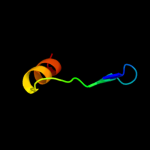
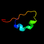
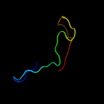
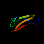
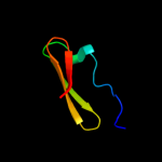
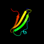
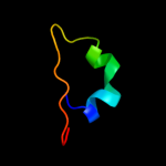
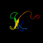
1 c3cgiD_
48.2
21
PDB header: unknown functionChain: D: PDB Molecule: propanediol utilization protein pduu;PDBTitle: crystal structure of the pduu shell protein from the pdu2 microcompartment
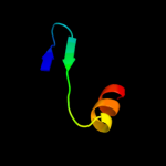
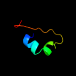
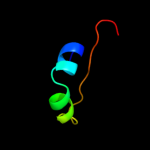
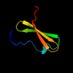
2 c3ia0c_
46.0
29
PDB header: structural proteinChain: C: PDB Molecule: ethanolamine utilization protein euts;PDBTitle: ethanolamine utilization microcompartment shell subunit,2 euts-g39v mutant
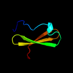
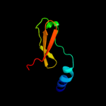
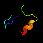
3 d1ulia1
42.1
19
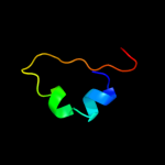
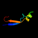
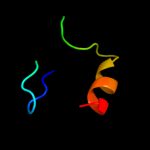
Fold: ISP domainSuperfamily: ISP domainFamily: Ring hydroxylating alpha subunit ISP domain4 c2odkD_
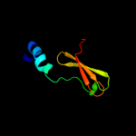
35.3
25
PDB header: structural genomics, unknown functionChain: D: PDB Molecule: hypothetical protein;PDBTitle: putative prevent-host-death protein from nitrosomonas europaea
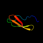
5 c3hryA_
31.4
29
PDB header: antitoxinChain: A: PDB Molecule: prevent host death protein;PDBTitle: crystal structure of phd in a trigonal space group and partially2 disordered
6 d2odka1
30.5
24
Fold: YefM-likeSuperfamily: YefM-likeFamily: YefM-like7 d2bmoa1
26.0
15
Fold: ISP domainSuperfamily: ISP domainFamily: Ring hydroxylating alpha subunit ISP domain8 d1o7na1
24.9
17
Fold: ISP domainSuperfamily: ISP domainFamily: Ring hydroxylating alpha subunit ISP domain9 c3c5pF_
24.6
30
PDB header: structural genomics, unknown functionChain: F: PDB Molecule: protein bas0735 of unknown function;PDBTitle: crystal structure of bas0735, a protein of unknown function from2 bacillus anthracis str. sterne
10 c3gkqB_
23.6
15
PDB header: oxidoreductaseChain: B: PDB Molecule: terminal oxygenase component of carbazole 1,9a-PDBTitle: terminal oxygenase of carbazole 1,9a-dioxygenase from2 novosphingobium sp. ka1
11 c3gcfC_
20.9
20
PDB header: oxidoreductaseChain: C: PDB Molecule: terminal oxygenase component of carbazole 1,9a-PDBTitle: terminal oxygenase of carbazole 1,9a-dioxygenase from2 nocardioides aromaticivorans ic177
12 d2b1xa1
20.1
32
Fold: ISP domainSuperfamily: ISP domainFamily: Ring hydroxylating alpha subunit ISP domain13 d2a6qb1
20.0
29
Fold: YefM-likeSuperfamily: YefM-likeFamily: YefM-like14 c3hs2H_
19.3
29
PDB header: antitoxinChain: H: PDB Molecule: prevent host death protein;PDBTitle: crystal structure of phd truncated to residue 57 in an orthorhombic2 space group
15 c2jx8A_
18.7
39
PDB header: transcriptionChain: A: PDB Molecule: phosphorylated ctd-interacting factor 1;PDBTitle: solution structure of hpcif1 ww domain
16 d2de6a1
17.6
17
Fold: ISP domainSuperfamily: ISP domainFamily: Ring hydroxylating alpha subunit ISP domain17 c2jsoA_
16.1
18
PDB header: signaling proteinChain: A: PDB Molecule: polymyxin resistance protein pmrd;PDBTitle: antimicrobial resistance protein
18 d1wqla1
13.5
17
Fold: ISP domainSuperfamily: ISP domainFamily: Ring hydroxylating alpha subunit ISP domain19 c2ckcA_
13.3
24
PDB header: hydrolaseChain: A: PDB Molecule: chromodomain-helicase-dna-binding protein 7;PDBTitle: solution structures of the brk domains of the human chromo2 helicase domain 7 and 8, reveals structural similarity3 with gyf domain suggesting a role in protein interaction
20 d2ckca1
13.3
24
Fold: GYF/BRK domain-likeSuperfamily: BRK domain-likeFamily: BRK domain-like21 d2a6qa1
not modelled
11.0
30
Fold: YefM-likeSuperfamily: YefM-likeFamily: YefM-like22 d2fbaa1
not modelled
10.8
16
Fold: alpha/alpha toroidSuperfamily: Six-hairpin glycosidasesFamily: Glucoamylase23 c1ncgA_
not modelled
10.3
27
PDB header: cell adhesion proteinChain: A: PDB Molecule: n-cadherin;PDBTitle: structural basis of cell-cell adhesion by cadherins
24 d1z01a1
not modelled
10.2
22
Fold: ISP domainSuperfamily: ISP domainFamily: Ring hydroxylating alpha subunit ISP domain25 c1zvnA_
not modelled
9.9
32
PDB header: ----Chain: A: PDB Molecule: mn-cadherin;PDBTitle: crystal structure of chick mn-cadherin ec1
26 d1tdha3
not modelled
9.9
28
Fold: Glucocorticoid receptor-like (DNA-binding domain)Superfamily: Glucocorticoid receptor-like (DNA-binding domain)Family: C-terminal, Zn-finger domain of MutM-like DNA repair proteins27 c2rqxA_
not modelled
9.5
21
PDB header: signaling proteinChain: A: PDB Molecule: polymyxin b resistance protein;PDBTitle: solution nmr structure of pmrd from klebsiella pneumoniae
28 c2e9qA_
not modelled
9.4
18
PDB header: plant proteinChain: A: PDB Molecule: 11s globulin subunit beta;PDBTitle: recombinant pro-11s globulin of pumpkin
29 d2idaa1
not modelled
9.0
14
Fold: RING/U-boxSuperfamily: RING/U-boxFamily: Zf-UBP30 d2pbka1
not modelled
9.0
18
Fold: Herpes virus serine proteinase, assemblinSuperfamily: Herpes virus serine proteinase, assemblinFamily: Herpes virus serine proteinase, assemblin31 d1njha_
not modelled
8.8
22
Fold: Hypothetical protein YojFSuperfamily: Hypothetical protein YojFFamily: Hypothetical protein YojF32 c3ff7B_
not modelled
8.1
27
PDB header: cell adhesion/immunue systemChain: B: PDB Molecule: epithelial cadherin;PDBTitle: structure of nk cell receptor klrg1 bound to e-cadherin
33 d1aa7a_
not modelled
8.1
35
Fold: Influenza virus matrix protein M1Superfamily: Influenza virus matrix protein M1Family: Influenza virus matrix protein M134 d1o6ea_
not modelled
8.0
13
Fold: Herpes virus serine proteinase, assemblinSuperfamily: Herpes virus serine proteinase, assemblinFamily: Herpes virus serine proteinase, assemblin35 c2b1xE_
not modelled
7.9
26
PDB header: oxidoreductaseChain: E: PDB Molecule: naphthalene dioxygenase large subunit;PDBTitle: crystal structure of naphthalene 1,2-dioxygenase from rhodococcus sp.
36 c3g5oA_
not modelled
7.4
21
PDB header: toxin/antitoxinChain: A: PDB Molecule: uncharacterized protein rv2865;PDBTitle: the crystal structure of the toxin-antitoxin complex relbe2 (rv2865-2 2866) from mycobacterium tuberculosis
37 d1y0pa3
not modelled
7.1
17
Fold: Succinate dehydrogenase/fumarate reductase flavoprotein, catalytic domainSuperfamily: Succinate dehydrogenase/fumarate reductase flavoprotein, catalytic domainFamily: Succinate dehydrogenase/fumarate reductase flavoprotein, catalytic domain38 c3entB_
not modelled
7.0
20
PDB header: structural proteinChain: B: PDB Molecule: putative uncharacterized protein;PDBTitle: crystal structure of nitrollin, a betagamma-crystallin from2 nitrosospira multiformis-in alternate space group (p65)
39 d1ynha1
not modelled
7.0
16
Fold: Pentein, beta/alpha-propellerSuperfamily: PenteinFamily: Succinylarginine dihydrolase-like40 d1r76a_
not modelled
6.9
33
Fold: alpha/alpha toroidSuperfamily: Family 10 polysaccharide lyaseFamily: Family 10 polysaccharide lyase41 d2omzb1
not modelled
6.9
29
Fold: Immunoglobulin-like beta-sandwichSuperfamily: Cadherin-likeFamily: Cadherin42 c3jr7A_
not modelled
6.5
9
PDB header: structural genomics, unknown functionChain: A: PDB Molecule: uncharacterized egv family protein cog1307;PDBTitle: the crystal structure of the protein of degv family cog1307 with2 unknown function from ruminococcus gnavus atcc 29149
43 d1vr5a1
not modelled
6.5
11
Fold: Periplasmic binding protein-like IISuperfamily: Periplasmic binding protein-like IIFamily: Phosphate binding protein-like44 c1uljA_
not modelled
6.5
19
PDB header: oxidoreductaseChain: A: PDB Molecule: biphenyl dioxygenase large subunit;PDBTitle: biphenyl dioxygenase (bpha1a2) in complex with the substrate
45 d1iega_
not modelled
6.1
19
Fold: Herpes virus serine proteinase, assemblinSuperfamily: Herpes virus serine proteinase, assemblinFamily: Herpes virus serine proteinase, assemblin46 c3gteB_
not modelled
6.0
21
PDB header: electron transport, oxidoreductaseChain: B: PDB Molecule: ddmc;PDBTitle: crystal structure of dicamba monooxygenase with non-heme2 iron
47 c2hjjA_
not modelled
6.0
15
PDB header: structural genomics, unknown functionChain: A: PDB Molecule: hypothetical protein ykff;PDBTitle: solution nmr structure of protein ykff from escherichia coli.2 northeast structural genomics target er397.
48 d2hjja1
not modelled
6.0
15
Fold: dsRBD-likeSuperfamily: YcfA/nrd intein domainFamily: YkfF-like49 c2o7jA_
not modelled
5.9
11
PDB header: sugar binding proteinChain: A: PDB Molecule: oligopeptide abc transporter, periplasmicPDBTitle: the x-ray crystal structure of a thermophilic cellobiose2 binding protein bound with cellopentaose
50 c2js3B_
not modelled
5.9
28
PDB header: structural genomics, unknown functionChain: B: PDB Molecule: uncharacterized protein;PDBTitle: nmr structure of protein q6n9a4_rhopa. northeast structural genomics2 consortium target rpt8
51 c2de7B_
not modelled
5.5
17
PDB header: oxidoreductaseChain: B: PDB Molecule: terminal oxygenase component of carbazole;PDBTitle: the substrate-bound complex between oxygenase and2 ferredoxin in carbazole 1,9a-dioxygenase
52 d1at3a_
not modelled
5.3
16
Fold: Herpes virus serine proteinase, assemblinSuperfamily: Herpes virus serine proteinase, assemblinFamily: Herpes virus serine proteinase, assemblin53 d1smye_
not modelled
5.3
30
Fold: RPB6/omega subunit-likeSuperfamily: RPB6/omega subunit-likeFamily: RNA polymerase omega subunit54 d1vkza1
not modelled
5.3
21
Fold: Barrel-sandwich hybridSuperfamily: Rudiment single hybrid motifFamily: BC C-terminal domain-like55 c1nchB_
not modelled
5.3
27
PDB header: cell adhesion proteinChain: B: PDB Molecule: n-cadherin;PDBTitle: structural basis of cell-cell adhesion by cadherins
56 c1nciB_
not modelled
5.2
30
PDB header: cell adhesion proteinChain: B: PDB Molecule: n-cadherin;PDBTitle: structural basis of cell-cell adhesion by cadherins


