Phyre Home Retrieve Phyre Job Id
Emaill.a.kelley@imperial.ac.uk
DescriptionP76146
DateThu Jan 5 12:19:40 GMT 2012
Unique Job ID4496a4e9330fb04a

Summary 

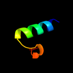
Top model
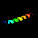
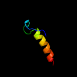
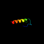
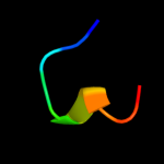
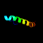
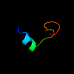
Image coloured by rainbow N → C terminus
Model (left) based on template d1kqfb2
Top template information
Fold:Single transmembrane helix
Superfamily:Iron-sulfur subunit of formate dehydrogenase N, transmembrane anchor
Family:Iron-sulfur subunit of formate dehydrogenase N, transmembrane anchor
Confidence and coverage
Confidence: 34.3% Coverage: 9%
27 residues ( 9% of your sequence) have been modelled with 34.3% confidence by the single highest scoring template.
You may wish to submit your sequence to Phyrealarm. This will automatically scan your sequence every week for new potential templates as they appear in the Phyre2 library.
Please note: You must be registered and logged in to use Phyrealarm.
3D viewing
Interactive 3D view in Jmol

Sequence analysis 

Secondary structure and disorder prediction 

   1........10.........20.........30.........40.........50.........60
Sequence  MIVRPQQHWLRRIFVWHGSVLSKISSRLLLNFLFSIAVIFMLPWYTHLGIKFTLAPFSIL
Secondary structure 


SS confidence 


Disorder  ???????


????
??


?


Disorder confidence 


   .........70.........80.........90.........100.........110.........120
Sequence  GVAIAIFLGFRNNAGYARYVEARKLWGQLMIASRSLLREVKTTLPDSASVREFARLQIAF
Secondary structure 

SS confidence 


Disorder 


Disorder confidence 


   .........130.........140.........150.........160.........170.........180
Sequence  AHCLRMTLRKQPQAEVLAHYLKTEDLQRVLASNSPANRILLIMGEWLAVQRRNGQLSDIL
Secondary structure 


SS confidence 


Disorder 


Disorder confidence 


   .........190.........200.........210.........220.........230.........240
Sequence  FISLNDRLNDISAVLAGCERIAYTPIPFAYTLILHRTVYLFCIMLPFALVVDLHYMTPFI
Secondary structure 


SS confidence 


Disorder 


Disorder confidence 


   .........250.........260.........270.........280.........290.........300
Sequence  SVLISYTFISLDCLAEELEDPFGTENNDLPLDAICNAIEIDLLQMNDEAEIPAKILPDRH
Secondary structure 


SS confidence 


Disorder 


?

????


???
Disorder confidence 


   ....
Sequence  YQLT
Secondary structure 


SS confidence 


Disorder  ????
Disorder confidence 


Confidence Key
High(9)                    Low (0)
?Disordered
Alpha helix
Beta strand

Domain analysis 

Hover over an aligned region to see model and summary info

Please note, only up to the top 20 hits are modelled to reduce computer load

RankAligned region

PDB 1kqf chain B domain 2

3D model

Region: 19 - 45
Aligned: 27
Modelled: 27
Confidence: 34.3%
Identity: 7%
Fold: Single transmembrane helix
Superfamily: Iron-sulfur subunit of formate dehydrogenase N, transmembrane anchor
Family: Iron-sulfur subunit of formate dehydrogenase N, transmembrane anchor

Phyre2

PDB 1h9a chain A domain 1

3D model

Region: 258 - 265
Aligned: 8
Modelled: 8
Confidence: 17.9%
Identity: 63%
Fold: NAD(P)-binding Rossmann-fold domains
Superfamily: NAD(P)-binding Rossmann-fold domains
Family: Glyceraldehyde-3-phosphate dehydrogenase-like, N-terminal domain

Phyre2

PDB 1qki chain A domain 1

3D model

Region: 258 - 265
Aligned: 8
Modelled: 8
Confidence: 17.9%
Identity: 50%
Fold: NAD(P)-binding Rossmann-fold domains
Superfamily: NAD(P)-binding Rossmann-fold domains
Family: Glyceraldehyde-3-phosphate dehydrogenase-like, N-terminal domain

Phyre2

PDB 1u84 chain A

3D model

Region: 260 - 295
Aligned: 30
Modelled: 36
Confidence: 12.5%
Identity: 33%
Fold: YugE-like
Superfamily: YugE-like
Family: YugE-like

Phyre2

PDB 1y1l chain A

3D model

Region: 258 - 288
Aligned: 29
Modelled: 31
Confidence: 12.3%
Identity: 21%
Fold: Phosphotyrosine protein phosphatases I-like
Superfamily: Phosphotyrosine protein phosphatases I
Family: Low-molecular-weight phosphotyrosine protein phosphatases

Phyre2

PDB 3rh0 chain A

3D model

Region: 258 - 286
Aligned: 27
Modelled: 29
Confidence: 11.3%
Identity: 11%
PDB header:oxidoreductase
Chain: A: PDB Molecule:arsenate reductase;
PDBTitle: corynebacterium glutamicum mycothiol/mycoredoxin1-dependent arsenate2 reductase cg_arsc2

Phyre2

PDB 2kx2 chain A

3D model

Region: 261 - 283
Aligned: 23
Modelled: 23
Confidence: 11.1%
Identity: 26%
PDB header:structural genomics, unknown function
Chain: A: PDB Molecule:putative uncharacterized protein;
PDBTitle: the solution structure of mth1821

Phyre2

PDB 1jf8 chain A

3D model

Region: 258 - 285
Aligned: 26
Modelled: 28
Confidence: 10.9%
Identity: 19%
Fold: Phosphotyrosine protein phosphatases I-like
Superfamily: Phosphotyrosine protein phosphatases I
Family: Low-molecular-weight phosphotyrosine protein phosphatases

Phyre2

PDB 1h9a chain A

3D model

Region: 258 - 265
Aligned: 8
Modelled: 8
Confidence: 9.6%
Identity: 63%
PDB header:oxidoreductase (choh(d) - nad(p))
Chain: A: PDB Molecule:glucose 6-phosphate 1-dehydrogenase;
PDBTitle: complex of active mutant (q365->c) of glucose 6-phosphate2 dehydrogenase from l. mesenteroides with coenzyme nadp

Phyre2

PDB 1d1q chain A

3D model

Region: 257 - 287
Aligned: 29
Modelled: 31
Confidence: 9.3%
Identity: 21%
Fold: Phosphotyrosine protein phosphatases I-like
Superfamily: Phosphotyrosine protein phosphatases I
Family: Low-molecular-weight phosphotyrosine protein phosphatases

Phyre2

PDB 1xvi chain A

3D model

Region: 262 - 271
Aligned: 10
Modelled: 10
Confidence: 9.0%
Identity: 50%
PDB header:hydrolase
Chain: A: PDB Molecule:putative mannosyl-3-phosphoglycerate phosphatase;
PDBTitle: crystal structure of yedp, phosphatase-like domain protein2 from escherichia coli k12

Phyre2

PDB 1xvi chain A

3D model

Region: 262 - 271
Aligned: 10
Modelled: 10
Confidence: 9.0%
Identity: 50%
Fold: HAD-like
Superfamily: HAD-like
Family: Predicted hydrolases Cof

Phyre2

PDB 1qki chain E

3D model

Region: 258 - 291
Aligned: 28
Modelled: 34
Confidence: 8.7%
Identity: 21%
PDB header:oxidoreductase
Chain: E: PDB Molecule:glucose-6-phosphate 1-dehydrogenase;
PDBTitle: x-ray structure of human glucose 6-phosphate dehydrogenase2 (variant canton r459l) complexed with structural nadp+

Phyre2

PDB 2bhl chain B

3D model

Region: 258 - 265
Aligned: 8
Modelled: 8
Confidence: 8.7%
Identity: 50%
PDB header:oxidoreductase (choh(d)-nadp)
Chain: B: PDB Molecule:glucose-6-phosphate 1-dehydrogenase;
PDBTitle: x-ray structure of human glucose-6-phosphate dehydrogenase2 (deletion variant) complexed with glucose-6-phosphate

Phyre2

PDB 1wzc chain A domain 1

3D model

Region: 262 - 271
Aligned: 10
Modelled: 10
Confidence: 8.2%
Identity: 40%
Fold: HAD-like
Superfamily: HAD-like
Family: Predicted hydrolases Cof

Phyre2

PDB 1p8a chain A

3D model

Region: 258 - 291
Aligned: 31
Modelled: 34
Confidence: 8.1%
Identity: 13%
Fold: Phosphotyrosine protein phosphatases I-like
Superfamily: Phosphotyrosine protein phosphatases I
Family: Low-molecular-weight phosphotyrosine protein phosphatases

Phyre2

PDB 3ns4 chain A

3D model

Region: 4 - 27
Aligned: 24
Modelled: 23
Confidence: 7.5%
Identity: 17%
PDB header:protein binding
Chain: A: PDB Molecule:vacuolar protein sorting-associated protein 53;
PDBTitle: structure of a c-terminal fragment of its vps53 subunit suggests2 similarity of garp to a family of tethering complexes

Phyre2

PDB 1gnt chain A

3D model

Region: 249 - 271
Aligned: 23
Modelled: 23
Confidence: 7.5%
Identity: 43%
Fold: Prismane protein-like
Superfamily: Prismane protein-like
Family: Hybrid cluster protein (prismane protein)

Phyre2

PDB 1s2o chain A domain 1

3D model

Region: 262 - 271
Aligned: 10
Modelled: 10
Confidence: 6.9%
Identity: 40%
Fold: HAD-like
Superfamily: HAD-like
Family: Predicted hydrolases Cof

Phyre2

PDB 2f76 chain X domain 1

3D model

Region: 251 - 284
Aligned: 34
Modelled: 34
Confidence: 6.7%
Identity: 15%
Fold: Retroviral matrix proteins
Superfamily: Retroviral matrix proteins
Family: Mason-Pfizer monkey virus matrix protein

Phyre2
1

d1kqfb2
2

d1h9aa1
3

d1qkia1
4

d1u84a_
5

d1y1la_
6

c3rh0A_
7

c2kx2A_
8

d1jf8a_
9

c1h9aA_
10

d1d1qa_
11

c1xviA_
12

d1xvia_
13

c1qkiE_
14

c2bhlB_
15

d1wzca1
16

d1p8aa_
17

c3ns4A_
18

d1gnta_
19

d1s2oa1
20

d2f76x1
21


22


23


24


25


26


27


28


29


30


31


32


33


34


35


36


37


38


39


40


41


42


43


44


45


46


47


48


Detailed template information 

#
Template Alignment Coverage3D Model Confidence
% i.d. Template Information
1d1kqfb2


34.3 7 Fold:Single transmembrane helix
Superfamily:Iron-sulfur subunit of formate dehydrogenase N, transmembrane anchor
Family:Iron-sulfur subunit of formate dehydrogenase N, transmembrane anchor
2d1h9aa1


17.9 63 Fold:NAD(P)-binding Rossmann-fold domains
Superfamily:NAD(P)-binding Rossmann-fold domains
Family:Glyceraldehyde-3-phosphate dehydrogenase-like, N-terminal domain
3d1qkia1


17.9 50 Fold:NAD(P)-binding Rossmann-fold domains
Superfamily:NAD(P)-binding Rossmann-fold domains
Family:Glyceraldehyde-3-phosphate dehydrogenase-like, N-terminal domain
4d1u84a_


12.5 33 Fold:YugE-like
Superfamily:YugE-like
Family:YugE-like
5d1y1la_


12.3 21 Fold:Phosphotyrosine protein phosphatases I-like
Superfamily:Phosphotyrosine protein phosphatases I
Family:Low-molecular-weight phosphotyrosine protein phosphatases
6c3rh0A_


11.3 11 PDB header:oxidoreductase
Chain: A: PDB Molecule:arsenate reductase;
PDBTitle: corynebacterium glutamicum mycothiol/mycoredoxin1-dependent arsenate2 reductase cg_arsc2
7c2kx2A_


11.1 26 PDB header:structural genomics, unknown function
Chain: A: PDB Molecule:putative uncharacterized protein;
PDBTitle: the solution structure of mth1821
8d1jf8a_


10.9 19 Fold:Phosphotyrosine protein phosphatases I-like
Superfamily:Phosphotyrosine protein phosphatases I
Family:Low-molecular-weight phosphotyrosine protein phosphatases
9c1h9aA_


9.6 63 PDB header:oxidoreductase (choh(d) - nad(p))
Chain: A: PDB Molecule:glucose 6-phosphate 1-dehydrogenase;
PDBTitle: complex of active mutant (q365->c) of glucose 6-phosphate2 dehydrogenase from l. mesenteroides with coenzyme nadp
10d1d1qa_


9.3 21 Fold:Phosphotyrosine protein phosphatases I-like
Superfamily:Phosphotyrosine protein phosphatases I
Family:Low-molecular-weight phosphotyrosine protein phosphatases
11c1xviA_


9.0 50 PDB header:hydrolase
Chain: A: PDB Molecule:putative mannosyl-3-phosphoglycerate phosphatase;
PDBTitle: crystal structure of yedp, phosphatase-like domain protein2 from escherichia coli k12
12d1xvia_


9.0 50 Fold:HAD-like
Superfamily:HAD-like
Family:Predicted hydrolases Cof
13c1qkiE_


8.7 21 PDB header:oxidoreductase
Chain: E: PDB Molecule:glucose-6-phosphate 1-dehydrogenase;
PDBTitle: x-ray structure of human glucose 6-phosphate dehydrogenase2 (variant canton r459l) complexed with structural nadp+
14c2bhlB_


8.7 50 PDB header:oxidoreductase (choh(d)-nadp)
Chain: B: PDB Molecule:glucose-6-phosphate 1-dehydrogenase;
PDBTitle: x-ray structure of human glucose-6-phosphate dehydrogenase2 (deletion variant) complexed with glucose-6-phosphate
15d1wzca1


8.2 40 Fold:HAD-like
Superfamily:HAD-like
Family:Predicted hydrolases Cof
16d1p8aa_


8.1 13 Fold:Phosphotyrosine protein phosphatases I-like
Superfamily:Phosphotyrosine protein phosphatases I
Family:Low-molecular-weight phosphotyrosine protein phosphatases
17c3ns4A_


7.5 17 PDB header:protein binding
Chain: A: PDB Molecule:vacuolar protein sorting-associated protein 53;
PDBTitle: structure of a c-terminal fragment of its vps53 subunit suggests2 similarity of garp to a family of tethering complexes
18d1gnta_


7.5 43 Fold:Prismane protein-like
Superfamily:Prismane protein-like
Family:Hybrid cluster protein (prismane protein)
19d1s2oa1


6.9 40 Fold:HAD-like
Superfamily:HAD-like
Family:Predicted hydrolases Cof
20d2f76x1


6.7 15 Fold:Retroviral matrix proteins
Superfamily:Retroviral matrix proteins
Family:Mason-Pfizer monkey virus matrix protein
21d1gs0a1


not modelled 6.5 12 Fold:Domain of poly(ADP-ribose) polymerase
Superfamily:Domain of poly(ADP-ribose) polymerase
Family:Domain of poly(ADP-ribose) polymerase
22d1biaa1


not modelled 6.4 22 Fold:DNA/RNA-binding 3-helical bundle
Superfamily:"Winged helix" DNA-binding domain
Family:Biotin repressor-like
23d2rbka1


not modelled 6.3 40 Fold:HAD-like
Superfamily:HAD-like
Family:Predicted hydrolases Cof
24c2cwdA_


not modelled 6.2 23 PDB header:hydrolase
Chain: A: PDB Molecule:low molecular weight phosphotyrosine protein phosphatase;
PDBTitle: crystal structure of tt1001 protein from thermus thermophilus hb8
25d2amya1


not modelled 6.1 40 Fold:HAD-like
Superfamily:HAD-like
Family:Predicted hydrolases Cof
26c3g9hA_


not modelled 6.1 42 PDB header:endocytosis
Chain: A: PDB Molecule:suppressor of yeast profilin deletion;
PDBTitle: crystal structure of the c-terminal mu homology domain of2 syp1
27c3fzqA_


not modelled 6.0 40 PDB header:hydrolase
Chain: A: PDB Molecule:putative hydrolase;
PDBTitle: crystal structure of putative haloacid dehalogenase-like hydrolase2 (yp_001086940.1) from clostridium difficile 630 at 2.10 a resolution
28c3niwA_


not modelled 5.9 29 PDB header:structural genomics, unknown function
Chain: A: PDB Molecule:haloacid dehalogenase-like hydrolase;
PDBTitle: crystal structure of a haloacid dehalogenase-like hydrolase from2 bacteroides thetaiotaomicron
29d2izva1


not modelled 5.9 25 Fold:SOCS box-like
Superfamily:SOCS box-like
Family:SOCS box-like
30d2rd6a1


not modelled 5.9 9 Fold:Domain of poly(ADP-ribose) polymerase
Superfamily:Domain of poly(ADP-ribose) polymerase
Family:Domain of poly(ADP-ribose) polymerase
31c3gygA_


not modelled 5.9 40 PDB header:hydrolase
Chain: A: PDB Molecule:ntd biosynthesis operon putative hydrolase ntdb;
PDBTitle: crystal structure of yhjk (haloacid dehalogenase-like hydrolase2 protein) from bacillus subtilis
32c3dnpA_


not modelled 5.8 29 PDB header:structural genomics, unknown function
Chain: A: PDB Molecule:stress response protein yhax;
PDBTitle: crystal structure of stress response protein yhax from bacillus2 subtilis
33d3blhb1


not modelled 5.8 13 Fold:Cyclin-like
Superfamily:Cyclin-like
Family:Cyclin
34c3jvcA_


not modelled 5.7 30 PDB header:lipid binding protein
Chain: A: PDB Molecule:conserved hypothetical membrane lipoprotein;
PDBTitle: crystal structure of the lipoprotein_17 domain from2 q9pra0_urepa protein of ureaplasma parvum. northeast3 structural genomics consortium target uur17a.
35d1z0kb1


not modelled 5.5 11 Fold:Long alpha-hairpin
Superfamily:Rabenosyn-5 Rab-binding domain-like
Family:Rabenosyn-5 Rab-binding domain-like
36c3iynO_


not modelled 5.5 25 PDB header:virus
Chain: O: PDB Molecule:hexon-associated protein;
PDBTitle: 3.6-angstrom cryoem structure of human adenovirus type 5
37d5pnta_


not modelled 5.5 14 Fold:Phosphotyrosine protein phosphatases I-like
Superfamily:Phosphotyrosine protein phosphatases I
Family:Low-molecular-weight phosphotyrosine protein phosphatases
38d1rlma_


not modelled 5.4 21 Fold:HAD-like
Superfamily:HAD-like
Family:Predicted hydrolases Cof
39c2l18A_


not modelled 5.4 17 PDB header:oxidoreductase
Chain: A: PDB Molecule:arsenate reductase;
PDBTitle: an arsenate reductase in the phosphate binding state
40c3jviA_


not modelled 5.3 19 PDB header:hydrolase
Chain: A: PDB Molecule:protein tyrosine phosphatase;
PDBTitle: product state mimic crystal structure of protein tyrosine phosphatase2 from entamoeba histolytica
41c1s6cB_


not modelled 5.3 42 PDB header:transport protein
Chain: B: PDB Molecule:potassium voltage-gated channel subfamily d member 2;
PDBTitle: crystal structure of the complex between kchip1 and kv4.2 n1-30
42d2aqea1


not modelled 5.3 14 Fold:DNA/RNA-binding 3-helical bundle
Superfamily:Homeodomain-like
Family:SWIRM domain
43d1aa7a_


not modelled 5.3 38 Fold:Influenza virus matrix protein M1
Superfamily:Influenza virus matrix protein M1
Family:Influenza virus matrix protein M1
44d1qjta_


not modelled 5.2 17 Fold:EF Hand-like
Superfamily:EF-hand
Family:Eps15 homology domain (EH domain)
45d1nrwa_


not modelled 5.2 21 Fold:HAD-like
Superfamily:HAD-like
Family:Predicted hydrolases Cof
46d1dg9a_


not modelled 5.2 14 Fold:Phosphotyrosine protein phosphatases I-like
Superfamily:Phosphotyrosine protein phosphatases I
Family:Low-molecular-weight phosphotyrosine protein phosphatases
47c2jvwA_


not modelled 5.1 10 PDB header:structural genomics, unknown function
Chain: A: PDB Molecule:uncharacterized protein;
PDBTitle: solution nmr structure of uncharacterized protein q5e7h1 from vibrio2 fischeri. northeast structural genomics target vfr117
48d1efya1


not modelled 5.1 12 Fold:Domain of poly(ADP-ribose) polymerase
Superfamily:Domain of poly(ADP-ribose) polymerase
Family:Domain of poly(ADP-ribose) polymerase

Binding site prediction 

Due to computational demand, binding site predictions are not run for batch jobs

If you want to predict binding sites, please manually submit your model of choice to 3DLigandSite


Phyre is for academic use only

Please cite: Protein structure prediction on the web: a case study using the Phyre server
Kelley LA and Sternberg MJE. Nature Protocols 4, 363 - 371 (2009) [pdf] [Import into BibTeX]
 
If you use the binding site predictions from 3DLigandSite, please also cite:
3DLigandSite: predicting ligand-binding sites using similar structures.
Wass MN, Kelley LA and Sternberg MJ Nucleic Acids Research 38, W469-73 (2010) [PubMed]
 
© Structural Bioinformatics Group
Imperial College London
Lawrence Kelley, Benjamin Jefferys
Disclaimer
Terms and Conditions
Component software
Template detection: HHpred 1.51
Secondary structure prediction: Psi-pred 2.5
Disorder prediction: Disopred 2.4
Transmembrane prediction: Memsat_SVM
Multi-template modelling and ab initio: Poing 1.0