Phyre Home Retrieve Phyre Job Id
Emaill.a.kelley@imperial.ac.uk
DescriptionP32694
DateThu Jan 5 11:50:16 GMT 2012
Unique Job ID38820bd8ae5a2616

Summary 

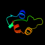
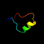
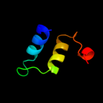
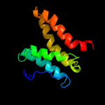
Top model
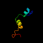
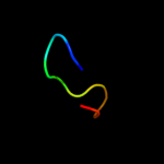
Image coloured by rainbow N → C terminus
Model (left) based on template c2k5eA_
Top template information
PDB header:structural genomics, unknown function
Chain: A: PDB Molecule:uncharacterized protein;
PDBTitle: solution structure of putative uncharacterized protein2 gsu1278 from methanocaldococcus jannaschii, northeast3 structural genomics consortium (nesg) target gsr195
Confidence and coverage
Confidence: 28.3% Coverage: 17%
40 residues ( 17% of your sequence) have been modelled with 28.3% confidence by the single highest scoring template.
You may wish to submit your sequence to Phyrealarm. This will automatically scan your sequence every week for new potential templates as they appear in the Phyre2 library.
Please note: You must be registered and logged in to use Phyrealarm.
3D viewing
Interactive 3D view in Jmol

Sequence analysis 

Secondary structure and disorder prediction 

   1........10.........20.........30.........40.........50.........60
Sequence  MWVNKYIDDCTDEDLNDRDFIASVVDRAIFHFAINSICNPGDNKDAMPIEQCTFDVETKN
Secondary structure 


SS confidence 


Disorder  ??


?
??


Disorder confidence 


   .........70.........80.........90.........100.........110.........120
Sequence  DLPSTVQLFYEESKDNEPLANIHFQAIGSGFLTFVNACQEHDDNSLKLFASLLISLSYSS
Secondary structure 


SS confidence 


Disorder 


?
??


Disorder confidence 


   .........130.........140.........150.........160.........170.........180
Sequence  AYADLSETVYINENNESYLKAQFEKLSQRDMKKYLGEMKRLADGGEMNFDGYLDKMSHLV
Secondary structure 


SS confidence 


Disorder 


?
?

?


??
Disorder confidence 


   .........190.........200.........210.........220.........230.....
Sequence  NEGTLDPDILSKMRDAAPQLISFAKSFDPTSKEEIKILTDTSKLIYDLFGVKSEK
Secondary structure 


SS confidence 


Disorder  ?????


??


?


?????
Disorder confidence 


Confidence Key
High(9)                    Low (0)
?Disordered
Alpha helix
Beta strand

Domain analysis 

Hover over an aligned region to see model and summary info

Please note, only up to the top 20 hits are modelled to reduce computer load

RankAligned region

PDB 2k5e chain A

3D model

Region: 63 - 112
Aligned: 40
Modelled: 50
Confidence: 28.3%
Identity: 18%
PDB header:structural genomics, unknown function
Chain: A: PDB Molecule:uncharacterized protein;
PDBTitle: solution structure of putative uncharacterized protein2 gsu1278 from methanocaldococcus jannaschii, northeast3 structural genomics consortium (nesg) target gsr195

Phyre2

PDB 2uub chain E domain 1

3D model

Region: 124 - 160
Aligned: 37
Modelled: 37
Confidence: 20.1%
Identity: 19%
Fold: Ribosomal protein S5 domain 2-like
Superfamily: Ribosomal protein S5 domain 2-like
Family: Translational machinery components

Phyre2

PDB 2pr1 chain B

3D model

Region: 194 - 207
Aligned: 14
Modelled: 14
Confidence: 18.0%
Identity: 50%
PDB header:transferase
Chain: B: PDB Molecule:uncharacterized n-acetyltransferase ylbp;
PDBTitle: crystal structure of the bacillus subtilis n-acetyltransferase ylbp2 protein in complex with coenzyme-a

Phyre2

PDB 1y6z chain A

3D model

Region: 74 - 195
Aligned: 86
Modelled: 86
Confidence: 13.6%
Identity: 16%
PDB header:structural genomics, unknown function
Chain: A: PDB Molecule:heat shock protein, putative;
PDBTitle: middle domain of plasmodium falciparum putative heat shock protein2 pf14_0417

Phyre2

PDB 2jug chain B

3D model

Region: 180 - 217
Aligned: 38
Modelled: 38
Confidence: 11.9%
Identity: 21%
PDB header:biosynthetic protein
Chain: B: PDB Molecule:tubc protein;
PDBTitle: multienzyme docking in hybrid megasynthetases

Phyre2

PDB 2gu2 chain A domain 1

3D model

Region: 180 - 214
Aligned: 35
Modelled: 35
Confidence: 11.1%
Identity: 23%
Fold: Phosphorylase/hydrolase-like
Superfamily: Zn-dependent exopeptidases
Family: AstE/AspA-like

Phyre2

PDB 1ci4 chain A

3D model

Region: 35 - 47
Aligned: 13
Modelled: 13
Confidence: 10.4%
Identity: 15%
Fold: SAM domain-like
Superfamily: Barrier-to-autointegration factor, BAF
Family: Barrier-to-autointegration factor, BAF

Phyre2

PDB 2f2i chain A

3D model

Region: 52 - 63
Aligned: 12
Modelled: 12
Confidence: 10.1%
Identity: 50%
PDB header:antimicrobial protein
Chain: A: PDB Molecule:kalata-b1;
PDBTitle: solution structure of [p20d,v21k]-kalata b1

Phyre2

PDB 2kn8 chain A

3D model

Region: 120 - 148
Aligned: 29
Modelled: 29
Confidence: 9.4%
Identity: 24%
PDB header:protein binding, dna binding protein
Chain: A: PDB Molecule:dna cleavage and packaging protein large subunit, ul89;
PDBTitle: nmr structure of the c-terminal domain of pul89

Phyre2

PDB 3elg chain A domain 1

3D model

Region: 55 - 82
Aligned: 28
Modelled: 28
Confidence: 9.1%
Identity: 29%
Fold: BLIP-like
Superfamily: BT0923-like
Family: BT0923-like

Phyre2

PDB 3mdj chain B

3D model

Region: 130 - 215
Aligned: 81
Modelled: 86
Confidence: 8.9%
Identity: 19%
PDB header:hydrolase/hydrolase inhibitor
Chain: B: PDB Molecule:endoplasmic reticulum aminopeptidase 1;
PDBTitle: er aminopeptidase, erap1, bound to the zinc aminopeptidase inhibitor,2 bestatin

Phyre2

PDB 1kdx chain B

3D model

Region: 144 - 158
Aligned: 15
Modelled: 15
Confidence: 8.4%
Identity: 40%
PDB header:transcription regulation complex
Chain: B: PDB Molecule:creb;
PDBTitle: kix domain of mouse cbp (creb binding protein) in complex2 with phosphorylated kinase inducible domain (pkid) of rat3 creb (cyclic amp response element binding protein), nmr 174 structures

Phyre2

PDB 1cuk chain A domain 1

3D model

Region: 113 - 144
Aligned: 32
Modelled: 32
Confidence: 8.2%
Identity: 19%
Fold: RuvA C-terminal domain-like
Superfamily: DNA helicase RuvA subunit, C-terminal domain
Family: DNA helicase RuvA subunit, C-terminal domain

Phyre2

PDB 3e4h chain A

3D model

Region: 52 - 63
Aligned: 12
Modelled: 12
Confidence: 7.8%
Identity: 42%
PDB header:plant protein
Chain: A: PDB Molecule:varv peptide f;
PDBTitle: crystal structure of the cyclotide varv f

Phyre2

PDB 2jwm chain A

3D model

Region: 52 - 63
Aligned: 12
Modelled: 12
Confidence: 7.6%
Identity: 33%
PDB header:plant protein
Chain: A: PDB Molecule:kalata-b7;
PDBTitle: nmr spatial srtucture of ternary complex kalata b7/mn2+/dpc2 micelle

Phyre2

PDB 3di3 chain A

3D model

Region: 81 - 112
Aligned: 32
Modelled: 32
Confidence: 7.5%
Identity: 19%
PDB header:cytokine/cytokine receptor
Chain: A: PDB Molecule:interleukin-7;
PDBTitle: crystal structure of the complex of human interleukin-7 with2 glycosylated human interleukin-7 receptor alpha ectodomain

Phyre2

PDB 3due chain A domain 1

3D model

Region: 56 - 83
Aligned: 28
Modelled: 25
Confidence: 6.8%
Identity: 25%
Fold: BLIP-like
Superfamily: BT0923-like
Family: BT0923-like

Phyre2

PDB 2qfa chain C

3D model

Region: 34 - 45
Aligned: 11
Modelled: 12
Confidence: 6.6%
Identity: 55%
PDB header:cell cycle/cell cycle/cell cycle
Chain: C: PDB Molecule:inner centromere protein;
PDBTitle: crystal structure of a survivin-borealin-incenp core complex

Phyre2

PDB 2kzv chain A

3D model

Region: 173 - 213
Aligned: 41
Modelled: 41
Confidence: 6.5%
Identity: 20%
PDB header:structural genomics, unknown function
Chain: A: PDB Molecule:uncharacterized protein;
PDBTitle: solution nmr structure of cv_0373(175-257) protein from2 chromobacterium violaceum, northeast structural genomics consortium3 target cvr118a

Phyre2

PDB 3rjo chain A

3D model

Region: 130 - 235
Aligned: 99
Modelled: 106
Confidence: 6.3%
Identity: 17%
PDB header:hydrolase
Chain: A: PDB Molecule:endoplasmic reticulum aminopeptidase 1;
PDBTitle: crystal structure of erap1 peptide binding domain

Phyre2
1

c2k5eA_
2

d2uube1
3

c2pr1B_
4

c1y6zA_
5

c2jugB_
6

d2gu2a1
7

d1ci4a_
8

c2f2iA_
9

c2kn8A_
10

d3elga1
11

c3mdjB_
12

c1kdxB_
13

d1cuka1
14

c3e4hA_
15

c2jwmA_
16

c3di3A_
17

d3duea1
18

c2qfaC_
19

c2kzvA_
20

c3rjoA_
21


22


23


24


25


Detailed template information 

#
Template Alignment Coverage3D Model Confidence
% i.d. Template Information
1c2k5eA_


28.3 18 PDB header:structural genomics, unknown function
Chain: A: PDB Molecule:uncharacterized protein;
PDBTitle: solution structure of putative uncharacterized protein2 gsu1278 from methanocaldococcus jannaschii, northeast3 structural genomics consortium (nesg) target gsr195
2d2uube1


20.1 19 Fold:Ribosomal protein S5 domain 2-like
Superfamily:Ribosomal protein S5 domain 2-like
Family:Translational machinery components
3c2pr1B_


18.0 50 PDB header:transferase
Chain: B: PDB Molecule:uncharacterized n-acetyltransferase ylbp;
PDBTitle: crystal structure of the bacillus subtilis n-acetyltransferase ylbp2 protein in complex with coenzyme-a
4c1y6zA_


13.6 16 PDB header:structural genomics, unknown function
Chain: A: PDB Molecule:heat shock protein, putative;
PDBTitle: middle domain of plasmodium falciparum putative heat shock protein2 pf14_0417
5c2jugB_


11.9 21 PDB header:biosynthetic protein
Chain: B: PDB Molecule:tubc protein;
PDBTitle: multienzyme docking in hybrid megasynthetases
6d2gu2a1


11.1 23 Fold:Phosphorylase/hydrolase-like
Superfamily:Zn-dependent exopeptidases
Family:AstE/AspA-like
7d1ci4a_


10.4 15 Fold:SAM domain-like
Superfamily:Barrier-to-autointegration factor, BAF
Family:Barrier-to-autointegration factor, BAF
8c2f2iA_


10.1 50 PDB header:antimicrobial protein
Chain: A: PDB Molecule:kalata-b1;
PDBTitle: solution structure of [p20d,v21k]-kalata b1
9c2kn8A_


9.4 24 PDB header:protein binding, dna binding protein
Chain: A: PDB Molecule:dna cleavage and packaging protein large subunit, ul89;
PDBTitle: nmr structure of the c-terminal domain of pul89
10d3elga1


9.1 29 Fold:BLIP-like
Superfamily:BT0923-like
Family:BT0923-like
11c3mdjB_


8.9 19 PDB header:hydrolase/hydrolase inhibitor
Chain: B: PDB Molecule:endoplasmic reticulum aminopeptidase 1;
PDBTitle: er aminopeptidase, erap1, bound to the zinc aminopeptidase inhibitor,2 bestatin
12c1kdxB_


8.4 40 PDB header:transcription regulation complex
Chain: B: PDB Molecule:creb;
PDBTitle: kix domain of mouse cbp (creb binding protein) in complex2 with phosphorylated kinase inducible domain (pkid) of rat3 creb (cyclic amp response element binding protein), nmr 174 structures
13d1cuka1


8.2 19 Fold:RuvA C-terminal domain-like
Superfamily:DNA helicase RuvA subunit, C-terminal domain
Family:DNA helicase RuvA subunit, C-terminal domain
14c3e4hA_


7.8 42 PDB header:plant protein
Chain: A: PDB Molecule:varv peptide f;
PDBTitle: crystal structure of the cyclotide varv f
15c2jwmA_


7.6 33 PDB header:plant protein
Chain: A: PDB Molecule:kalata-b7;
PDBTitle: nmr spatial srtucture of ternary complex kalata b7/mn2+/dpc2 micelle
16c3di3A_


7.5 19 PDB header:cytokine/cytokine receptor
Chain: A: PDB Molecule:interleukin-7;
PDBTitle: crystal structure of the complex of human interleukin-7 with2 glycosylated human interleukin-7 receptor alpha ectodomain
17d3duea1


6.8 25 Fold:BLIP-like
Superfamily:BT0923-like
Family:BT0923-like
18c2qfaC_


6.6 55 PDB header:cell cycle/cell cycle/cell cycle
Chain: C: PDB Molecule:inner centromere protein;
PDBTitle: crystal structure of a survivin-borealin-incenp core complex
19c2kzvA_


6.5 20 PDB header:structural genomics, unknown function
Chain: A: PDB Molecule:uncharacterized protein;
PDBTitle: solution nmr structure of cv_0373(175-257) protein from2 chromobacterium violaceum, northeast structural genomics consortium3 target cvr118a
20c3rjoA_


6.3 17 PDB header:hydrolase
Chain: A: PDB Molecule:endoplasmic reticulum aminopeptidase 1;
PDBTitle: crystal structure of erap1 peptide binding domain
21d1pt4a_


not modelled 6.3 42 Fold:Knottins (small inhibitors, toxins, lectins)
Superfamily:Cyclotides
Family:Kalata B1
22d1tyka_


not modelled 5.7 44 Fold:Knottins (small inhibitors, toxins, lectins)
Superfamily:omega toxin-like
Family:Spider toxins
23d1nb1a_


not modelled 5.5 50 Fold:Knottins (small inhibitors, toxins, lectins)
Superfamily:Cyclotides
Family:Kalata B1
24d2e1ba1


not modelled 5.4 24 Fold:Reductase/isomerase/elongation factor common domain
Superfamily:Translation proteins
Family:AlaX-M N-terminal domain-like
25c1k0rB_


not modelled 5.3 13 PDB header:transcription
Chain: B: PDB Molecule:nusa;
PDBTitle: crystal structure of mycobacterium tuberculosis nusa

Binding site prediction 

Due to computational demand, binding site predictions are not run for batch jobs

If you want to predict binding sites, please manually submit your model of choice to 3DLigandSite

Transmembrane helix prediction 

Transmembrane helices have been predicted in your sequence to adopt the topology shown below


Phyre is for academic use only

Please cite: Protein structure prediction on the web: a case study using the Phyre server
Kelley LA and Sternberg MJE. Nature Protocols 4, 363 - 371 (2009) [pdf] [Import into BibTeX]
 
If you use the binding site predictions from 3DLigandSite, please also cite:
3DLigandSite: predicting ligand-binding sites using similar structures.
Wass MN, Kelley LA and Sternberg MJ Nucleic Acids Research 38, W469-73 (2010) [PubMed]
 
© Structural Bioinformatics Group
Imperial College London
Lawrence Kelley, Benjamin Jefferys
Disclaimer
Terms and Conditions
Component software
Template detection: HHpred 1.51
Secondary structure prediction: Psi-pred 2.5
Disorder prediction: Disopred 2.4
Transmembrane prediction: Memsat_SVM
Multi-template modelling and ab initio: Poing 1.0