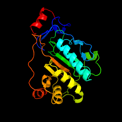
Phyre Home Retrieve Phyre Job Id
Emaill.a.kelley@imperial.ac.uk
DescriptionP0C0T5
DateThu Jan 5 11:29:59 GMT 2012
Unique Job ID2f630f96423e5520

Summary 

Top model
Image coloured by rainbow N → C terminus
Model (left) based on template d1tzpa_
Top template information
Fold:Hedgehog/DD-peptidase
Superfamily:Hedgehog/DD-peptidase
Family:MepA-like
Confidence and coverage
Confidence:100.0% Coverage: 88%
240 residues ( 88% of your sequence) have been modelled with 100.0% confidence by the single highest scoring template.
3D viewing
Interactive 3D view in Jmol

Sequence analysis 

Secondary structure and disorder prediction 

   1........10.........20.........30.........40.........50.........60
Sequence  MNKTAIALLALLASSASLAATPWQKITQPVPGSAQSIGSFSNGCIVGADTLPIQSEHYQV
Secondary structure 


SS confidence 


Disorder  ???


??????????????????????


?

??
????


Disorder confidence 


   .........70.........80.........90.........100.........110.........120
Sequence  MRTDQRRYFGHPDLVMFIQRLSSQVSNLGMGTVLIGDMGMPAGGRFNGGHASHQTGLDVD
Secondary structure 


SS confidence 


Disorder 


?
?


?
?????
??


Disorder confidence 


   .........130.........140.........150.........160.........170.........180
Sequence  IFLQLPKTRWTSAQLLRPQALDLVSRDGKHVVSTLWKPEIFSLIKLAAQDKDVTRIFVNP
Secondary structure 


SS confidence 


Disorder 


????????????


?


???


Disorder confidence 


   .........190.........200.........210.........220.........230.........240
Sequence  AIKQQLCLDAGTDRDWLRKVRPWFQHRAHMHVRLRCPADSLECEDQPLPPSGDGCGAELQ
Secondary structure 


SS confidence 


Disorder 


????
?

?


?


?????????????????


Disorder confidence 


   .........250.........260.........270....
Sequence  SWFEPPKPGTTKPEKKTPPPLPPSCQALLDEHVI
Secondary structure 


SS confidence 


Disorder 


???????????????????


?????
Disorder confidence 


Confidence Key
High(9)                    Low (0)
?Disordered
Alpha helix
Beta strand

Domain analysis 

Hover over an aligned region to see model and summary info

Please note, only up to the top 20 hits are modelled to reduce computer load

RankAligned region

PDB 1tzp chain A

3D model

Region: 20 - 274
Aligned: 240
Modelled: 240
Confidence: 100.0%
Identity: 100%
Fold: Hedgehog/DD-peptidase
Superfamily: Hedgehog/DD-peptidase
Family: MepA-like

Phyre2

PDB 2vo9 chain C

3D model

Region: 71 - 129
Aligned: 59
Modelled: 59
Confidence: 80.3%
Identity: 17%
PDB header:hydrolase
Chain: C: PDB Molecule:l-alanyl-d-glutamate peptidase;
PDBTitle: crystal structure of the enzymatically active domain of the2 listeria monocytogenes bacteriophage 500 endolysin ply500

Phyre2

PDB 2vo9 chain A domain 1

3D model

Region: 66 - 129
Aligned: 64
Modelled: 64
Confidence: 41.6%
Identity: 16%
Fold: Hedgehog/DD-peptidase
Superfamily: Hedgehog/DD-peptidase
Family: VanY-like

Phyre2

PDB 1r44 chain A

3D model

Region: 107 - 171
Aligned: 65
Modelled: 65
Confidence: 39.2%
Identity: 17%
Fold: Hedgehog/DD-peptidase
Superfamily: Hedgehog/DD-peptidase
Family: VanX-like

Phyre2

PDB 1lbu chain A

3D model

Region: 203 - 213
Aligned: 11
Modelled: 11
Confidence: 33.4%
Identity: 36%
PDB header:hydrolase
Chain: A: PDB Molecule:muramoyl-pentapeptide carboxypeptidase;
PDBTitle: hydrolase metallo (zn) dd-peptidase

Phyre2

PDB 3ag5 chain A

3D model

Region: 158 - 180
Aligned: 23
Modelled: 23
Confidence: 25.2%
Identity: 22%
PDB header:ligase
Chain: A: PDB Molecule:pantothenate synthetase;
PDBTitle: crystal structure of pantothenate synthetase from staphylococcus2 aureus

Phyre2

PDB 1iho chain A

3D model

Region: 158 - 180
Aligned: 23
Modelled: 23
Confidence: 23.3%
Identity: 39%
Fold: Adenine nucleotide alpha hydrolase-like
Superfamily: Nucleotidylyl transferase
Family: Pantothenate synthetase (Pantoate-beta-alanine ligase, PanC)

Phyre2

PDB 1v8f chain A

3D model

Region: 158 - 180
Aligned: 23
Modelled: 23
Confidence: 22.4%
Identity: 30%
Fold: Adenine nucleotide alpha hydrolase-like
Superfamily: Nucleotidylyl transferase
Family: Pantothenate synthetase (Pantoate-beta-alanine ligase, PanC)

Phyre2

PDB 2fao chain B

3D model

Region: 55 - 147
Aligned: 90
Modelled: 93
Confidence: 20.9%
Identity: 10%
PDB header:hydrolase/transferase
Chain: B: PDB Molecule:probable atp-dependent dna ligase;
PDBTitle: crystal structure of pseudomonas aeruginosa ligd polymerase2 domain

Phyre2

PDB 3inn chain B

3D model

Region: 158 - 180
Aligned: 23
Modelled: 23
Confidence: 20.7%
Identity: 35%
PDB header:ligase
Chain: B: PDB Molecule:pantothenate synthetase;
PDBTitle: crystal structure of pantoate-beta-alanine-ligase in complex2 with atp at low occupancy at 2.1 a resolution

Phyre2

PDB 3uk2 chain B

3D model

Region: 158 - 180
Aligned: 23
Modelled: 23
Confidence: 19.6%
Identity: 48%
PDB header:ligase
Chain: B: PDB Molecule:pantothenate synthetase;
PDBTitle: the structure of pantothenate synthetase from burkholderia2 thailandensis

Phyre2

PDB 2ejc chain A

3D model

Region: 158 - 180
Aligned: 23
Modelled: 23
Confidence: 19.3%
Identity: 39%
PDB header:ligase
Chain: A: PDB Molecule:pantoate--beta-alanine ligase;
PDBTitle: crystal structure of pantoate--beta-alanine ligase (panc)2 from thermotoga maritima

Phyre2

PDB 2a84 chain A domain 1

3D model

Region: 158 - 180
Aligned: 23
Modelled: 23
Confidence: 18.6%
Identity: 35%
Fold: Adenine nucleotide alpha hydrolase-like
Superfamily: Nucleotidylyl transferase
Family: Pantothenate synthetase (Pantoate-beta-alanine ligase, PanC)

Phyre2

PDB 3n8h chain A

3D model

Region: 158 - 180
Aligned: 23
Modelled: 23
Confidence: 18.4%
Identity: 48%
PDB header:ligase
Chain: A: PDB Molecule:pantothenate synthetase;
PDBTitle: crystal structure of pantoate-beta-alanine ligase from francisella2 tularensis

Phyre2

PDB 3mxt chain A

3D model

Region: 158 - 193
Aligned: 36
Modelled: 36
Confidence: 15.5%
Identity: 44%
PDB header:ligase
Chain: A: PDB Molecule:pantothenate synthetase;
PDBTitle: crystal structure of pantoate-beta-alanine ligase from campylobacter2 jejuni

Phyre2

PDB 3c7b chain A

3D model

Region: 208 - 219
Aligned: 12
Modelled: 12
Confidence: 15.0%
Identity: 33%
PDB header:oxidoreductase
Chain: A: PDB Molecule:sulfite reductase, dissimilatory-type subunit alpha;
PDBTitle: structure of the dissimilatory sulfite reductase from archaeoglobus2 fulgidus

Phyre2

PDB 2iru chain A

3D model

Region: 55 - 142
Aligned: 85
Modelled: 88
Confidence: 14.8%
Identity: 8%
PDB header:transferase
Chain: A: PDB Molecule:putative dna ligase-like protein rv0938/mt0965;
PDBTitle: crystal structure of the polymerase domain from mycobacterium2 tuberculosis ligase d

Phyre2

PDB 2cwz chain A domain 1

3D model

Region: 69 - 92
Aligned: 24
Modelled: 24
Confidence: 14.8%
Identity: 4%
Fold: Thioesterase/thiol ester dehydrase-isomerase
Superfamily: Thioesterase/thiol ester dehydrase-isomerase
Family: TTHA0967-like

Phyre2

PDB 1nkg chain A domain 3

3D model

Region: 103 - 125
Aligned: 23
Modelled: 23
Confidence: 12.7%
Identity: 22%
Fold: Supersandwich
Superfamily: Galactose mutarotase-like
Family: Rhamnogalacturonase B, RhgB, N-terminal domain

Phyre2

PDB 1x9v chain A

3D model

Region: 70 - 105
Aligned: 36
Modelled: 36
Confidence: 12.1%
Identity: 19%
PDB header:viral protein
Chain: A: PDB Molecule:vpr protein;
PDBTitle: dimeric structure of the c-terminal domain of vpr

Phyre2
1

d1tzpa_
2

c2vo9C_
3

d2vo9a1
4

d1r44a_
5

c1lbuA_
6

c3ag5A_
7

d1ihoa_
8

d1v8fa_
9

c2faoB_
10

c3innB_
11

c3uk2B_
12

c2ejcA_
13

d2a84a1
14

c3n8hA_
15

c3mxtA_
16

c3c7bA_
17

c2iruA_
18

d2cwza1
19

d1nkga3
20

c1x9vA_
21


22


23


24


25


26


27


28


29


30


31


32


33


34


35


36


37


38


39


40


41


42


43


Detailed template information 

#
Template Alignment Coverage3D Model Confidence
% i.d. Template Information
1d1tzpa_


100.0 100 Fold:Hedgehog/DD-peptidase
Superfamily:Hedgehog/DD-peptidase
Family:MepA-like
2c2vo9C_


80.3 17 PDB header:hydrolase
Chain: C: PDB Molecule:l-alanyl-d-glutamate peptidase;
PDBTitle: crystal structure of the enzymatically active domain of the2 listeria monocytogenes bacteriophage 500 endolysin ply500
3d2vo9a1


41.6 16 Fold:Hedgehog/DD-peptidase
Superfamily:Hedgehog/DD-peptidase
Family:VanY-like
4d1r44a_


39.2 17 Fold:Hedgehog/DD-peptidase
Superfamily:Hedgehog/DD-peptidase
Family:VanX-like
5c1lbuA_


33.4 36 PDB header:hydrolase
Chain: A: PDB Molecule:muramoyl-pentapeptide carboxypeptidase;
PDBTitle: hydrolase metallo (zn) dd-peptidase
6c3ag5A_


25.2 22 PDB header:ligase
Chain: A: PDB Molecule:pantothenate synthetase;
PDBTitle: crystal structure of pantothenate synthetase from staphylococcus2 aureus
7d1ihoa_


23.3 39 Fold:Adenine nucleotide alpha hydrolase-like
Superfamily:Nucleotidylyl transferase
Family:Pantothenate synthetase (Pantoate-beta-alanine ligase, PanC)
8d1v8fa_


22.4 30 Fold:Adenine nucleotide alpha hydrolase-like
Superfamily:Nucleotidylyl transferase
Family:Pantothenate synthetase (Pantoate-beta-alanine ligase, PanC)
9c2faoB_


20.9 10 PDB header:hydrolase/transferase
Chain: B: PDB Molecule:probable atp-dependent dna ligase;
PDBTitle: crystal structure of pseudomonas aeruginosa ligd polymerase2 domain
10c3innB_


20.7 35 PDB header:ligase
Chain: B: PDB Molecule:pantothenate synthetase;
PDBTitle: crystal structure of pantoate-beta-alanine-ligase in complex2 with atp at low occupancy at 2.1 a resolution
11c3uk2B_


19.6 48 PDB header:ligase
Chain: B: PDB Molecule:pantothenate synthetase;
PDBTitle: the structure of pantothenate synthetase from burkholderia2 thailandensis
12c2ejcA_


19.3 39 PDB header:ligase
Chain: A: PDB Molecule:pantoate--beta-alanine ligase;
PDBTitle: crystal structure of pantoate--beta-alanine ligase (panc)2 from thermotoga maritima
13d2a84a1


18.6 35 Fold:Adenine nucleotide alpha hydrolase-like
Superfamily:Nucleotidylyl transferase
Family:Pantothenate synthetase (Pantoate-beta-alanine ligase, PanC)
14c3n8hA_


18.4 48 PDB header:ligase
Chain: A: PDB Molecule:pantothenate synthetase;
PDBTitle: crystal structure of pantoate-beta-alanine ligase from francisella2 tularensis
15c3mxtA_


15.5 44 PDB header:ligase
Chain: A: PDB Molecule:pantothenate synthetase;
PDBTitle: crystal structure of pantoate-beta-alanine ligase from campylobacter2 jejuni
16c3c7bA_


15.0 33 PDB header:oxidoreductase
Chain: A: PDB Molecule:sulfite reductase, dissimilatory-type subunit alpha;
PDBTitle: structure of the dissimilatory sulfite reductase from archaeoglobus2 fulgidus
17c2iruA_


14.8 8 PDB header:transferase
Chain: A: PDB Molecule:putative dna ligase-like protein rv0938/mt0965;
PDBTitle: crystal structure of the polymerase domain from mycobacterium2 tuberculosis ligase d
18d2cwza1


14.8 4 Fold:Thioesterase/thiol ester dehydrase-isomerase
Superfamily:Thioesterase/thiol ester dehydrase-isomerase
Family:TTHA0967-like
19d1nkga3


12.7 22 Fold:Supersandwich
Superfamily:Galactose mutarotase-like
Family:Rhamnogalacturonase B, RhgB, N-terminal domain
20c1x9vA_


12.1 19 PDB header:viral protein
Chain: A: PDB Molecule:vpr protein;
PDBTitle: dimeric structure of the c-terminal domain of vpr
21d1ujpa_


not modelled 12.0 19 Fold:TIM beta/alpha-barrel
Superfamily:Ribulose-phoshate binding barrel
Family:Tryptophan biosynthesis enzymes
22d2o8ra3


not modelled 11.8 32 Fold:Phospholipase D/nuclease
Superfamily:Phospholipase D/nuclease
Family:Polyphosphate kinase C-terminal domain
23c2jksA_


not modelled 11.4 17 PDB header:immune system
Chain: A: PDB Molecule:bradyzoite surface antigen bsr4;
PDBTitle: crystal structure of the the bradyzoite specific antigen2 bsr4 from toxoplasma gondii.
24c2zuuA_


not modelled 9.8 18 PDB header:transferase
Chain: A: PDB Molecule:lacto-n-biose phosphorylase;
PDBTitle: crystal structure of galacto-n-biose/lacto-n-biose i phosphorylase in2 complex with glcnac
25c3kuvB_


not modelled 9.7 8 PDB header:hydrolase
Chain: B: PDB Molecule:fluoroacetyl coenzyme a thioesterase;
PDBTitle: structural basis of the activity and substrate specificity of the2 fluoroacetyl-coa thioesterase flk - t42s mutant in complex with3 acetate.
26d2v4ja2


not modelled 9.3 50 Fold:Ferredoxin-like
Superfamily:Nitrite/Sulfite reductase N-terminal domain-like
Family:DsrA/DsrB N-terminal-domain-like
27c3qooA_


not modelled 8.6 14 PDB header:structural genomics, unknown function
Chain: A: PDB Molecule:uncharacterized protein;
PDBTitle: crystal structure of hot-dog-like taci_0573 protein from2 thermanaerovibrio acidaminovorans
28d1gvia1


not modelled 7.3 19 Fold:Immunoglobulin-like beta-sandwich
Superfamily:E set domains
Family:E-set domains of sugar-utilizing enzymes
29c2x2zE_


not modelled 6.6 42 PDB header:membrane protein
Chain: E: PDB Molecule:apical membrane antigen 1, putative;
PDBTitle: crystal structure ama1 from toxoplasma gondii
30c3navB_


not modelled 6.5 18 PDB header:lyase
Chain: B: PDB Molecule:tryptophan synthase alpha chain;
PDBTitle: crystal structure of an alpha subunit of tryptophan synthase from2 vibrio cholerae o1 biovar el tor str. n16961
31d1wzla1


not modelled 6.4 33 Fold:Immunoglobulin-like beta-sandwich
Superfamily:E set domains
Family:E-set domains of sugar-utilizing enzymes
32d1pvta_


not modelled 6.0 17 Fold:AraD/HMP-PK domain-like
Superfamily:AraD/HMP-PK domain-like
Family:AraD-like aldolase/epimerase
33d1j0ha1


not modelled 6.0 31 Fold:Immunoglobulin-like beta-sandwich
Superfamily:E set domains
Family:E-set domains of sugar-utilizing enzymes
34d1c25a_


not modelled 5.8 25 Fold:Rhodanese/Cell cycle control phosphatase
Superfamily:Rhodanese/Cell cycle control phosphatase
Family:Cell cycle control phosphatase, catalytic domain
35d1txla_


not modelled 5.6 29 Fold:Lipocalins
Superfamily:Lipocalins
Family:Hypothetical protein YodA
36c1txlA_


not modelled 5.6 29 PDB header:structural genomics, unknown function
Chain: A: PDB Molecule:metal-binding protein yoda;
PDBTitle: crystal structure of metal-binding protein yoda from e.2 coli, pfam duf149
37c2q8aA_


not modelled 5.5 33 PDB header:immune system
Chain: A: PDB Molecule:apical membrane antigen 1;
PDBTitle: structure of the malaria antigen ama1 in complex with a growth-2 inhibitory antibody
38c2w82C_


not modelled 5.4 45 PDB header:replication inhibitor
Chain: C: PDB Molecule:orf18;
PDBTitle: the structure of arda
39d1dqaa1


not modelled 5.4 8 Fold:Ferredoxin-like
Superfamily:NAD-binding domain of HMG-CoA reductase
Family:NAD-binding domain of HMG-CoA reductase
40d1gqpa_


not modelled 5.3 27 Fold:Galactose-binding domain-like
Superfamily:Galactose-binding domain-like
Family:APC10-like
41c1h4uA_


not modelled 5.1 17 PDB header:extracellular matrix protein
Chain: A: PDB Molecule:nidogen-1;
PDBTitle: domain g2 of mouse nidogen-1
42c2l57A_


not modelled 5.1 22 PDB header:structural genomics, unknown function
Chain: A: PDB Molecule:uncharacterized protein;
PDBTitle: solution structure of an uncharacterized thioredoin-like protein from2 clostridium perfringens
43d1xuba1


not modelled 5.0 17 Fold:Diaminopimelate epimerase-like
Superfamily:Diaminopimelate epimerase-like
Family:PhzC/PhzF-like

Binding site prediction 

Due to computational demand, binding site predictions are not run for batch jobs

If you want to predict binding sites, please manually submit your model of choice to 3DLigandSite


Phyre is for academic use only

Please cite: Protein structure prediction on the web: a case study using the Phyre server
Kelley LA and Sternberg MJE. Nature Protocols 4, 363 - 371 (2009) [pdf] [Import into BibTeX]
 
If you use the binding site predictions from 3DLigandSite, please also cite:
3DLigandSite: predicting ligand-binding sites using similar structures.
Wass MN, Kelley LA and Sternberg MJ Nucleic Acids Research 38, W469-73 (2010) [PubMed]
 
© Structural Bioinformatics Group
Imperial College London
Lawrence Kelley, Benjamin Jefferys
Disclaimer
Terms and Conditions
Component software
Template detection: HHpred 1.51
Secondary structure prediction: Psi-pred 2.5
Disorder prediction: Disopred 2.4
Transmembrane prediction: Memsat_SVM
Multi-template modelling and ab initio: Poing 1.0