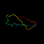
1 c2izpB_
20.1
29
PDB header: toxinChain: B: PDB Molecule: putative membrane antigen;PDBTitle: bipd - an invasion prtein associated with the type-iii2 secretion system of burkholderia pseudomallei.
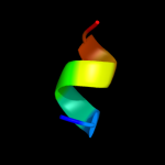
2 d2izpa1
17.2
29
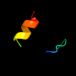
Fold: IpaD-likeSuperfamily: IpaD-likeFamily: IpaD-like3 c3hc7A_
16.6
20
PDB header: cell adhesionChain: A: PDB Molecule: gene 12 protein;PDBTitle: crystal structure of lysin b from mycobacteriophage d29
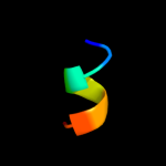
4 d1oh4a_
14.8
28
Fold: Galactose-binding domain-likeSuperfamily: Galactose-binding domain-likeFamily: Family 27 carbohydrate binding module, CBM275 c1msvB_
13.5
19
PDB header: lyaseChain: B: PDB Molecule: s-adenosylmethionine decarboxylase proenzyme;PDBTitle: the s68a s-adenosylmethionine decarboxylase proenzyme2 processing mutant.
6 c3ep3A_
12.9
19
PDB header: lyaseChain: A: PDB Molecule: s-adenosylmethionine decarboxylase alpha chain;PDBTitle: human adometdc d174n mutant with no putrescine bound
7 d1upka_
10.8
18
Fold: alpha-alpha superhelixSuperfamily: ARM repeatFamily: Mo25 protein8 d1jl0a_
10.8
19
Fold: S-adenosylmethionine decarboxylaseSuperfamily: S-adenosylmethionine decarboxylaseFamily: S-adenosylmethionine decarboxylase9 d1phpa_
10.7
21
Fold: Phosphoglycerate kinaseSuperfamily: Phosphoglycerate kinaseFamily: Phosphoglycerate kinase10 c3q2tC_
9.5
32
PDB header: rna binding protein/rnaChain: C: PDB Molecule: cleavage and polyadenylation specificity factor subunit 6;PDBTitle: crystal structure of cfim68 rrm/cfim25/rna complex
11 c2hh9A_
9.3
26
PDB header: transferaseChain: A: PDB Molecule: thiamin pyrophosphokinase;PDBTitle: thiamin pyrophosphokinase from candida albicans
12 d1ejia_
9.2
38
Fold: PLP-dependent transferase-likeSuperfamily: PLP-dependent transferasesFamily: GABA-aminotransferase-like13 d1pmhx_
8.6
26
Fold: Galactose-binding domain-likeSuperfamily: Galactose-binding domain-likeFamily: Family 27 carbohydrate binding module, CBM2714 c2k6rA_
8.3
75
PDB header: de novo proteinChain: A: PDB Molecule: full sequence design 1 synthetic superstable;PDBTitle: protein folding on a highly rugged landscape: experimental2 observation of glassy dynamics and structural frustration
15 c2hjqA_
8.2
47
PDB header: structural genomicsChain: A: PDB Molecule: hypothetical protein yqbf;PDBTitle: nmr structure of bacillus subtilis protein yqbf, northeast2 structural genomics target sr449
16 c1fmeA_
8.1
75
PDB header: de novo proteinChain: A: PDB Molecule: fsd-ey peptide;PDBTitle: solution structure of fsd-ey, a novel peptide assuming a2 beta-beta-alpha fold
17 c1fsvA_
7.9
75
PDB header: beta beta alpha motifChain: A: PDB Molecule: full sequence design 1 of beta beta alpha motif;PDBTitle: full sequence design 1 (fsd-1) of beta beta alpha motif,2 nmr, minimized average structure
18 c1fsdA_
7.9
75
PDB header: novel sequenceChain: A: PDB Molecule: full sequence design 1 of beta beta alpha motif;PDBTitle: full sequence design 1 (fsd-1) of beta beta alpha motif,2 nmr, 41 structures
19 c3da7G_
7.8
41
PDB header: protein bindingChain: G: PDB Molecule: barnase circular permutant;PDBTitle: a conformationally strained, circular permutant of barnase
20 c2l5gB_
7.6
50
PDB header: transcription regulatorChain: B: PDB Molecule: putative uncharacterized protein ncor2;PDBTitle: co-ordinates and 1h, 13c and 15n chemical shift assignments for the2 complex of gps2 53-90 and smrt 167-207
21 c2hvzA_
not modelled
7.6
24
PDB header: rna binding proteinChain: A: PDB Molecule: splicing factor, arginine/serine-rich 7;PDBTitle: solution structure of the rrm domain of sr rich factor 9g8
22 c1zmrA_
not modelled
7.3
19
PDB header: transferaseChain: A: PDB Molecule: phosphoglycerate kinase;PDBTitle: crystal structure of the e. coli phosphoglycerate kinase
23 c3cvzA_
not modelled
7.3
25
PDB header: structural proteinChain: A: PDB Molecule: s-layer protein;PDBTitle: structural insights into the molecular organization of the2 s-layer from clostridium difficile
24 c2rhqA_
not modelled
7.1
21
PDB header: ligaseChain: A: PDB Molecule: phenylalanyl-trna synthetase alpha chain;PDBTitle: phers from staphylococcus haemolyticus- rational protein2 engineering and inhibitor studies
25 d2arha1
not modelled
7.0
35
Fold: Acyl-CoA N-acyltransferases (Nat)Superfamily: Acyl-CoA N-acyltransferases (Nat)Family: Aq 1966-like26 d1fiua_
not modelled
7.0
54
Fold: Restriction endonuclease-likeSuperfamily: Restriction endonuclease-likeFamily: Restriction endonuclease NgoIV27 d2hjqa2
not modelled
6.9
27
Fold: GINS/PriA/YqbF domainSuperfamily: PriA/YqbF domainFamily: YqbF N-terminal domain-like28 d1z1za1
not modelled
6.7
25
Fold: Phage tail protein-likeSuperfamily: Phage tail protein-likeFamily: Lambda phage gpU-like29 d1pgsa1
not modelled
6.6
53
Fold: Nucleoplasmin-like/VP (viral coat and capsid proteins)Superfamily: PHM/PNGase FFamily: Glycosyl-asparaginase30 c2o8kA_
not modelled
6.6
47
PDB header: transcription/dnaChain: A: PDB Molecule: rna polymerase sigma factor rpon;PDBTitle: nmr structure of the sigma-54 rpon domain bound to the-242 promoter element
31 c2ytuA_
not modelled
6.6
38
PDB header: signaling proteinChain: A: PDB Molecule: friend leukemia integration 1 transcriptionPDBTitle: solution structure of the sam_pnt-domain of the human2 friend leukemiaintegration 1 transcription factor
32 c3hlzA_
not modelled
6.6
33
PDB header: structural genomics, unknown functionChain: A: PDB Molecule: uncharacterized protein bt_1490;PDBTitle: crystal structure of bt_1490 (np_810393.1) from bacteroides2 thetaiotaomicron vpi-5482 at 1.50 a resolution
33 d1hd0a_
not modelled
6.5
28
Fold: Ferredoxin-likeSuperfamily: RNA-binding domain, RBDFamily: Canonical RBD34 c1v0sA_
not modelled
6.3
31
PDB header: hydrolaseChain: A: PDB Molecule: phospholipase d;PDBTitle: uninhibited form of phospholipase d from streptomyces sp.2 strain pmf
35 d1z3xa1
not modelled
6.2
25
Fold: alpha-alpha superhelixSuperfamily: ARM repeatFamily: GUN4-associated domain36 d1mn4a_
not modelled
6.2
23
Fold: Common fold of diphtheria toxin/transcription factors/cytochrome fSuperfamily: p53-like transcription factorsFamily: DNA-binding domain from NDT8037 d1k3ea_
not modelled
6.2
16
Fold: Secretion chaperone-likeSuperfamily: Type III secretory system chaperone-likeFamily: Type III secretory system chaperone38 c2c4bB_
not modelled
6.2
41
PDB header: fusion proteinChain: B: PDB Molecule: barnase mcoeeti fusion;PDBTitle: inhibitor cystine knot protein mcoeeti fused to the2 catalytically inactive barnase mutant h102a
39 d2bida_
not modelled
5.9
21
Fold: Toxins' membrane translocation domainsSuperfamily: Bcl-2 inhibitors of programmed cell deathFamily: Bcl-2 inhibitors of programmed cell death40 c2e5iA_
not modelled
5.8
24
PDB header: gene regulationChain: A: PDB Molecule: heterogeneous nuclear ribonucleoprotein l-like;PDBTitle: solution structure of rna binding domain 2 in heterogeneous2 nuclear ribonucleoprotein l-like
41 c2dkxA_
not modelled
5.7
38
PDB header: signaling proteinChain: A: PDB Molecule: sam pointed domain-containing ets transcriptionPDBTitle: solution structure of the sam_pnt-domain of ets2 transcription factor pdef (prostate ets)
42 d1jyoa_
not modelled
5.4
13
Fold: Secretion chaperone-likeSuperfamily: Type III secretory system chaperone-likeFamily: Type III secretory system chaperone43 c3md1B_
not modelled
5.4
35
PDB header: rna binding proteinChain: B: PDB Molecule: nuclear and cytoplasmic polyadenylated rna-binding proteinPDBTitle: crystal structure of the second rrm domain of yeast poly(u)-binding2 protein (pub1)
44 d1x9ba_
not modelled
5.4
44
Fold: Protozoan pheromone-likeSuperfamily: Hypothetical membrane protein Ta0354, soluble domainFamily: Hypothetical membrane protein Ta0354, soluble domain45 c3lgbB_
not modelled
5.4
36
PDB header: transferaseChain: B: PDB Molecule: dna primase large subunit;PDBTitle: crystal structure of the fe-s domain of the yeast dna primase
46 c3if8A_
not modelled
5.2
20
PDB header: cell cycleChain: A: PDB Molecule: protein zwilch homolog;PDBTitle: crystal structure of zwilch, a member of the rzz kinetochore complex
47 d1erpa_
not modelled
5.0
47
Fold: Protozoan pheromone-likeSuperfamily: Protozoan pheromone proteinsFamily: Protozoan pheromone proteins