1 c2zt9H_
62.0
38
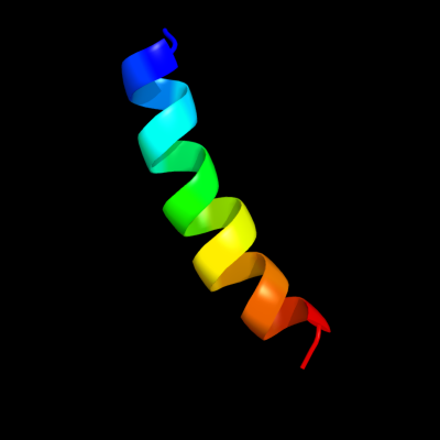
PDB header: photosynthesisChain: H: PDB Molecule: cytochrome b6-f complex subunit 8;PDBTitle: crystal structure of the cytochrome b6f complex from nostoc sp. pcc2 7120
2 c2e76H_
42.9
35
PDB header: photosynthesisChain: H: PDB Molecule: cytochrome b6-f complex subunit 8;PDBTitle: crystal structure of the cytochrome b6f complex with tridecyl-2 stigmatellin (tds) from m.laminosus
3 c2e74H_
42.9
35
PDB header: photosynthesisChain: H: PDB Molecule: cytochrome b6-f complex subunit 8;PDBTitle: crystal structure of the cytochrome b6f complex from m.laminosus
4 c2e75H_
42.9
35
PDB header: photosynthesisChain: H: PDB Molecule: cytochrome b6-f complex subunit 8;PDBTitle: crystal structure of the cytochrome b6f complex with 2-nonyl-4-2 hydroxyquinoline n-oxide (nqno) from m.laminosus
5 d2e74h1
38.4
35
Fold: Single transmembrane helixSuperfamily: PetN subunit of the cytochrome b6f complexFamily: PetN subunit of the cytochrome b6f complex6 c2pnvA_
35.1
29
PDB header: membrane proteinChain: A: PDB Molecule: small conductance calcium-activated potassiumPDBTitle: crystal structure of the leucine zipper domain of small-2 conductance ca2+-activated k+ (skca) channel from rattus3 norvegicus
7 c3m9hB_
34.9
23
PDB header: chaperoneChain: B: PDB Molecule: proteasome-associated atpase;PDBTitle: crystal structure of the amino terminal coiled coil domain of the2 mycobacterium tuberculosis proteasomal atpase mpa
8 c1vf5H_
30.6
35
PDB header: photosynthesisChain: H: PDB Molecule: protein pet n;PDBTitle: crystal structure of cytochrome b6f complex from m.laminosus
9 c1vf5U_
27.5
35
PDB header: photosynthesisChain: U: PDB Molecule: protein pet n;PDBTitle: crystal structure of cytochrome b6f complex from m.laminosus
10 c2d2cU_
23.6
35
PDB header: photosynthesisChain: U: PDB Molecule: cytochrome b6-f complex subunit viii;PDBTitle: crystal structure of cytochrome b6f complex with dbmib from2 m. laminosus
11 c2d2cH_
23.6
35
PDB header: photosynthesisChain: H: PDB Molecule: cytochrome b6-f complex subunit viii;PDBTitle: crystal structure of cytochrome b6f complex with dbmib from2 m. laminosus
12 c3swfA_
21.8
37
PDB header: transport proteinChain: A: PDB Molecule: cgmp-gated cation channel alpha-1;PDBTitle: cnga1 621-690 containing clz domain
13 c1ce0B_
19.9
19
PDB header: hiv-1 envelope proteinChain: B: PDB Molecule: protein (leucine zipper model h38-p1);PDBTitle: trimerization specificity in hiv-1 gp41: analysis with a2 gcn4 leucine zipper model
14 c1ztaA_
19.4
25
PDB header: dna-binding motifChain: A: PDB Molecule: leucine zipper monomer;PDBTitle: the solution structure of a leucine-zipper motif peptide
15 c1zmeD_
18.0
16
PDB header: transcription/dnaChain: D: PDB Molecule: proline utilization transcription activator;PDBTitle: crystal structure of put3/dna complex
16 c1ij2C_
17.2
25
PDB header: transcriptionChain: C: PDB Molecule: general control protein gcn4;PDBTitle: gcn4-pvtl coiled-coil trimer with threonine at the a(16)2 position
17 c1rb1B_
16.9
25
PDB header: dna binding proteinChain: B: PDB Molecule: general control protein gcn4;PDBTitle: gcn4-leucine zipper core mutant as n16a trigonal automatic2 solution
18 c3k7zA_
16.9
25
PDB header: dna binding proteinChain: A: PDB Molecule: general control protein gcn4;PDBTitle: gcn4-leucine zipper core mutant as n16a trigonal automatic2 solution
19 c3k7zB_
16.9
25
PDB header: dna binding proteinChain: B: PDB Molecule: general control protein gcn4;PDBTitle: gcn4-leucine zipper core mutant as n16a trigonal automatic2 solution
20 c1rb1A_
16.9
25
PDB header: dna binding proteinChain: A: PDB Molecule: general control protein gcn4;PDBTitle: gcn4-leucine zipper core mutant as n16a trigonal automatic2 solution
21 c1rb6C_
not modelled
16.9
25
PDB header: dna binding proteinChain: C: PDB Molecule: general control protein gcn4;PDBTitle: antiparallel trimer of gcn4-leucine zipper core mutant as2 n16a tetragonal form
22 c1swiA_
not modelled
16.9
25
PDB header: leucine zipperChain: A: PDB Molecule: gcn4p1;PDBTitle: gcn4-leucine zipper core mutant as n16a complexed with2 benzene
23 c2akfB_
not modelled
16.5
35
PDB header: protein bindingChain: B: PDB Molecule: coronin-1a;PDBTitle: crystal structure of the coiled-coil domain of coronin 1
24 c2akfC_
not modelled
16.5
35
PDB header: protein bindingChain: C: PDB Molecule: coronin-1a;PDBTitle: crystal structure of the coiled-coil domain of coronin 1
25 c2akfA_
not modelled
16.5
35
PDB header: protein bindingChain: A: PDB Molecule: coronin-1a;PDBTitle: crystal structure of the coiled-coil domain of coronin 1
26 c1ij3C_
not modelled
16.3
25
PDB header: transcriptionChain: C: PDB Molecule: general control protein gcn4;PDBTitle: gcn4-pvsl coiled-coil trimer with serine at the a(16)2 position
27 c1ij3B_
not modelled
16.3
25
PDB header: transcriptionChain: B: PDB Molecule: general control protein gcn4;PDBTitle: gcn4-pvsl coiled-coil trimer with serine at the a(16)2 position
28 c1ij2B_
not modelled
15.7
25
PDB header: transcriptionChain: B: PDB Molecule: general control protein gcn4;PDBTitle: gcn4-pvtl coiled-coil trimer with threonine at the a(16)2 position
29 c1p9iA_
not modelled
15.6
32
PDB header: unknown functionChain: A: PDB Molecule: cortexillin i/gcn4 hybrid peptide;PDBTitle: coiled-coil x-ray structure at 1.17 a resolution
30 c1qcrD_
not modelled
14.1
17
PDB header: PDB COMPND: 31 c1junB_
not modelled
13.2
40
PDB header: transcription regulationChain: B: PDB Molecule: c-jun homodimer;PDBTitle: nmr study of c-jun homodimer
32 c2ergA_
not modelled
12.5
13
PDB header: transcription activator/dnaChain: A: PDB Molecule: regulatory protein leu3;PDBTitle: crystal structure of leu3 dna-binding domain with a single2 h50c mutation complexed with a 15mer dna duplex
33 c2k21A_
not modelled
12.0
22
PDB header: membrane proteinChain: A: PDB Molecule: potassium voltage-gated channel subfamily ePDBTitle: nmr structure of human kcne1 in lmpg micelles at ph 6.0 and2 40 degree c
34 c3ci9B_
not modelled
11.2
43
PDB header: transcriptionChain: B: PDB Molecule: heat shock factor-binding protein 1;PDBTitle: crystal structure of the human hsbp1
35 c3he5A_
not modelled
11.2
37
PDB header: de novo proteinChain: A: PDB Molecule: synzip1;PDBTitle: heterospecific coiled-coil pair synzip2:synzip1
36 c2y69Z_
not modelled
11.2
25
PDB header: electron transportChain: Z: PDB Molecule: cytochrome c oxidase polypeptide 8h;PDBTitle: bovine heart cytochrome c oxidase re-refined with molecular2 oxygen
37 c1gk6B_
not modelled
10.4
15
PDB header: vimentinChain: B: PDB Molecule: vimentin;PDBTitle: human vimentin coil 2b fragment linked to gcn4 leucine2 zipper (z2b)
38 c2o7hF_
not modelled
10.3
21
PDB header: transcriptionChain: F: PDB Molecule: general control protein gcn4;PDBTitle: crystal structure of trimeric coiled coil gcn4 leucine zipper
39 c3p8cE_
not modelled
9.9
22
PDB header: protein bindingChain: E: PDB Molecule: probable protein brick1;PDBTitle: structure and control of the actin regulatory wave complex
40 d1v54m_
not modelled
9.9
25
Fold: Single transmembrane helixSuperfamily: Mitochondrial cytochrome c oxidase subunit VIIIb (aka IX)Family: Mitochondrial cytochrome c oxidase subunit VIIIb (aka IX)41 c1u0iA_
not modelled
9.4
37
PDB header: de novo proteinChain: A: PDB Molecule: iaal-e3;PDBTitle: iaal-e3/k3 heterodimer
42 c3rylB_
not modelled
8.8
45
PDB header: protein bindingChain: B: PDB Molecule: protein vpa1370;PDBTitle: dimerization domain of vibrio parahemolyticus vopl
43 c3nmdA_
not modelled
8.7
26
PDB header: transferaseChain: A: PDB Molecule: cgmp dependent protein kinase;PDBTitle: crystal structure of the leucine zipper domain of cgmp dependent2 protein kinase i beta
44 c2xzrA_
not modelled
7.8
16
PDB header: cell adhesionChain: A: PDB Molecule: immunoglobulin-binding protein eibd;PDBTitle: escherichia coli immunoglobulin-binding protein eibd 391-438 fused2 to gcn4 adaptors
45 c2ke4A_
not modelled
7.8
15
PDB header: membrane proteinChain: A: PDB Molecule: cdc42-interacting protein 4;PDBTitle: the nmr structure of the tc10 and cdc42 interacting domain2 of cip4
46 c2xv5A_
not modelled
7.5
13
PDB header: structural proteinChain: A: PDB Molecule: lamin-a/c;PDBTitle: human lamin a coil 2b fragment
47 c1u2uA_
not modelled
7.4
40
PDB header: transcriptionChain: A: PDB Molecule: general control protein gcn4;PDBTitle: nmr solution structure of a designed heterodimeric leucine2 zipper
48 d1grja1
not modelled
7.1
20
Fold: Long alpha-hairpinSuperfamily: GreA transcript cleavage protein, N-terminal domainFamily: GreA transcript cleavage protein, N-terminal domain49 c1hwtC_
not modelled
7.1
39
PDB header: gene regulation/dnaChain: C: PDB Molecule: protein (heme activator protein);PDBTitle: structure of a hap1/dna complex reveals dramatically2 asymmetric dna binding by a homodimeric protein
50 c2yy0D_
not modelled
7.1
3
PDB header: transcriptionChain: D: PDB Molecule: c-myc-binding protein;PDBTitle: crystal structure of ms0802, c-myc-1 binding protein domain2 from homo sapiens
51 c2lf0A_
not modelled
6.6
20
PDB header: structural genomics, unknown functionChain: A: PDB Molecule: uncharacterized protein yibl;PDBTitle: solution structure of sf3636, a two-domain unknown function protein2 from shigella flexneri 2a, determined by joint refinement of nmr,3 residual dipolar couplings and small-angle x-ray scatting, nesg4 target sfr339/ocsp target sf3636
52 c1dipA_
not modelled
6.5
18
PDB header: acetylationChain: A: PDB Molecule: delta-sleep-inducing peptide immunoreactivePDBTitle: the solution structure of porcine delta-sleep-inducing2 peptide immunoreactive peptide, nmr, 10 structures
53 d2f23a1
not modelled
6.0
25
Fold: Long alpha-hairpinSuperfamily: GreA transcript cleavage protein, N-terminal domainFamily: GreA transcript cleavage protein, N-terminal domain54 c3n4xB_
not modelled
5.9
3
PDB header: replicationChain: B: PDB Molecule: monopolin complex subunit csm1;PDBTitle: structure of csm1 full-length
55 c3riaC_
not modelled
5.8
11
PDB header: transport protein/immune systemChain: C: PDB Molecule: avermectin-sensitive glutamate-gated chloride channel gluclPDBTitle: c. elegans glutamate-gated chloride channel (glucl) in complex with2 fab, ivermectin and iodide.
56 d1ioka2
not modelled
5.4
15
Fold: The "swivelling" beta/beta/alpha domainSuperfamily: GroEL apical domain-likeFamily: GroEL-like chaperone, apical domain57 c2zfcB_
not modelled
5.4
44
PDB header: viral proteinChain: B: PDB Molecule: hiv-1 gp41;PDBTitle: x-ray crystal structure of an engineered n-terminal hiv-12 gp41 trimer with enhanced stability and potency