Phyre Home Retrieve Phyre Job Id
Emaill.a.kelley@imperial.ac.uk
DescriptionP42589
DateThu Jan 5 12:01:36 GMT 2012
Unique Job ID2c408aa3bd3e7f5a

Summary 

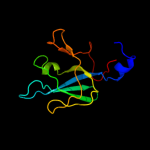
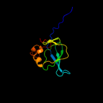
Top model
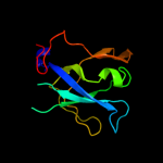
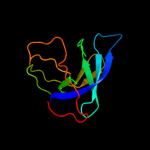
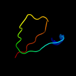
Image coloured by rainbow N → C terminus
Model (left) based on template d1pxfa_
Top template information
Fold:OB-fold
Superfamily:Nucleic acid-binding proteins
Family:Myf domain
Confidence and coverage
Confidence:100.0% Coverage:100%
110 residues (100% of your sequence) have been modelled with 100.0% confidence by the single highest scoring template.
3D viewing
Interactive 3D view in Jmol

Sequence analysis 

Secondary structure and disorder prediction 

   1........10.........20.........30.........40.........50.........60
Sequence  METVAYADFARLEMRVGKIVEVKRHENADKLYIVQVDVGQKTLQTVTSLVPYYSEEELMG
Secondary structure 


SS confidence 


Disorder  ??????


Disorder confidence 


   .........70.........80.........90.........100.........110
Sequence  KTVVVLCNLQKAKMRGETSECMLLCAETDDGSESVLLTPERMMPAGVRVV
Secondary structure 


SS confidence 


Disorder 


??????


???????
Disorder confidence 


Confidence Key
High(9)                    Low (0)
?Disordered
Alpha helix
Beta strand

Domain analysis 

Hover over an aligned region to see model and summary info

Please note, only up to the top 20 hits are modelled to reduce computer load

RankAligned region

PDB 1pxf chain A

3D model

Region: 1 - 110
Aligned: 110
Modelled: 110
Confidence: 100.0%
Identity: 100%
Fold: OB-fold
Superfamily: Nucleic acid-binding proteins
Family: Myf domain

Phyre2

PDB 2cwp chain A

3D model

Region: 1 - 109
Aligned: 108
Modelled: 109
Confidence: 100.0%
Identity: 32%
PDB header:ligase
Chain: A: PDB Molecule:metrs related protein;
PDBTitle: crystal structure of metrs related protein from pyrococcus horikoshii

Phyre2

PDB 1pyb chain A

3D model

Region: 2 - 109
Aligned: 106
Modelled: 108
Confidence: 100.0%
Identity: 28%
Fold: OB-fold
Superfamily: Nucleic acid-binding proteins
Family: Myf domain

Phyre2

PDB 1gd7 chain A

3D model

Region: 3 - 109
Aligned: 106
Modelled: 107
Confidence: 100.0%
Identity: 33%
Fold: OB-fold
Superfamily: Nucleic acid-binding proteins
Family: Myf domain

Phyre2

PDB 2q2i chain A

3D model

Region: 3 - 109
Aligned: 106
Modelled: 107
Confidence: 100.0%
Identity: 30%
PDB header:chaperone
Chain: A: PDB Molecule:secretion chaperone;
PDBTitle: crystal structure of the protein secretion chaperone csaa from2 agrobacterium tumefaciens.

Phyre2

PDB 1fl0 chain A

3D model

Region: 8 - 110
Aligned: 101
Modelled: 103
Confidence: 100.0%
Identity: 37%
Fold: OB-fold
Superfamily: Nucleic acid-binding proteins
Family: Myf domain

Phyre2

PDB 1mkh chain A

3D model

Region: 3 - 109
Aligned: 104
Modelled: 104
Confidence: 100.0%
Identity: 49%
Fold: OB-fold
Superfamily: Nucleic acid-binding proteins
Family: Myf domain

Phyre2

PDB 1ntg chain A

3D model

Region: 9 - 109
Aligned: 101
Modelled: 101
Confidence: 100.0%
Identity: 43%
Fold: OB-fold
Superfamily: Nucleic acid-binding proteins
Family: Myf domain

Phyre2

PDB 2nzo chain D

3D model

Region: 2 - 109
Aligned: 107
Modelled: 108
Confidence: 100.0%
Identity: 27%
PDB header:chaperone
Chain: D: PDB Molecule:protein csaa;
PDBTitle: crystal structure of a secretion chaperone csaa from bacillus subtilis2 in the space group p 32 2 1

Phyre2

PDB 3bu2 chain B

3D model

Region: 3 - 110
Aligned: 108
Modelled: 108
Confidence: 99.9%
Identity: 19%
PDB header:rna binding protein
Chain: B: PDB Molecule:putative trna-binding protein;
PDBTitle: crystal structure of a trna-binding protein from2 staphylococcus saprophyticus subsp. saprophyticus.3 northeast structural genomics consortium target syr77

Phyre2

PDB 3pco chain D

3D model

Region: 13 - 109
Aligned: 97
Modelled: 97
Confidence: 99.9%
Identity: 29%
PDB header:ligase
Chain: D: PDB Molecule:phenylalanyl-trna synthetase, beta chain;
PDBTitle: crystal structure of e. coli phenylalanine-trna synthetase complexed2 with phenylalanine and amp

Phyre2

PDB 2e8g chain B

3D model

Region: 7 - 107
Aligned: 95
Modelled: 101
Confidence: 99.9%
Identity: 21%
PDB header:rna binding protein
Chain: B: PDB Molecule:hypothetical protein ph0536;
PDBTitle: the structure of protein from p. horikoshii at 1.7 angstrom2 resolution

Phyre2

PDB 1jjc chain B domain 3

3D model

Region: 13 - 109
Aligned: 95
Modelled: 97
Confidence: 99.9%
Identity: 18%
Fold: OB-fold
Superfamily: Nucleic acid-binding proteins
Family: Myf domain

Phyre2

PDB 2akw chain B

3D model

Region: 13 - 109
Aligned: 95
Modelled: 96
Confidence: 99.9%
Identity: 17%
PDB header:ligase
Chain: B: PDB Molecule:phenylalanyl-trna synthetase beta chain;
PDBTitle: crystal structure of t.thermophilus phenylalanyl-trna synthetase2 complexed with p-cl-phenylalanine

Phyre2

PDB 2rhs chain B

3D model

Region: 13 - 109
Aligned: 86
Modelled: 97
Confidence: 99.7%
Identity: 28%
PDB header:ligase
Chain: B: PDB Molecule:phenylalanyl-trna synthetase beta chain;
PDBTitle: phers from staphylococcus haemolyticus- rational protein2 engineering and inhibitor studies

Phyre2

PDB 3l4g chain L

3D model

Region: 70 - 110
Aligned: 41
Modelled: 41
Confidence: 89.2%
Identity: 7%
PDB header:ligase
Chain: L: PDB Molecule:phenylalanyl-trna synthetase beta chain;
PDBTitle: crystal structure of homo sapiens cytoplasmic phenylalanyl-trna2 synthetase

Phyre2

PDB 1jjc chain B domain 1

3D model

Region: 18 - 31
Aligned: 14
Modelled: 14
Confidence: 15.2%
Identity: 21%
Fold: Putative DNA-binding domain
Superfamily: Putative DNA-binding domain
Family: Domains B1 and B5 of PheRS-beta, PheT

Phyre2

PDB 1vjn chain A

3D model

Region: 5 - 37
Aligned: 33
Modelled: 33
Confidence: 10.6%
Identity: 21%
Fold: Metallo-hydrolase/oxidoreductase
Superfamily: Metallo-hydrolase/oxidoreductase
Family: Hypothetical protein TM0207

Phyre2

PDB 2fhd chain A

3D model

Region: 2 - 33
Aligned: 32
Modelled: 32
Confidence: 8.3%
Identity: 19%
PDB header:cell cycle
Chain: A: PDB Molecule:dna repair protein rhp9/crb2;
PDBTitle: crystal structure of crb2 tandem tudor domains

Phyre2

PDB 1qmg chain A domain 2

3D model

Region: 27 - 66
Aligned: 40
Modelled: 40
Confidence: 6.8%
Identity: 13%
Fold: NAD(P)-binding Rossmann-fold domains
Superfamily: NAD(P)-binding Rossmann-fold domains
Family: 6-phosphogluconate dehydrogenase-like, N-terminal domain

Phyre2
1

d1pxfa_
2

c2cwpA_
3

d1pyba_
4

d1gd7a_
5

c2q2iA_
6

d1fl0a_
7

d1mkha_
8

d1ntga_
9

c2nzoD_
10

c3bu2B_
11

c3pcoD_
12

c2e8gB_
13

d1jjcb3
14

c2akwB_
15

c2rhsB_
16

c3l4gL_
17

d1jjcb1
18

d1vjna_
19

c2fhdA_
20

d1qmga2
21


22


23


24


25


Detailed template information 

#
Template Alignment Coverage3D Model Confidence
% i.d. Template Information
1d1pxfa_


100.0 100 Fold:OB-fold
Superfamily:Nucleic acid-binding proteins
Family:Myf domain
2c2cwpA_


100.0 32 PDB header:ligase
Chain: A: PDB Molecule:metrs related protein;
PDBTitle: crystal structure of metrs related protein from pyrococcus horikoshii
3d1pyba_


100.0 28 Fold:OB-fold
Superfamily:Nucleic acid-binding proteins
Family:Myf domain
4d1gd7a_


100.0 33 Fold:OB-fold
Superfamily:Nucleic acid-binding proteins
Family:Myf domain
5c2q2iA_


100.0 30 PDB header:chaperone
Chain: A: PDB Molecule:secretion chaperone;
PDBTitle: crystal structure of the protein secretion chaperone csaa from2 agrobacterium tumefaciens.
6d1fl0a_


100.0 37 Fold:OB-fold
Superfamily:Nucleic acid-binding proteins
Family:Myf domain
7d1mkha_


100.0 49 Fold:OB-fold
Superfamily:Nucleic acid-binding proteins
Family:Myf domain
8d1ntga_


100.0 43 Fold:OB-fold
Superfamily:Nucleic acid-binding proteins
Family:Myf domain
9c2nzoD_


100.0 27 PDB header:chaperone
Chain: D: PDB Molecule:protein csaa;
PDBTitle: crystal structure of a secretion chaperone csaa from bacillus subtilis2 in the space group p 32 2 1
10c3bu2B_


99.9 19 PDB header:rna binding protein
Chain: B: PDB Molecule:putative trna-binding protein;
PDBTitle: crystal structure of a trna-binding protein from2 staphylococcus saprophyticus subsp. saprophyticus.3 northeast structural genomics consortium target syr77
11c3pcoD_


99.9 29 PDB header:ligase
Chain: D: PDB Molecule:phenylalanyl-trna synthetase, beta chain;
PDBTitle: crystal structure of e. coli phenylalanine-trna synthetase complexed2 with phenylalanine and amp
12c2e8gB_


99.9 21 PDB header:rna binding protein
Chain: B: PDB Molecule:hypothetical protein ph0536;
PDBTitle: the structure of protein from p. horikoshii at 1.7 angstrom2 resolution
13d1jjcb3


99.9 18 Fold:OB-fold
Superfamily:Nucleic acid-binding proteins
Family:Myf domain
14c2akwB_


99.9 17 PDB header:ligase
Chain: B: PDB Molecule:phenylalanyl-trna synthetase beta chain;
PDBTitle: crystal structure of t.thermophilus phenylalanyl-trna synthetase2 complexed with p-cl-phenylalanine
15c2rhsB_


99.7 28 PDB header:ligase
Chain: B: PDB Molecule:phenylalanyl-trna synthetase beta chain;
PDBTitle: phers from staphylococcus haemolyticus- rational protein2 engineering and inhibitor studies
16c3l4gL_


89.2 7 PDB header:ligase
Chain: L: PDB Molecule:phenylalanyl-trna synthetase beta chain;
PDBTitle: crystal structure of homo sapiens cytoplasmic phenylalanyl-trna2 synthetase
17d1jjcb1


15.2 21 Fold:Putative DNA-binding domain
Superfamily:Putative DNA-binding domain
Family:Domains B1 and B5 of PheRS-beta, PheT
18d1vjna_


10.6 21 Fold:Metallo-hydrolase/oxidoreductase
Superfamily:Metallo-hydrolase/oxidoreductase
Family:Hypothetical protein TM0207
19c2fhdA_


8.3 19 PDB header:cell cycle
Chain: A: PDB Molecule:dna repair protein rhp9/crb2;
PDBTitle: crystal structure of crb2 tandem tudor domains
20d1qmga2


6.8 13 Fold:NAD(P)-binding Rossmann-fold domains
Superfamily:NAD(P)-binding Rossmann-fold domains
Family:6-phosphogluconate dehydrogenase-like, N-terminal domain
21d2gyci1


not modelled 6.6 16 Fold:Ribosomal protein L14
Superfamily:Ribosomal protein L14
Family:Ribosomal protein L14
22c3iz6D_


not modelled 6.3 26 PDB header:ribosome
Chain: D: PDB Molecule:40s ribosomal protein s4 (s4e);
PDBTitle: localization of the small subunit ribosomal proteins into a 5.5 a2 cryo-em map of triticum aestivum translating 80s ribosome
23c2xzmW_


not modelled 6.1 23 PDB header:ribosome
Chain: W: PDB Molecule:40s ribosomal protein s4;
PDBTitle: crystal structure of the eukaryotic 40s ribosomal2 subunit in complex with initiation factor 1. this file3 contains the 40s subunit and initiation factor for4 molecule 1
24d1rbla2


not modelled 5.8 14 Fold:Ferredoxin-like
Superfamily:RuBisCO, large subunit, small (N-terminal) domain
Family:Ribulose 1,5-bisphosphate carboxylase-oxygenase
25c3si5X_


not modelled 5.6 20 PDB header:cell cycle
Chain: X: PDB Molecule:protein casc5;
PDBTitle: kinetochore-bubr1 kinase complex

Binding site prediction 

Due to computational demand, binding site predictions are not run for batch jobs

If you want to predict binding sites, please manually submit your model of choice to 3DLigandSite


Phyre is for academic use only

Please cite: Protein structure prediction on the web: a case study using the Phyre server
Kelley LA and Sternberg MJE. Nature Protocols 4, 363 - 371 (2009) [pdf] [Import into BibTeX]
 
If you use the binding site predictions from 3DLigandSite, please also cite:
3DLigandSite: predicting ligand-binding sites using similar structures.
Wass MN, Kelley LA and Sternberg MJ Nucleic Acids Research 38, W469-73 (2010) [PubMed]
 
© Structural Bioinformatics Group
Imperial College London
Lawrence Kelley, Benjamin Jefferys
Disclaimer
Terms and Conditions
Component software
Template detection: HHpred 1.51
Secondary structure prediction: Psi-pred 2.5
Disorder prediction: Disopred 2.4
Transmembrane prediction: Memsat_SVM
Multi-template modelling and ab initio: Poing 1.0