Phyre Home Retrieve Phyre Job Id
Emaill.a.kelley@imperial.ac.uk
DescriptionP46856
DateThu Jan 5 12:04:28 GMT 2012
Unique Job ID2b263dbb77c05392

Summary 

Top model
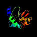
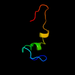
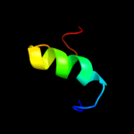
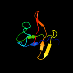
Image coloured by rainbow N → C terminus
Model (left) based on template d2paga1
Top template information
Fold:SMI1/KNR4-like
Superfamily:SMI1/KNR4-like
Family:SMI1/KNR4-like
Confidence and coverage
Confidence: 66.1% Coverage: 72%
100 residues ( 72% of your sequence) have been modelled with 66.1% confidence by the single highest scoring template.
You may wish to submit your sequence to Phyrealarm. This will automatically scan your sequence every week for new potential templates as they appear in the Phyre2 library.
Please note: You must be registered and logged in to use Phyrealarm.
3D viewing
Interactive 3D view in Jmol

Sequence analysis 

Secondary structure and disorder prediction 

   1........10.........20.........30.........40.........50.........60
Sequence  MMTKTQINKLIKMMNDLDYPFEAPLKESFIESIIQIEFNSNSTNCLEKLCNEVSILFKNQ
Secondary structure 


SS confidence 


Disorder  ???


???

???


Disorder confidence 


   .........70.........80.........90.........100.........110.........120
Sequence  PDYLTFLRAMDGFEVNGLRLFSLSIPEPSVKNLFAVNEFYRNNDDFINPDLQERLVIGDY
Secondary structure 


SS confidence 


Disorder 


??


?


Disorder confidence 


   .........130........
Sequence  SISIFTYDIKGDAANLLI
Secondary structure 


SS confidence 


Disorder 


??????
Disorder confidence 


Confidence Key
High(9)                    Low (0)
?Disordered
Alpha helix
Beta strand

Domain analysis 

Hover over an aligned region to see model and summary info

Please note, only up to the top 20 hits are modelled to reduce computer load

RankAligned region

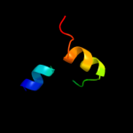
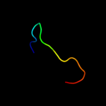
PDB 2pag chain A domain 1

3D model

Region: 7 - 131
Aligned: 100
Modelled: 107
Confidence: 66.1%
Identity: 19%
Fold: SMI1/KNR4-like
Superfamily: SMI1/KNR4-like
Family: SMI1/KNR4-like

Phyre2

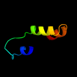
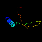
PDB 1yfn chain A domain 1

3D model

Region: 76 - 138
Aligned: 59
Modelled: 63
Confidence: 60.1%
Identity: 19%
Fold: SspB-like
Superfamily: SspB-like
Family: Stringent starvation protein B, SspB

Phyre2

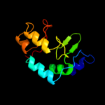
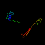
PDB 1ou9 chain A

3D model

Region: 76 - 138
Aligned: 59
Modelled: 63
Confidence: 55.2%
Identity: 20%
Fold: SspB-like
Superfamily: SspB-like
Family: Stringent starvation protein B, SspB

Phyre2

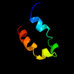
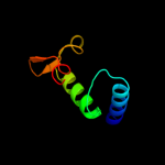
PDB 1zsz chain C domain 1

3D model

Region: 76 - 138
Aligned: 59
Modelled: 63
Confidence: 45.4%
Identity: 20%
Fold: SspB-like
Superfamily: SspB-like
Family: Stringent starvation protein B, SspB

Phyre2

PDB 1ou8 chain A

3D model

Region: 76 - 131
Aligned: 52
Modelled: 56
Confidence: 42.3%
Identity: 19%
Fold: SspB-like
Superfamily: SspB-like
Family: Stringent starvation protein B, SspB

Phyre2

PDB 1pon chain B

3D model

Region: 51 - 74
Aligned: 24
Modelled: 24
Confidence: 23.7%
Identity: 33%
PDB header:calcium-binding protein
Chain: B: PDB Molecule:troponin c;
PDBTitle: site iii-site iv troponin c heterodimer, nmr

Phyre2

PDB 1e7l chain A domain 1

3D model

Region: 59 - 76
Aligned: 18
Modelled: 18
Confidence: 20.7%
Identity: 28%
Fold: LEM/SAP HeH motif
Superfamily: Recombination endonuclease VII, C-terminal and dimerization domains
Family: Recombination endonuclease VII, C-terminal and dimerization domains

Phyre2

PDB 2f05 chain A domain 1

3D model

Region: 28 - 69
Aligned: 42
Modelled: 42
Confidence: 19.4%
Identity: 19%
Fold: PAH2 domain
Superfamily: PAH2 domain
Family: PAH2 domain

Phyre2

PDB 2icg chain A domain 1

3D model

Region: 7 - 130
Aligned: 102
Modelled: 106
Confidence: 13.7%
Identity: 21%
Fold: SMI1/KNR4-like
Superfamily: SMI1/KNR4-like
Family: SMI1/KNR4-like

Phyre2

PDB 1jvr chain A

3D model

Region: 23 - 60
Aligned: 31
Modelled: 38
Confidence: 11.9%
Identity: 29%
Fold: Retroviral matrix proteins
Superfamily: Retroviral matrix proteins
Family: HTLV-II matrix protein

Phyre2

PDB 2hw2 chain A

3D model

Region: 108 - 123
Aligned: 16
Modelled: 16
Confidence: 11.9%
Identity: 19%
PDB header:transferase
Chain: A: PDB Molecule:rifampin adp-ribosyl transferase;
PDBTitle: crystal structure of rifampin adp-ribosyl transferase in2 complex with rifampin

Phyre2

PDB 1zd1 chain B

3D model

Region: 73 - 107
Aligned: 31
Modelled: 35
Confidence: 11.8%
Identity: 29%
PDB header:transferase
Chain: B: PDB Molecule:sulfotransferase 4a1;
PDBTitle: human sulfortransferase sult4a1

Phyre2

PDB 2prv chain A domain 1

3D model

Region: 61 - 132
Aligned: 72
Modelled: 72
Confidence: 10.0%
Identity: 11%
Fold: SMI1/KNR4-like
Superfamily: SMI1/KNR4-like
Family: SMI1/KNR4-like

Phyre2

PDB 2a8v chain A

3D model

Region: 94 - 114
Aligned: 21
Modelled: 21
Confidence: 9.3%
Identity: 19%
PDB header:protein/rna
Chain: A: PDB Molecule:rna binding domain of rho transcription
PDBTitle: rho transcription termination factor/rna complex

Phyre2

PDB 2i5h chain A domain 1

3D model

Region: 107 - 121
Aligned: 15
Modelled: 15
Confidence: 9.3%
Identity: 27%
Fold: AF1531-like
Superfamily: AF1531-like
Family: AF1531-like

Phyre2

PDB 2i5h chain A

3D model

Region: 107 - 121
Aligned: 15
Modelled: 15
Confidence: 9.3%
Identity: 27%
PDB header:structural genomics, unknown function
Chain: A: PDB Molecule:hypothetical protein af1531;
PDBTitle: crystal structure of af1531 from archaeoglobus fulgidus,2 pfam duf655

Phyre2

PDB 3lfm chain A

3D model

Region: 64 - 122
Aligned: 42
Modelled: 42
Confidence: 9.1%
Identity: 14%
PDB header:oxidoreductase
Chain: A: PDB Molecule:protein fto;
PDBTitle: crystal structure of the fat mass and obesity associated (fto) protein2 reveals basis for its substrate specificity

Phyre2

PDB 1w63 chain P

3D model

Region: 7 - 132
Aligned: 77
Modelled: 83
Confidence: 8.9%
Identity: 22%
PDB header:endocytosis
Chain: P: PDB Molecule:adaptor-related protein complex 1, mu 1 subunit;
PDBTitle: ap1 clathrin adaptor core

Phyre2

PDB 2r1f chain B

3D model

Region: 1 - 62
Aligned: 56
Modelled: 62
Confidence: 8.7%
Identity: 18%
PDB header:structural genomics, unknown function
Chain: B: PDB Molecule:predicted aminodeoxychorismate lyase;
PDBTitle: crystal structure of predicted aminodeoxychorismate lyase from2 escherichia coli

Phyre2

PDB 1vaz chain A

3D model

Region: 19 - 44
Aligned: 26
Modelled: 26
Confidence: 8.2%
Identity: 23%
Fold: NSFL1 (p97 ATPase) cofactor p47, SEP domain
Superfamily: NSFL1 (p97 ATPase) cofactor p47, SEP domain
Family: NSFL1 (p97 ATPase) cofactor p47, SEP domain

Phyre2
1

d2paga1
2

d1yfna1
3

d1ou9a_
4

d1zszc1
5

d1ou8a_
6

c1ponB_
7

d1e7la1
8

d2f05a1
9

d2icga1
10

d1jvra_
11

c2hw2A_
12

c1zd1B_
13

d2prva1
14

c2a8vA_
15

d2i5ha1
16

c2i5hA_
17

c3lfmA_
18

c1w63P_
19

c2r1fB_
20

d1vaza_
21


22


23


24


25


26


27


28


29


30


31


32


33


34


35


36


37


38


39


40


41


42


Detailed template information 

#
Template Alignment Coverage3D Model Confidence
% i.d. Template Information
1d2paga1


66.1 19 Fold:SMI1/KNR4-like
Superfamily:SMI1/KNR4-like
Family:SMI1/KNR4-like
2d1yfna1


60.1 19 Fold:SspB-like
Superfamily:SspB-like
Family:Stringent starvation protein B, SspB
3d1ou9a_


55.2 20 Fold:SspB-like
Superfamily:SspB-like
Family:Stringent starvation protein B, SspB
4d1zszc1


45.4 20 Fold:SspB-like
Superfamily:SspB-like
Family:Stringent starvation protein B, SspB
5d1ou8a_


42.3 19 Fold:SspB-like
Superfamily:SspB-like
Family:Stringent starvation protein B, SspB
6c1ponB_


23.7 33 PDB header:calcium-binding protein
Chain: B: PDB Molecule:troponin c;
PDBTitle: site iii-site iv troponin c heterodimer, nmr
7d1e7la1


20.7 28 Fold:LEM/SAP HeH motif
Superfamily:Recombination endonuclease VII, C-terminal and dimerization domains
Family:Recombination endonuclease VII, C-terminal and dimerization domains
8d2f05a1


19.4 19 Fold:PAH2 domain
Superfamily:PAH2 domain
Family:PAH2 domain
9d2icga1


13.7 21 Fold:SMI1/KNR4-like
Superfamily:SMI1/KNR4-like
Family:SMI1/KNR4-like
10d1jvra_


11.9 29 Fold:Retroviral matrix proteins
Superfamily:Retroviral matrix proteins
Family:HTLV-II matrix protein
11c2hw2A_


11.9 19 PDB header:transferase
Chain: A: PDB Molecule:rifampin adp-ribosyl transferase;
PDBTitle: crystal structure of rifampin adp-ribosyl transferase in2 complex with rifampin
12c1zd1B_


11.8 29 PDB header:transferase
Chain: B: PDB Molecule:sulfotransferase 4a1;
PDBTitle: human sulfortransferase sult4a1
13d2prva1


10.0 11 Fold:SMI1/KNR4-like
Superfamily:SMI1/KNR4-like
Family:SMI1/KNR4-like
14c2a8vA_


9.3 19 PDB header:protein/rna
Chain: A: PDB Molecule:rna binding domain of rho transcription
PDBTitle: rho transcription termination factor/rna complex
15d2i5ha1


9.3 27 Fold:AF1531-like
Superfamily:AF1531-like
Family:AF1531-like
16c2i5hA_


9.3 27 PDB header:structural genomics, unknown function
Chain: A: PDB Molecule:hypothetical protein af1531;
PDBTitle: crystal structure of af1531 from archaeoglobus fulgidus,2 pfam duf655
17c3lfmA_


9.1 14 PDB header:oxidoreductase
Chain: A: PDB Molecule:protein fto;
PDBTitle: crystal structure of the fat mass and obesity associated (fto) protein2 reveals basis for its substrate specificity
18c1w63P_


8.9 22 PDB header:endocytosis
Chain: P: PDB Molecule:adaptor-related protein complex 1, mu 1 subunit;
PDBTitle: ap1 clathrin adaptor core
19c2r1fB_


8.7 18 PDB header:structural genomics, unknown function
Chain: B: PDB Molecule:predicted aminodeoxychorismate lyase;
PDBTitle: crystal structure of predicted aminodeoxychorismate lyase from2 escherichia coli
20d1vaza_


8.2 23 Fold:NSFL1 (p97 ATPase) cofactor p47, SEP domain
Superfamily:NSFL1 (p97 ATPase) cofactor p47, SEP domain
Family:NSFL1 (p97 ATPase) cofactor p47, SEP domain
21d1vpza_


not modelled 8.1 24 Fold:CsrA-like
Superfamily:CsrA-like
Family:CsrA-like
22c1qmoG_


not modelled 7.8 30 PDB header:lectin
Chain: G: PDB Molecule:mannose binding lectin, fril;
PDBTitle: structure of fril, a legume lectin that delays2 hematopoietic progenitor maturation
23d1a62a2


not modelled 7.8 19 Fold:OB-fold
Superfamily:Nucleic acid-binding proteins
Family:Cold shock DNA-binding domain-like
24d1tdja2


not modelled 7.7 22 Fold:Ferredoxin-like
Superfamily:ACT-like
Family:Allosteric threonine deaminase C-terminal domain
25c3csqC_


not modelled 7.5 31 PDB header:hydrolase
Chain: C: PDB Molecule:morphogenesis protein 1;
PDBTitle: crystal and cryoem structural studies of a cell wall2 degrading enzyme in the bacteriophage phi29 tail
26d1a9xa3


not modelled 7.4 22 Fold:PreATP-grasp domain
Superfamily:PreATP-grasp domain
Family:BC N-terminal domain-like
27d1kshb_


not modelled 7.3 31 Fold:Immunoglobulin-like beta-sandwich
Superfamily:E set domains
Family:RhoGDI-like
28d2f2ab1


not modelled 7.1 11 Fold:GatB/YqeY motif
Superfamily:GatB/YqeY motif
Family:GatB/GatE C-terminal domain-like
29d1qtma1


not modelled 6.9 47 Fold:Ribonuclease H-like motif
Superfamily:Ribonuclease H-like
Family:DnaQ-like 3'-5' exonuclease
30d1ss6a_


not modelled 6.6 23 Fold:NSFL1 (p97 ATPase) cofactor p47, SEP domain
Superfamily:NSFL1 (p97 ATPase) cofactor p47, SEP domain
Family:NSFL1 (p97 ATPase) cofactor p47, SEP domain
31c2f4qA_


not modelled 6.5 23 PDB header:isomerase
Chain: A: PDB Molecule:type i topoisomerase, putative;
PDBTitle: crystal structure of deinococcus radiodurans topoisomerase ib
32c3fbyC_


not modelled 6.3 26 PDB header:cell adhesion
Chain: C: PDB Molecule:cartilage oligomeric matrix protein;
PDBTitle: the crystal structure of the signature domain of cartilage oligomeric2 matrix protein.
33d1o66a_


not modelled 6.3 14 Fold:TIM beta/alpha-barrel
Superfamily:Phosphoenolpyruvate/pyruvate domain
Family:Ketopantoate hydroxymethyltransferase PanB
34c1vpzB_


not modelled 6.1 24 PDB header:rna binding protein
Chain: B: PDB Molecule:carbon storage regulator homolog;
PDBTitle: crystal structure of a putative carbon storage regulator protein2 (csra, pa0905) from pseudomonas aeruginosa at 2.05 a resolution
35c1c8m2_


not modelled 6.1 25 PDB header:virus
Chain: 2: PDB Molecule:human rhinovirus 16 coat protein;
PDB Fragment:residues 2-78; PDBTitle: refined crystal structure of human rhinovirus 16 complexed2 with vp63843 (pleconaril), an anti-picornaviral drug3 currently in clinical trials
36d1elka_


not modelled 6.0 16 Fold:alpha-alpha superhelix
Superfamily:ENTH/VHS domain
Family:VHS domain
37c2b1uA_


not modelled 5.9 10 PDB header:metal binding protein
Chain: A: PDB Molecule:calmodulin-like protein 5;
PDBTitle: solution structure of calmodulin-like skin protein c2 terminal domain
38d1d8ua_


not modelled 5.6 6 Fold:Globin-like
Superfamily:Globin-like
Family:Globins
39c2jppB_


not modelled 5.4 32 PDB header:translation/rna
Chain: B: PDB Molecule:translational repressor;
PDBTitle: structural basis of rsma/csra rna recognition: structure of2 rsme bound to the shine-dalgarno sequence of hcna mrna
40d1hdsa_


not modelled 5.2 13 Fold:Globin-like
Superfamily:Globin-like
Family:Globins
41c2amiA_


not modelled 5.2 13 PDB header:cell cycle
Chain: A: PDB Molecule:caltractin;
PDBTitle: solution structure of the calcium-loaded n-terminal sensor2 domain of centrin
42d1zgha1


not modelled 5.1 22 Fold:FMT C-terminal domain-like
Superfamily:FMT C-terminal domain-like
Family:Post formyltransferase domain

Binding site prediction 

Due to computational demand, binding site predictions are not run for batch jobs

If you want to predict binding sites, please manually submit your model of choice to 3DLigandSite


Phyre is for academic use only

Please cite: Protein structure prediction on the web: a case study using the Phyre server
Kelley LA and Sternberg MJE. Nature Protocols 4, 363 - 371 (2009) [pdf] [Import into BibTeX]
 
If you use the binding site predictions from 3DLigandSite, please also cite:
3DLigandSite: predicting ligand-binding sites using similar structures.
Wass MN, Kelley LA and Sternberg MJ Nucleic Acids Research 38, W469-73 (2010) [PubMed]
 
© Structural Bioinformatics Group
Imperial College London
Lawrence Kelley, Benjamin Jefferys
Disclaimer
Terms and Conditions
Component software
Template detection: HHpred 1.51
Secondary structure prediction: Psi-pred 2.5
Disorder prediction: Disopred 2.4
Transmembrane prediction: Memsat_SVM
Multi-template modelling and ab initio: Poing 1.0