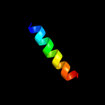
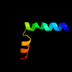
Phyre Home Retrieve Phyre Job Id
Emaill.a.kelley@imperial.ac.uk
DescriptionP21365
DateThu Jan 5 11:38:11 GMT 2012
Unique Job ID2a8dc698076db141

Summary 

Top model
Image coloured by rainbow N → C terminus
Model (left) based on template c2kvlA_
Top template information
PDB header:viral protein
Chain: A: PDB Molecule:major outer capsid protein vp7;
PDBTitle: nmr structure of the c-terminal domain of vp7
Confidence and coverage
Confidence: 23.2% Coverage: 8%
19 residues ( 8% of your sequence) have been modelled with 23.2% confidence by the single highest scoring template.
You may wish to submit your sequence to Phyrealarm. This will automatically scan your sequence every week for new potential templates as they appear in the Phyre2 library.
Please note: You must be registered and logged in to use Phyrealarm.
3D viewing
Interactive 3D view in Jmol

Sequence analysis 

Secondary structure and disorder prediction 

   1........10.........20.........30.........40.........50.........60
Sequence  MSITAQSVYRDTGNFFRNQFMTILLVSLLCAFITVVLGHVFSPSDAQLAQLNDGVPVSGS
Secondary structure 


SS confidence 


Disorder  ????


??????
Disorder confidence 


   .........70.........80.........90.........100.........110.........120
Sequence  SGLFDLVQNMSPEQQQILLQASAASTFSGLIGNAILAGGVILIIQLVSAGQRVSALRAIG
Secondary structure 


SS confidence 


Disorder 


???


??
????


Disorder confidence 


   .........130.........140.........150.........160.........170.........180
Sequence  ASAPILPKLFILIFLTTLLVQIGIMLVVVPGIIMAILLALAPVMLVQDKMGVFASMRSSM
Secondary structure 


SS confidence 


Disorder 


????


Disorder confidence 


   .........190.........200.........210.........220.........230.........240
Sequence  RLTWANMRLVAPAVLSWLLAKTLLLLFASSFAALTPEIGAVLANTLSNLISAILLIYLFR
Secondary structure 


SS confidence 


Disorder 


??????


Disorder confidence 


   .......
Sequence  LYMLIRQ
Secondary structure 

SS confidence 


Disorder 


???
Disorder confidence 


Confidence Key
High(9)                    Low (0)
?Disordered
Alpha helix
Beta strand

Domain analysis 

Hover over an aligned region to see model and summary info

Please note, only up to the top 20 hits are modelled to reduce computer load

RankAligned region

PDB 2kvl chain A

3D model

Region: 177 - 195
Aligned: 19
Modelled: 19
Confidence: 23.2%
Identity: 26%
PDB header:viral protein
Chain: A: PDB Molecule:major outer capsid protein vp7;
PDBTitle: nmr structure of the c-terminal domain of vp7

Phyre2

PDB 3hd7 chain A

3D model

Region: 177 - 212
Aligned: 36
Modelled: 36
Confidence: 17.8%
Identity: 8%
PDB header:exocytosis
Chain: A: PDB Molecule:vesicle-associated membrane protein 2;
PDBTitle: helical extension of the neuronal snare complex into the membrane,2 spacegroup c 1 2 1

Phyre2

PDB 1cii chain A

3D model

Region: 185 - 234
Aligned: 50
Modelled: 50
Confidence: 13.5%
Identity: 18%
PDB header:transmembrane protein
Chain: A: PDB Molecule:colicin ia;
PDBTitle: colicin ia

Phyre2

PDB 1k8r chain B

3D model

Region: 173 - 182
Aligned: 10
Modelled: 10
Confidence: 9.5%
Identity: 30%
Fold: beta-Grasp (ubiquitin-like)
Superfamily: Ubiquitin-like
Family: Ras-binding domain, RBD

Phyre2

PDB 2hgc chain A domain 1

3D model

Region: 6 - 19
Aligned: 14
Modelled: 14
Confidence: 9.0%
Identity: 21%
Fold: DNA/RNA-binding 3-helical bundle
Superfamily: "Winged helix" DNA-binding domain
Family: YjcQ-like

Phyre2

PDB 2hgc chain A

3D model

Region: 6 - 19
Aligned: 14
Modelled: 14
Confidence: 9.0%
Identity: 21%
PDB header:structural genomics, unknown function
Chain: A: PDB Molecule:yjcq protein;
PDBTitle: solution nmr structure of the yjcq protein from bacillus2 subtilis. northeast structural genomics target sr346.

Phyre2

PDB 1k8k chain E

3D model

Region: 7 - 19
Aligned: 13
Modelled: 13
Confidence: 8.7%
Identity: 8%
Fold: Arp2/3 complex 21 kDa subunit ARPC3
Superfamily: Arp2/3 complex 21 kDa subunit ARPC3
Family: Arp2/3 complex 21 kDa subunit ARPC3

Phyre2

PDB 1rh5 chain C

3D model

Region: 11 - 39
Aligned: 29
Modelled: 29
Confidence: 8.6%
Identity: 17%
PDB header:protein transport
Chain: C: PDB Molecule:secbeta;
PDBTitle: the structure of a protein conducting channel

Phyre2

PDB 1rh5 chain C

3D model

Region: 11 - 39
Aligned: 29
Modelled: 29
Confidence: 8.6%
Identity: 17%
Fold: Single transmembrane helix
Superfamily: Sec-beta subunit
Family: Sec-beta subunit

Phyre2

PDB 3go5 chain A

3D model

Region: 167 - 195
Aligned: 29
Modelled: 28
Confidence: 8.2%
Identity: 0%
PDB header:gene regulation
Chain: A: PDB Molecule:multidomain protein with s1 rna-binding domains;
PDBTitle: crystal structure of a multidomain protein with nucleic acid binding2 domains (sp_0946) from streptococcus pneumoniae tigr4 at 1.40 a3 resolution

Phyre2

PDB 1pf4 chain A domain 2

3D model

Region: 173 - 216
Aligned: 44
Modelled: 44
Confidence: 8.2%
Identity: 5%
Fold: ABC transporter transmembrane region
Superfamily: ABC transporter transmembrane region
Family: ABC transporter transmembrane region

Phyre2

PDB 1m45 chain B

3D model

Region: 9 - 24
Aligned: 16
Modelled: 16
Confidence: 8.2%
Identity: 6%
PDB header:cell cycle protein
Chain: B: PDB Molecule:iq2 motif from myo2p, a class v myosin;
PDBTitle: crystal structure of mlc1p bound to iq2 of myo2p, a class v2 myosin

Phyre2

PDB 1q2i chain A

3D model

Region: 176 - 188
Aligned: 13
Modelled: 13
Confidence: 7.7%
Identity: 0%
PDB header:antitumor protein
Chain: A: PDB Molecule:pnc27;
PDBTitle: nmr solution structure of a peptide from the mdm-2 binding2 domain of the p53 protein that is selectively cytotoxic to3 cancer cells

Phyre2

PDB 1unh chain D

3D model

Region: 241 - 247
Aligned: 7
Modelled: 7
Confidence: 7.4%
Identity: 71%
PDB header:cell cycle
Chain: D: PDB Molecule:cyclin-dependent kinase 5 activator 1;
PDBTitle: structural mechanism for the inhibition of cdk5-p25 by2 roscovitine, aloisine and indirubin.

Phyre2

PDB 1unl chain D

3D model

Region: 241 - 247
Aligned: 7
Modelled: 7
Confidence: 7.4%
Identity: 71%
Fold: Cyclin-like
Superfamily: Cyclin-like
Family: Cyclin

Phyre2

PDB 1kq8 chain A

3D model

Region: 97 - 127
Aligned: 31
Modelled: 31
Confidence: 7.3%
Identity: 19%
PDB header:transcription
Chain: A: PDB Molecule:hepatocyte nuclear factor 3 forkhead homolog 1;
PDBTitle: solution structure of winged helix protein hfh-1

Phyre2

PDB 1kq8 chain A

3D model

Region: 97 - 127
Aligned: 31
Modelled: 31
Confidence: 7.3%
Identity: 19%
Fold: DNA/RNA-binding 3-helical bundle
Superfamily: "Winged helix" DNA-binding domain
Family: Forkhead DNA-binding domain

Phyre2

PDB 3jqo chain V

3D model

Region: 183 - 201
Aligned: 19
Modelled: 14
Confidence: 7.0%
Identity: 11%
PDB header:transport protein
Chain: V: PDB Molecule:traf protein;
PDBTitle: crystal structure of the outer membrane complex of a type iv2 secretion system

Phyre2

PDB 4a1a chain M

3D model

Region: 2 - 20
Aligned: 19
Modelled: 19
Confidence: 7.0%
Identity: 5%
PDB header:ribosome
Chain: M: PDB Molecule:60s ribosomal protein l5;
PDBTitle: t.thermophila 60s ribosomal subunit in complex with2 initiation factor 6. this file contains 5s rrna,3 5.8s rrna and proteins of molecule 3.

Phyre2

PDB 1zp0 chain D

3D model

Region: 151 - 182
Aligned: 32
Modelled: 32
Confidence: 6.9%
Identity: 13%
PDB header:oxidoreductase
Chain: D: PDB Molecule:small cytochrome binding protein;
PDBTitle: crystal structure of mitochondrial respiratory complex ii2 bound with 3-nitropropionate and 2-thenoyltrifluoroacetone

Phyre2
1

c2kvlA_
2

c3hd7A_
3

c1ciiA_
4

d1k8rb_
5

d2hgca1
6

c2hgcA_
7

d1k8ke_
8

c1rh5C_
9

d1rh5c_
10

c3go5A_
11

d1pf4a2
12

c1m45B_
13

c1q2iA_
14

c1unhD_
15

d1unld_
16

c1kq8A_
17

d1kq8a_
18

c3jqoV_
19

c4a1aM_
20

c1zp0D_
21


22


23


24


25


26


27


28


29


30


31


32


33


34


35


36


37


38


39


40


41


42


43


44


45


46


47


48


49


50


51


52


53


54


55


Detailed template information 

#
Template Alignment Coverage3D Model Confidence
% i.d. Template Information
1c2kvlA_


23.2 26 PDB header:viral protein
Chain: A: PDB Molecule:major outer capsid protein vp7;
PDBTitle: nmr structure of the c-terminal domain of vp7
2c3hd7A_


17.8 8 PDB header:exocytosis
Chain: A: PDB Molecule:vesicle-associated membrane protein 2;
PDBTitle: helical extension of the neuronal snare complex into the membrane,2 spacegroup c 1 2 1
3c1ciiA_


13.5 18 PDB header:transmembrane protein
Chain: A: PDB Molecule:colicin ia;
PDBTitle: colicin ia
4d1k8rb_


9.5 30 Fold:beta-Grasp (ubiquitin-like)
Superfamily:Ubiquitin-like
Family:Ras-binding domain, RBD
5d2hgca1


9.0 21 Fold:DNA/RNA-binding 3-helical bundle
Superfamily:"Winged helix" DNA-binding domain
Family:YjcQ-like
6c2hgcA_


9.0 21 PDB header:structural genomics, unknown function
Chain: A: PDB Molecule:yjcq protein;
PDBTitle: solution nmr structure of the yjcq protein from bacillus2 subtilis. northeast structural genomics target sr346.
7d1k8ke_


8.7 8 Fold:Arp2/3 complex 21 kDa subunit ARPC3
Superfamily:Arp2/3 complex 21 kDa subunit ARPC3
Family:Arp2/3 complex 21 kDa subunit ARPC3
8c1rh5C_


8.6 17 PDB header:protein transport
Chain: C: PDB Molecule:secbeta;
PDBTitle: the structure of a protein conducting channel
9d1rh5c_


8.6 17 Fold:Single transmembrane helix
Superfamily:Sec-beta subunit
Family:Sec-beta subunit
10c3go5A_


8.2 0 PDB header:gene regulation
Chain: A: PDB Molecule:multidomain protein with s1 rna-binding domains;
PDBTitle: crystal structure of a multidomain protein with nucleic acid binding2 domains (sp_0946) from streptococcus pneumoniae tigr4 at 1.40 a3 resolution
11d1pf4a2


8.2 5 Fold:ABC transporter transmembrane region
Superfamily:ABC transporter transmembrane region
Family:ABC transporter transmembrane region
12c1m45B_


8.2 6 PDB header:cell cycle protein
Chain: B: PDB Molecule:iq2 motif from myo2p, a class v myosin;
PDBTitle: crystal structure of mlc1p bound to iq2 of myo2p, a class v2 myosin
13c1q2iA_


7.7 0 PDB header:antitumor protein
Chain: A: PDB Molecule:pnc27;
PDBTitle: nmr solution structure of a peptide from the mdm-2 binding2 domain of the p53 protein that is selectively cytotoxic to3 cancer cells
14c1unhD_


7.4 71 PDB header:cell cycle
Chain: D: PDB Molecule:cyclin-dependent kinase 5 activator 1;
PDBTitle: structural mechanism for the inhibition of cdk5-p25 by2 roscovitine, aloisine and indirubin.
15d1unld_


7.4 71 Fold:Cyclin-like
Superfamily:Cyclin-like
Family:Cyclin
16c1kq8A_


7.3 19 PDB header:transcription
Chain: A: PDB Molecule:hepatocyte nuclear factor 3 forkhead homolog 1;
PDBTitle: solution structure of winged helix protein hfh-1
17d1kq8a_


7.3 19 Fold:DNA/RNA-binding 3-helical bundle
Superfamily:"Winged helix" DNA-binding domain
Family:Forkhead DNA-binding domain
18c3jqoV_


7.0 11 PDB header:transport protein
Chain: V: PDB Molecule:traf protein;
PDBTitle: crystal structure of the outer membrane complex of a type iv2 secretion system
19c4a1aM_


7.0 5 PDB header:ribosome
Chain: M: PDB Molecule:60s ribosomal protein l5;
PDBTitle: t.thermophila 60s ribosomal subunit in complex with2 initiation factor 6. this file contains 5s rrna,3 5.8s rrna and proteins of molecule 3.
20c1zp0D_


6.9 13 PDB header:oxidoreductase
Chain: D: PDB Molecule:small cytochrome binding protein;
PDBTitle: crystal structure of mitochondrial respiratory complex ii2 bound with 3-nitropropionate and 2-thenoyltrifluoroacetone
21d1cg5a_


not modelled 6.8 21 Fold:Globin-like
Superfamily:Globin-like
Family:Globins
22c3mkuA_


not modelled 6.6 13 PDB header:transport protein
Chain: A: PDB Molecule:multi antimicrobial extrusion protein (na(+)/drug
PDBTitle: structure of a cation-bound multidrug and toxin compound extrusion2 (mate) transporter
23c1e17A_


not modelled 6.4 10 PDB header:dna binding domain
Chain: A: PDB Molecule:afx;
PDBTitle: solution structure of the dna binding domain of the human2 forkhead transcription factor afx (foxo4)
24c3co7C_


not modelled 6.2 16 PDB header:transcription/dna
Chain: C: PDB Molecule:forkhead box protein o1;
PDBTitle: crystal structure of foxo1 dbd bound to dbe2 dna
25d1myta_


not modelled 6.2 19 Fold:Globin-like
Superfamily:Globin-like
Family:Globins
26d2fx0a2


not modelled 6.1 0 Fold:Tetracyclin repressor-like, C-terminal domain
Superfamily:Tetracyclin repressor-like, C-terminal domain
Family:Tetracyclin repressor-like, C-terminal domain
27d1hdsa_


not modelled 6.1 7 Fold:Globin-like
Superfamily:Globin-like
Family:Globins
28c2lkeA_


not modelled 5.9 30 PDB header:cell adhesion
Chain: A: PDB Molecule:integrin alpha-m;
PDBTitle: structures and interaction analyses of the integrin alpha-m beta-22 cytoplasmic tails
29c3g73A_


not modelled 5.9 10 PDB header:transcription/dna
Chain: A: PDB Molecule:forkhead box protein m1;
PDBTitle: structure of the foxm1 dna binding
30c1geaA_


not modelled 5.9 25 PDB header:neuropeptide
Chain: A: PDB Molecule:pituitary adenylate cyclase activating
PDBTitle: receptor-bound conformation of pacap21
31c2ciuA_


not modelled 5.9 0 PDB header:protein transport
Chain: A: PDB Molecule:import inner membrane translocase subunit tim21
PDBTitle: structure of the ims domain of the mitochondrial import2 protein tim21 from s. cerevisiae
32c1n2dC_


not modelled 5.8 6 PDB header:cell cycle
Chain: C: PDB Molecule:iq2 and iq3 motifs from myo2p, a class v myosin;
PDBTitle: ternary complex of mlc1p bound to iq2 and iq3 of myo2p, a2 class v myosin
33c2lkjA_


not modelled 5.6 33 PDB header:cell adhesion
Chain: A: PDB Molecule:integrin alpha-m;
PDBTitle: structures and interaction analyses of the integrin alpha-m beta-22 cytoplasmic tails
34d2lhba_


not modelled 5.6 7 Fold:Globin-like
Superfamily:Globin-like
Family:Globins
35d1d5va_


not modelled 5.6 6 Fold:DNA/RNA-binding 3-helical bundle
Superfamily:"Winged helix" DNA-binding domain
Family:Forkhead DNA-binding domain
36c2wwbB_


not modelled 5.6 7 PDB header:ribosome
Chain: B: PDB Molecule:protein transport protein sec61 subunit gamma;
PDBTitle: cryo-em structure of the mammalian sec61 complex bound to the2 actively translating wheat germ 80s ribosome
37c2rlwA_


not modelled 5.6 14 PDB header:toxin
Chain: A: PDB Molecule:plnf;
PDBTitle: three-dimensional structure of the two peptides that2 constitute the two-peptide bacteriocin plantaracin ef
38c2qq4A_


not modelled 5.5 20 PDB header:metal binding protein
Chain: A: PDB Molecule:iron-sulfur cluster biosynthesis protein iscu;
PDBTitle: crystal structure of iron-sulfur cluster biosynthesis2 protein iscu (ttha1736) from thermus thermophilus hb8
39c1yhuW_


not modelled 5.5 14 PDB header:oxygen storage/transport
Chain: W: PDB Molecule:hemoglobin b1a chain;
PDBTitle: crystal structure of riftia pachyptila c1 hemoglobin reveals novel2 assembly of 24 subunits.
40d3bpya1


not modelled 5.5 10 Fold:DNA/RNA-binding 3-helical bundle
Superfamily:"Winged helix" DNA-binding domain
Family:Forkhead DNA-binding domain
41d1scta_


not modelled 5.5 23 Fold:Globin-like
Superfamily:Globin-like
Family:Globins
42d2hfha_


not modelled 5.4 13 Fold:DNA/RNA-binding 3-helical bundle
Superfamily:"Winged helix" DNA-binding domain
Family:Forkhead DNA-binding domain
43c2zs1D_


not modelled 5.4 8 PDB header:oxygen storage, oxygen transport
Chain: D: PDB Molecule:extracellular giant hemoglobin major globin subunit b1;
PDBTitle: structural basis for the heterotropic and homotropic interactions of2 invertebrate giant hemoglobin
44d1sxja1


not modelled 5.4 9 Fold:post-AAA+ oligomerization domain-like
Superfamily:post-AAA+ oligomerization domain-like
Family:DNA polymerase III clamp loader subunits, C-terminal domain
45d1c17m_


not modelled 5.4 11 Fold:F1F0 ATP synthase subunit A
Superfamily:F1F0 ATP synthase subunit A
Family:F1F0 ATP synthase subunit A
46c1g2cN_


not modelled 5.3 13 PDB header:viral protein
Chain: N: PDB Molecule:fusion protein (f);
PDBTitle: human respiratory syncytial virus fusion protein core
47d2a07f1


not modelled 5.3 6 Fold:DNA/RNA-binding 3-helical bundle
Superfamily:"Winged helix" DNA-binding domain
Family:Forkhead DNA-binding domain
48c2wukD_


not modelled 5.3 17 PDB header:cell cycle
Chain: D: PDB Molecule:septum site-determining protein diviva;
PDBTitle: diviva n-terminal domain, f17a mutant
49c2nq2A_


not modelled 5.3 16 PDB header:metal transport
Chain: A: PDB Molecule:hypothetical abc transporter permease protein
PDBTitle: an inward-facing conformation of a putative metal-chelate2 type abc transporter.
50d1hbra_


not modelled 5.3 0 Fold:Globin-like
Superfamily:Globin-like
Family:Globins
51c2c0kB_


not modelled 5.3 11 PDB header:oxygen transport
Chain: B: PDB Molecule:hemoglobin;
PDBTitle: the structure of hemoglobin from the botfly gasterophilus2 intestinalis
52c3gztF_


not modelled 5.2 28 PDB header:virus
Chain: F: PDB Molecule:outer capsid glycoprotein vp7;
PDBTitle: vp7 recoated rotavirus dlp
53c3bjqA_


not modelled 5.2 14 PDB header:viral protein
Chain: A: PDB Molecule:phage-related protein;
PDBTitle: crystal structure of a phage-related protein (bb3626) from bordetella2 bronchiseptica rb50 at 2.05 a resolution
54d2gdma_


not modelled 5.2 9 Fold:Globin-like
Superfamily:Globin-like
Family:Globins
55c3h8aF_


not modelled 5.1 40 PDB header:lyase/protein binding
Chain: F: PDB Molecule:rnase e;
PDBTitle: crystal structure of e. coli enolase bound to its cognate rnase e2 recognition domain

Binding site prediction 

Due to computational demand, binding site predictions are not run for batch jobs

If you want to predict binding sites, please manually submit your model of choice to 3DLigandSite


Phyre is for academic use only

Please cite: Protein structure prediction on the web: a case study using the Phyre server
Kelley LA and Sternberg MJE. Nature Protocols 4, 363 - 371 (2009) [pdf] [Import into BibTeX]
 
If you use the binding site predictions from 3DLigandSite, please also cite:
3DLigandSite: predicting ligand-binding sites using similar structures.
Wass MN, Kelley LA and Sternberg MJ Nucleic Acids Research 38, W469-73 (2010) [PubMed]
 
© Structural Bioinformatics Group
Imperial College London
Lawrence Kelley, Benjamin Jefferys
Disclaimer
Terms and Conditions
Component software
Template detection: HHpred 1.51
Secondary structure prediction: Psi-pred 2.5
Disorder prediction: Disopred 2.4
Transmembrane prediction: Memsat_SVM
Multi-template modelling and ab initio: Poing 1.0