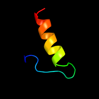
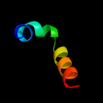
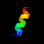
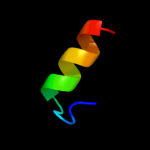
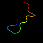
1 c2gvsA_
27.5
24
PDB header: lipid binding proteinChain: A: PDB Molecule: chemosensory protein csp-sg4;PDBTitle: nmr solution structure of cspsg4
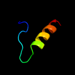
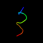
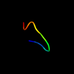
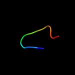
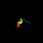
2 d1kx9b_
25.3
19
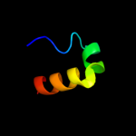
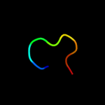
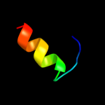
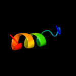
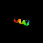
Fold: alpha-alpha superhelixSuperfamily: Chemosensory protein Csp2Family: Chemosensory protein Csp23 d1n8va_
22.5
19
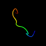
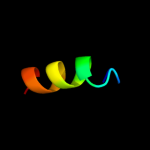
Fold: alpha-alpha superhelixSuperfamily: Chemosensory protein Csp2Family: Chemosensory protein Csp24 d1wm7a_
17.7
56
Fold: Knottins (small inhibitors, toxins, lectins)Superfamily: Scorpion toxin-likeFamily: Short-chain scorpion toxins5 d1b25a1
16.3
33
Fold: Aldehyde ferredoxin oxidoreductase, C-terminal domainsSuperfamily: Aldehyde ferredoxin oxidoreductase, C-terminal domainsFamily: Aldehyde ferredoxin oxidoreductase, C-terminal domains6 d1aora1
16.0
44
Fold: Aldehyde ferredoxin oxidoreductase, C-terminal domainsSuperfamily: Aldehyde ferredoxin oxidoreductase, C-terminal domainsFamily: Aldehyde ferredoxin oxidoreductase, C-terminal domains7 c2emcA_
12.4
18
PDB header: transcriptionChain: A: PDB Molecule: zinc finger protein 473;PDBTitle: solution structure of the c2h2 type zinc finger (region 641-2 673) of human zinc finger protein 473
8 c2z2qF_
12.0
23
PDB header: virus/rnaChain: F: PDB Molecule: coat protein gamma;PDBTitle: crystal structure of flock house virus
9 c1aorB_
11.5
44
PDB header: oxidoreductaseChain: B: PDB Molecule: aldehyde ferredoxin oxidoreductase;PDBTitle: structure of a hyperthermophilic tungstopterin enzyme,2 aldehyde ferredoxin oxidoreductase
10 d1x6va2
10.9
31
Fold: Adenine nucleotide alpha hydrolase-likeSuperfamily: Nucleotidylyl transferaseFamily: ATP sulfurylase catalytic domain11 c2a05A_
10.7
44
PDB header: signaling proteinChain: A: PDB Molecule: cysteine-rich secretory protein-2;PDBTitle: the cysteine-rich secretory protein domain of tpx-1 is2 related to ion channel toxins and regulates ryanodine3 receptor ca2+ signaling
12 d1jhda2
10.2
38
Fold: Adenine nucleotide alpha hydrolase-likeSuperfamily: Nucleotidylyl transferaseFamily: ATP sulfurylase catalytic domain13 c1b4nD_
8.9
33
PDB header: oxidoreductaseChain: D: PDB Molecule: formaldehyde ferredoxin oxidoreductase;PDBTitle: formaldehyde ferredoxin oxidoreductase from pyrococcus furiosus,2 complexed with glutarate
14 c1xx5B_
8.8
29
PDB header: toxinChain: B: PDB Molecule: natrin 1;PDBTitle: crystal structure of natrin from naja atra snake venom
15 d1qasa3
8.8
22
Fold: TIM beta/alpha-barrelSuperfamily: PLC-like phosphodiesterasesFamily: Mammalian PLC16 d1v47a2
8.7
38
Fold: Adenine nucleotide alpha hydrolase-likeSuperfamily: Nucleotidylyl transferaseFamily: ATP sulfurylase catalytic domain17 c2qjfB_
8.2
31
PDB header: transferaseChain: B: PDB Molecule: bifunctional 3'-phosphoadenosine 5'-PDBTitle: crystal structure of atp-sulfurylase domain of human paps2 synthetase 1
18 d2abka_
7.1
38
Fold: DNA-glycosylaseSuperfamily: DNA-glycosylaseFamily: Endonuclease III19 c2kdnA_
6.7
21
PDB header: unknown functionChain: A: PDB Molecule: putative uncharacterized protein pfe0790c;PDBTitle: solution structure of pfe0790c, a putative bola-like2 protein from the protozoan parasite plasmodium falciparum.
20 c1jhdA_
6.5
38
PDB header: transferaseChain: A: PDB Molecule: sulfate adenylyltransferase;PDBTitle: crystal structure of bacterial atp sulfurylase from the2 riftia pachyptila symbiont
21 c1xnjB_
not modelled
6.4
31
PDB header: transferaseChain: B: PDB Molecule: bifunctional 3'-phosphoadenosine 5'-phosphosulfatePDBTitle: aps complex of human paps synthetase 1
22 c1du9A_
not modelled
6.4
63
PDB header: toxinChain: A: PDB Molecule: bmp02 neurotoxin;PDBTitle: solution structure of bmp02, a natural scorpion toxin which2 blocks apamin-sensitive calcium-activated potassium3 channels, 25 structures
23 c1v47B_
not modelled
6.1
38
PDB header: transferaseChain: B: PDB Molecule: atp sulfurylase;PDBTitle: crystal structure of atp sulfurylase from thermus2 thermophillus hb8 in complex with aps
24 d1rrqa1
not modelled
5.9
50
Fold: DNA-glycosylaseSuperfamily: DNA-glycosylaseFamily: Mismatch glycosylase25 d1acwa_
not modelled
5.9
56
Fold: Knottins (small inhibitors, toxins, lectins)Superfamily: Scorpion toxin-likeFamily: Short-chain scorpion toxins26 d2zjr41
not modelled
5.9
44
Fold: Ribosomal protein L36Superfamily: Ribosomal protein L36Family: Ribosomal protein L3627 c3pm2A_
not modelled
5.7
31
PDB header: transport proteinChain: A: PDB Molecule: odorant binding protein (agap007287-pa);PDBTitle: crystal structure of a novel type of odorant binding protein from2 anopheles gambiae belonging to the c+ class
28 d2zkmx4
not modelled
5.2
24
Fold: TIM beta/alpha-barrelSuperfamily: PLC-like phosphodiesterasesFamily: Mammalian PLC