Phyre Home Retrieve Phyre Job Id
Emaill.a.kelley@imperial.ac.uk
DescriptionP76323
DateThu Jan 5 12:21:50 GMT 2012
Unique Job ID0cf5c745b06c8fb8

Summary 

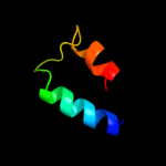
Top model
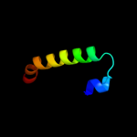
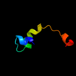
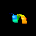
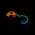
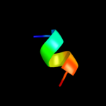
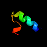
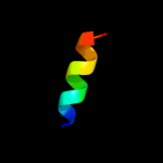
Image coloured by rainbow N → C terminus
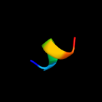
Model (left) based on template c3nkhB_
Top template information
PDB header:recombination
Chain: B: PDB Molecule:integrase;
PDBTitle: crystal structure of integrase from mrsa strain staphylococcus aureus
Confidence and coverage
Confidence: 94.2% Coverage: 39%
36 residues ( 39% of your sequence) have been modelled with 94.2% confidence by the single highest scoring template.
You may wish to submit your sequence to Phyrealarm. This will automatically scan your sequence every week for new potential templates as they appear in the Phyre2 library.
3D viewing
Interactive 3D view in Jmol

Sequence analysis 

Secondary structure and disorder prediction 

   1........10.........20.........30.........40.........50.........60
Sequence  MVVTTSDVVMCQMRRSDVQGGYRVYGSWMAENVQDQVSILNQKLSEFAPSMPHAVRSDVI
Secondary structure 


SS confidence 


Disorder  ?????


???


???


Disorder confidence 


   .........70.........80.........90..
Sequence  NNRLQNLHLHAHHFLIRRHQLITHLNPHLHRN
Secondary structure 


SS confidence 


Disorder 


??????
Disorder confidence 


Confidence Key
High(9)                    Low (0)
?Disordered
Alpha helix
Beta strand

Domain analysis 

Hover over an aligned region to see model and summary info

Please note, only up to the top 20 hits are modelled to reduce computer load

RankAligned region

PDB 3nkh chain B

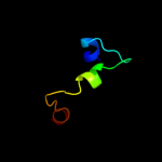
3D model

Region: 7 - 42
Aligned: 36
Modelled: 36
Confidence: 94.2%
Identity: 17%
PDB header:recombination
Chain: B: PDB Molecule:integrase;
PDBTitle: crystal structure of integrase from mrsa strain staphylococcus aureus

Phyre2

PDB 1ma7 chain A

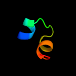
3D model

Region: 7 - 41
Aligned: 34
Modelled: 35
Confidence: 72.7%
Identity: 15%
PDB header:hydrolase, ligase/dna
Chain: A: PDB Molecule:cre recombinase;
PDBTitle: crystal structure of cre site-specific recombinase2 complexed with a mutant dna substrate, loxp-a8/t27

Phyre2

PDB 1f44 chain A domain 2

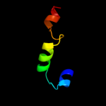
3D model

Region: 7 - 41
Aligned: 34
Modelled: 35
Confidence: 65.5%
Identity: 12%
Fold: DNA breaking-rejoining enzymes
Superfamily: DNA breaking-rejoining enzymes
Family: Lambda integrase-like, catalytic core

Phyre2

PDB 2a3v chain A

3D model

Region: 7 - 25
Aligned: 18
Modelled: 19
Confidence: 38.4%
Identity: 28%
PDB header:recombination
Chain: A: PDB Molecule:site-specific recombinase inti4;
PDBTitle: structural basis for broad dna-specificity in integron2 recombination

Phyre2

PDB 1crx chain A

3D model

Region: 7 - 41
Aligned: 34
Modelled: 35
Confidence: 37.3%
Identity: 15%
PDB header:replication/dna
Chain: A: PDB Molecule:cre recombinase;
PDBTitle: cre recombinase/dna complex reaction intermediate i

Phyre2

PDB 1aih chain A

3D model

Region: 7 - 39
Aligned: 32
Modelled: 33
Confidence: 35.4%
Identity: 13%
Fold: DNA breaking-rejoining enzymes
Superfamily: DNA breaking-rejoining enzymes
Family: Lambda integrase-like, catalytic core

Phyre2

PDB 1wii chain A

3D model

Region: 23 - 31
Aligned: 9
Modelled: 9
Confidence: 20.8%
Identity: 33%
Fold: Rubredoxin-like
Superfamily: Zinc beta-ribbon
Family: Putative zinc binding domain

Phyre2

PDB 1q46 chain A domain 1

3D model

Region: 3 - 24
Aligned: 22
Modelled: 22
Confidence: 13.8%
Identity: 14%
Fold: SAM domain-like
Superfamily: eIF2alpha middle domain-like
Family: eIF2alpha middle domain-like

Phyre2

PDB 1z1b chain A

3D model

Region: 7 - 21
Aligned: 15
Modelled: 15
Confidence: 12.4%
Identity: 0%
PDB header:dna binding protein/dna
Chain: A: PDB Molecule:integrase;
PDBTitle: crystal structure of a lambda integrase dimer bound to a2 coc' core site

Phyre2

PDB 1v54 chain L

3D model

Region: 74 - 81
Aligned: 8
Modelled: 8
Confidence: 10.4%
Identity: 75%
Fold: Single transmembrane helix
Superfamily: Mitochondrial cytochrome c oxidase subunit VIIc (aka VIIIa)
Family: Mitochondrial cytochrome c oxidase subunit VIIc (aka VIIIa)

Phyre2

PDB 1a0p chain A

3D model

Region: 7 - 22
Aligned: 16
Modelled: 16
Confidence: 10.2%
Identity: 19%
PDB header:dna recombination
Chain: A: PDB Molecule:site-specific recombinase xerd;
PDBTitle: site-specific recombinase, xerd

Phyre2

PDB 1p7d chain A

3D model

Region: 7 - 20
Aligned: 14
Modelled: 14
Confidence: 9.6%
Identity: 0%
Fold: DNA breaking-rejoining enzymes
Superfamily: DNA breaking-rejoining enzymes
Family: Lambda integrase-like, catalytic core

Phyre2

PDB 2y69 chain Y

3D model

Region: 74 - 81
Aligned: 8
Modelled: 8
Confidence: 9.5%
Identity: 75%
PDB header:electron transport
Chain: Y: PDB Molecule:cytochrome c oxidase subunit 7c;
PDBTitle: bovine heart cytochrome c oxidase re-refined with molecular2 oxygen

Phyre2

PDB 1gxh chain A

3D model

Region: 38 - 60
Aligned: 23
Modelled: 23
Confidence: 8.9%
Identity: 26%
Fold: Acyl carrier protein-like
Superfamily: Colicin E immunity proteins
Family: Colicin E immunity proteins

Phyre2

PDB 1kzv chain A

3D model

Region: 15 - 26
Aligned: 12
Modelled: 12
Confidence: 8.4%
Identity: 33%
PDB header:viral protein
Chain: A: PDB Molecule:vpr protein;
PDBTitle: structure of human immunodeficiency virus type 1 vpr(34-51)2 peptide in chloroform methanol

Phyre2

PDB 1kzt chain A

3D model

Region: 15 - 26
Aligned: 12
Modelled: 12
Confidence: 8.4%
Identity: 33%
PDB header:viral protein
Chain: A: PDB Molecule:vpr protein;
PDBTitle: structure of human immunodeficiency virus type 1 vpr(34-51)2 peptide in dpc micelle containing aqueous solution

Phyre2

PDB 1kzs chain A

3D model

Region: 15 - 26
Aligned: 12
Modelled: 12
Confidence: 8.4%
Identity: 33%
PDB header:viral protein
Chain: A: PDB Molecule:vpr protein;
PDBTitle: structure of human immunodeficiency virus type 1 vpr(34-51)2 peptide in aqueous tfe solution

Phyre2

PDB 1ytr chain A

3D model

Region: 12 - 28
Aligned: 17
Modelled: 17
Confidence: 7.2%
Identity: 18%
PDB header:antibiotic
Chain: A: PDB Molecule:bacteriocin plantaricin a;
PDBTitle: nmr structure of plantaricin a in dpc micelles, 202 structures

Phyre2

PDB 2hi3 chain A domain 1

3D model

Region: 33 - 64
Aligned: 31
Modelled: 32
Confidence: 7.1%
Identity: 23%
Fold: DNA/RNA-binding 3-helical bundle
Superfamily: Homeodomain-like
Family: Homeodomain

Phyre2

PDB 2k8j chain X

3D model

Region: 22 - 28
Aligned: 7
Modelled: 7
Confidence: 6.7%
Identity: 57%
PDB header:viral protein
Chain: X: PDB Molecule:p7tm2;
PDBTitle: solution structure of hcv p7 tm2

Phyre2
1

c3nkhB_
2

c1ma7A_
3

d1f44a2
4

c2a3vA_
5

c1crxA_
6

d1aiha_
7

d1wiia_
8

d1q46a1
9

c1z1bA_
10

d1v54l_
11

c1a0pA_
12

d1p7da_
13

c2y69Y_
14

d1gxha_
15

c1kzvA_
16

c1kztA_
17

c1kzsA_
18

c1ytrA_
19

d2hi3a1
20

c2k8jX_
21


22


23


24


25


26


27


28


29


30


31


32


33


Detailed template information 

#
Template Alignment Coverage3D Model Confidence
% i.d. Template Information
1c3nkhB_


94.2 17 PDB header:recombination
Chain: B: PDB Molecule:integrase;
PDBTitle: crystal structure of integrase from mrsa strain staphylococcus aureus
2c1ma7A_


72.7 15 PDB header:hydrolase, ligase/dna
Chain: A: PDB Molecule:cre recombinase;
PDBTitle: crystal structure of cre site-specific recombinase2 complexed with a mutant dna substrate, loxp-a8/t27
3d1f44a2


65.5 12 Fold:DNA breaking-rejoining enzymes
Superfamily:DNA breaking-rejoining enzymes
Family:Lambda integrase-like, catalytic core
4c2a3vA_


38.4 28 PDB header:recombination
Chain: A: PDB Molecule:site-specific recombinase inti4;
PDBTitle: structural basis for broad dna-specificity in integron2 recombination
5c1crxA_


37.3 15 PDB header:replication/dna
Chain: A: PDB Molecule:cre recombinase;
PDBTitle: cre recombinase/dna complex reaction intermediate i
6d1aiha_


35.4 13 Fold:DNA breaking-rejoining enzymes
Superfamily:DNA breaking-rejoining enzymes
Family:Lambda integrase-like, catalytic core
7d1wiia_


20.8 33 Fold:Rubredoxin-like
Superfamily:Zinc beta-ribbon
Family:Putative zinc binding domain
8d1q46a1


13.8 14 Fold:SAM domain-like
Superfamily:eIF2alpha middle domain-like
Family:eIF2alpha middle domain-like
9c1z1bA_


12.4 0 PDB header:dna binding protein/dna
Chain: A: PDB Molecule:integrase;
PDBTitle: crystal structure of a lambda integrase dimer bound to a2 coc' core site
10d1v54l_


10.4 75 Fold:Single transmembrane helix
Superfamily:Mitochondrial cytochrome c oxidase subunit VIIc (aka VIIIa)
Family:Mitochondrial cytochrome c oxidase subunit VIIc (aka VIIIa)
11c1a0pA_


10.2 19 PDB header:dna recombination
Chain: A: PDB Molecule:site-specific recombinase xerd;
PDBTitle: site-specific recombinase, xerd
12d1p7da_


9.6 0 Fold:DNA breaking-rejoining enzymes
Superfamily:DNA breaking-rejoining enzymes
Family:Lambda integrase-like, catalytic core
13c2y69Y_


9.5 75 PDB header:electron transport
Chain: Y: PDB Molecule:cytochrome c oxidase subunit 7c;
PDBTitle: bovine heart cytochrome c oxidase re-refined with molecular2 oxygen
14d1gxha_


8.9 26 Fold:Acyl carrier protein-like
Superfamily:Colicin E immunity proteins
Family:Colicin E immunity proteins
15c1kzvA_


8.4 33 PDB header:viral protein
Chain: A: PDB Molecule:vpr protein;
PDBTitle: structure of human immunodeficiency virus type 1 vpr(34-51)2 peptide in chloroform methanol
16c1kztA_


8.4 33 PDB header:viral protein
Chain: A: PDB Molecule:vpr protein;
PDBTitle: structure of human immunodeficiency virus type 1 vpr(34-51)2 peptide in dpc micelle containing aqueous solution
17c1kzsA_


8.4 33 PDB header:viral protein
Chain: A: PDB Molecule:vpr protein;
PDBTitle: structure of human immunodeficiency virus type 1 vpr(34-51)2 peptide in aqueous tfe solution
18c1ytrA_


7.2 18 PDB header:antibiotic
Chain: A: PDB Molecule:bacteriocin plantaricin a;
PDBTitle: nmr structure of plantaricin a in dpc micelles, 202 structures
19d2hi3a1


7.1 23 Fold:DNA/RNA-binding 3-helical bundle
Superfamily:Homeodomain-like
Family:Homeodomain
20c2k8jX_


6.7 57 PDB header:viral protein
Chain: X: PDB Molecule:p7tm2;
PDBTitle: solution structure of hcv p7 tm2
21d2gy9s1


not modelled 6.6 46 Fold:Ribosomal protein S19
Superfamily:Ribosomal protein S19
Family:Ribosomal protein S19
22d1uhsa_


not modelled 6.6 23 Fold:DNA/RNA-binding 3-helical bundle
Superfamily:Homeodomain-like
Family:Homeodomain
23c2wl8D_


not modelled 6.5 15 PDB header:protein transport
Chain: D: PDB Molecule:peroxisomal biogenesis factor 19;
PDBTitle: x-ray crystal structure of pex19p
24c1zd1B_


not modelled 6.0 10 PDB header:transferase
Chain: B: PDB Molecule:sulfotransferase 4a1;
PDBTitle: human sulfortransferase sult4a1
25d1f15b_


not modelled 5.7 35 Fold:Nucleoplasmin-like/VP (viral coat and capsid proteins)
Superfamily:Positive stranded ssRNA viruses
Family:Bromoviridae-like VP
26c2wvmA_


not modelled 5.7 55 PDB header:transferase
Chain: A: PDB Molecule:mannosyl-3-phosphoglycerate synthase;
PDBTitle: h309a mutant of mannosyl-3-phosphoglycerate synthase from2 thermus thermophilus hb27 in complex with3 gdp-alpha-d-mannose and mg(ii)
27d1f06a2


not modelled 5.6 29 Fold:FwdE/GAPDH domain-like
Superfamily:Glyceraldehyde-3-phosphate dehydrogenase-like, C-terminal domain
Family:Dihydrodipicolinate reductase-like
28c1f15C_


not modelled 5.6 35 PDB header:virus
Chain: C: PDB Molecule:coat protein;
PDBTitle: cucumber mosaic virus (strain fny)
29c2l5gA_


not modelled 5.6 29 PDB header:transcription regulator
Chain: A: PDB Molecule:g protein pathway suppressor 2;
PDBTitle: co-ordinates and 1h, 13c and 15n chemical shift assignments for the2 complex of gps2 53-90 and smrt 167-207
30d5csma_


not modelled 5.5 17 Fold:Chorismate mutase II
Superfamily:Chorismate mutase II
Family:Allosteric chorismate mutase
31d1i94m_


not modelled 5.4 9 Fold:S13-like H2TH domain
Superfamily:S13-like H2TH domain
Family:Ribosomal protein S13
32d2uubs1


not modelled 5.2 46 Fold:Ribosomal protein S19
Superfamily:Ribosomal protein S19
Family:Ribosomal protein S19
33d1ayia_


not modelled 5.2 21 Fold:Acyl carrier protein-like
Superfamily:Colicin E immunity proteins
Family:Colicin E immunity proteins

Binding site prediction 

Due to computational demand, binding site predictions are not run for batch jobs

If you want to predict binding sites, please manually submit your model of choice to 3DLigandSite


Phyre is for academic use only

Please cite: Protein structure prediction on the web: a case study using the Phyre server
Kelley LA and Sternberg MJE. Nature Protocols 4, 363 - 371 (2009) [pdf] [Import into BibTeX]
 
If you use the binding site predictions from 3DLigandSite, please also cite:
3DLigandSite: predicting ligand-binding sites using similar structures.
Wass MN, Kelley LA and Sternberg MJ Nucleic Acids Research 38, W469-73 (2010) [PubMed]
 
© Structural Bioinformatics Group
Imperial College London
Lawrence Kelley, Benjamin Jefferys
Disclaimer
Terms and Conditions
Component software
Template detection: HHpred 1.51
Secondary structure prediction: Psi-pred 2.5
Disorder prediction: Disopred 2.4
Transmembrane prediction: Memsat_SVM
Multi-template modelling and ab initio: Poing 1.0