Phyre Home Retrieve Phyre Job Id
Emaill.a.kelley@imperial.ac.uk
DescriptionP0A9H1
DateThu Jan 5 11:10:11 GMT 2012
Unique Job ID0b654156fca3c0dd

Summary 

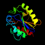
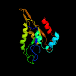
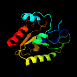
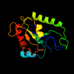
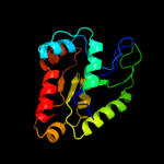
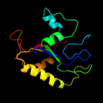
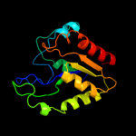
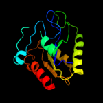
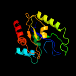
Top model
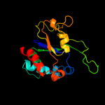
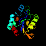
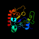
Image coloured by rainbow N → C terminus
Model (left) based on template d1muga_
Top template information
Fold:Uracil-DNA glycosylase-like
Superfamily:Uracil-DNA glycosylase-like
Family:Mug-like
Confidence and coverage
Confidence:100.0% Coverage: 98%
164 residues ( 98% of your sequence) have been modelled with 100.0% confidence by the single highest scoring template.
3D viewing
Interactive 3D view in Jmol

Sequence analysis 

Secondary structure and disorder prediction 

   1........10.........20.........30.........40.........50.........60
Sequence  MVEDILAPGLRVVFCGINPGLSSAGTGFPFAHPANRFWKVIYQAGFTDRQLKPQEAQHLL
Secondary structure 


SS confidence 


Disorder  ?????


???
?


Disorder confidence 


   .........70.........80.........90.........100.........110.........120
Sequence  DYRCGVTKLVDRPTVQANEVSKQELHAGGRKLIEKIEDYQPQALAILGKQAYEQGFSQRG
Secondary structure 


SS confidence 


Disorder 


?????????


Disorder confidence 


   .........130.........140.........150.........160........
Sequence  AQWGKQTLTIGSTQIWVLPNPSGLSRVSLEKLVEAYRELDQALVVRGR
Secondary structure 


SS confidence 


Disorder 


?


????
Disorder confidence 


Confidence Key
High(9)                    Low (0)
?Disordered
Alpha helix
Beta strand

Domain analysis 

Hover over an aligned region to see model and summary info

Please note, only up to the top 20 hits are modelled to reduce computer load

RankAligned region

PDB 1mug chain A

3D model

Region: 1 - 164
Aligned: 164
Modelled: 164
Confidence: 100.0%
Identity: 100%
Fold: Uracil-DNA glycosylase-like
Superfamily: Uracil-DNA glycosylase-like
Family: Mug-like

Phyre2

PDB 2d07 chain A

3D model

Region: 2 - 168
Aligned: 164
Modelled: 167
Confidence: 100.0%
Identity: 33%
PDB header:hydrolase
Chain: A: PDB Molecule:g/t mismatch-specific thymine dna glycosylase;
PDBTitle: crystal structure of sumo-3-modified thymine-dna glycosylase

Phyre2

PDB 2rba chain B

3D model

Region: 2 - 168
Aligned: 167
Modelled: 167
Confidence: 100.0%
Identity: 32%
PDB header:hydrolase/dna
Chain: B: PDB Molecule:g/t mismatch-specific thymine dna glycosylase;
PDBTitle: structure of human thymine dna glycosylase bound to abasic and2 undamaged dna

Phyre2

PDB 2c2p chain A

3D model

Region: 1 - 165
Aligned: 161
Modelled: 165
Confidence: 100.0%
Identity: 29%
PDB header:hydrolase
Chain: A: PDB Molecule:g/u mismatch-specific dna glycosylase;
PDBTitle: the crystal structure of mismatch specific uracil-dna2 glycosylase (mug) from deinococcus radiodurans

Phyre2

PDB 2l3f chain A

3D model

Region: 2 - 163
Aligned: 151
Modelled: 162
Confidence: 100.0%
Identity: 16%
PDB header:structural genomics, unknown function
Chain: A: PDB Molecule:uncharacterized protein;
PDBTitle: solution nmr structure of a putative uracil dna glycosylase from2 methanosarcina acetivorans, northeast structural genomics consortium3 target mvr76

Phyre2

PDB 1vk2 chain A

3D model

Region: 1 - 162
Aligned: 149
Modelled: 149
Confidence: 100.0%
Identity: 23%
Fold: Uracil-DNA glycosylase-like
Superfamily: Uracil-DNA glycosylase-like
Family: Mug-like

Phyre2

PDB 2d3y chain A

3D model

Region: 2 - 162
Aligned: 159
Modelled: 161
Confidence: 99.9%
Identity: 21%
PDB header:hydrolase
Chain: A: PDB Molecule:uracil-dna glycosylase;
PDBTitle: crystal structure of uracil-dna glycosylase from thermus thermophilus2 hb8

Phyre2

PDB 1ui0 chain A

3D model

Region: 1 - 163
Aligned: 150
Modelled: 150
Confidence: 99.9%
Identity: 19%
Fold: Uracil-DNA glycosylase-like
Superfamily: Uracil-DNA glycosylase-like
Family: Mug-like

Phyre2

PDB 3ikb chain B

3D model

Region: 2 - 167
Aligned: 157
Modelled: 164
Confidence: 99.9%
Identity: 12%
PDB header:structural genomics, unknown function
Chain: B: PDB Molecule:uncharacterized conserved protein;
PDBTitle: the structure of a conserved protein from streptococcus2 mutans ua159.

Phyre2

PDB 1oe4 chain A

3D model

Region: 5 - 158
Aligned: 138
Modelled: 148
Confidence: 99.5%
Identity: 21%
Fold: Uracil-DNA glycosylase-like
Superfamily: Uracil-DNA glycosylase-like
Family: Single-strand selective monofunctional uracil-DNA glycosylase SMUG1

Phyre2

PDB 2owr chain D

3D model

Region: 8 - 168
Aligned: 140
Modelled: 161
Confidence: 96.5%
Identity: 19%
PDB header:hydrolase
Chain: D: PDB Molecule:uracil-dna glycosylase;
PDBTitle: crystal structure of vaccinia virus uracil-dna glycosylase

Phyre2

PDB 3cxm chain A

3D model

Region: 8 - 168
Aligned: 152
Modelled: 152
Confidence: 95.8%
Identity: 16%
PDB header:hydrolase
Chain: A: PDB Molecule:uracil-dna glycosylase;
PDBTitle: leishmania naiffi uracil-dna glycosylase in complex with 5-bromouracil

Phyre2

PDB 3tr7 chain A

3D model

Region: 8 - 168
Aligned: 150
Modelled: 158
Confidence: 95.5%
Identity: 19%
PDB header:hydrolase
Chain: A: PDB Molecule:uracil-dna glycosylase;
PDBTitle: structure of a uracil-dna glycosylase (ung) from coxiella burnetii

Phyre2

PDB 3eug chain A

3D model

Region: 9 - 168
Aligned: 150
Modelled: 151
Confidence: 95.3%
Identity: 13%
Fold: Uracil-DNA glycosylase-like
Superfamily: Uracil-DNA glycosylase-like
Family: Uracil-DNA glycosylase

Phyre2

PDB 2hxm chain A domain 1

3D model

Region: 8 - 168
Aligned: 151
Modelled: 160
Confidence: 94.6%
Identity: 11%
Fold: Uracil-DNA glycosylase-like
Superfamily: Uracil-DNA glycosylase-like
Family: Uracil-DNA glycosylase

Phyre2

PDB 2zhx chain G

3D model

Region: 8 - 168
Aligned: 149
Modelled: 161
Confidence: 94.5%
Identity: 17%
PDB header:hydrolase/hydrolase inhibitor
Chain: G: PDB Molecule:uracil-dna glycosylase;
PDBTitle: crystal structure of uracil-dna glycosylase from mycobacterium2 tuberculosis in complex with a proteinaceous inhibitor

Phyre2

PDB 2j8x chain A domain 1

3D model

Region: 9 - 144
Aligned: 127
Modelled: 136
Confidence: 94.0%
Identity: 10%
Fold: Uracil-DNA glycosylase-like
Superfamily: Uracil-DNA glycosylase-like
Family: Uracil-DNA glycosylase

Phyre2

PDB 2boo chain A

3D model

Region: 9 - 168
Aligned: 150
Modelled: 151
Confidence: 93.5%
Identity: 14%
PDB header:hydrolase
Chain: A: PDB Molecule:uracil-dna glycosylase;
PDBTitle: the crystal structure of uracil-dna n-glycosylase (ung)2 from deinococcus radiodurans.

Phyre2

PDB 1okb chain A

3D model

Region: 8 - 168
Aligned: 150
Modelled: 150
Confidence: 93.1%
Identity: 14%
Fold: Uracil-DNA glycosylase-like
Superfamily: Uracil-DNA glycosylase-like
Family: Uracil-DNA glycosylase

Phyre2

PDB 1lau chain E

3D model

Region: 8 - 168
Aligned: 148
Modelled: 150
Confidence: 93.1%
Identity: 17%
Fold: Uracil-DNA glycosylase-like
Superfamily: Uracil-DNA glycosylase-like
Family: Uracil-DNA glycosylase

Phyre2
1

d1muga_
2

c2d07A_
3

c2rbaB_
4

c2c2pA_
5

c2l3fA_
6

d1vk2a_
7

c2d3yA_
8

d1ui0a_
9

c3ikbB_
10

d1oe4a_
11

c2owrD_
12

c3cxmA_
13

c3tr7A_
14

d3euga_
15

d2hxma1
16

c2zhxG_
17

d2j8xa1
18

c2booA_
19

d1okba_
20

d1laue_
21


22


23


24


25


26


27


28


29


30


31


32


33


34


35


36


37


38


Detailed template information 

#
Template Alignment Coverage3D Model Confidence
% i.d. Template Information
1d1muga_


100.0 100 Fold:Uracil-DNA glycosylase-like
Superfamily:Uracil-DNA glycosylase-like
Family:Mug-like
2c2d07A_


100.0 33 PDB header:hydrolase
Chain: A: PDB Molecule:g/t mismatch-specific thymine dna glycosylase;
PDBTitle: crystal structure of sumo-3-modified thymine-dna glycosylase
3c2rbaB_


100.0 32 PDB header:hydrolase/dna
Chain: B: PDB Molecule:g/t mismatch-specific thymine dna glycosylase;
PDBTitle: structure of human thymine dna glycosylase bound to abasic and2 undamaged dna
4c2c2pA_


100.0 29 PDB header:hydrolase
Chain: A: PDB Molecule:g/u mismatch-specific dna glycosylase;
PDBTitle: the crystal structure of mismatch specific uracil-dna2 glycosylase (mug) from deinococcus radiodurans
5c2l3fA_


100.0 16 PDB header:structural genomics, unknown function
Chain: A: PDB Molecule:uncharacterized protein;
PDBTitle: solution nmr structure of a putative uracil dna glycosylase from2 methanosarcina acetivorans, northeast structural genomics consortium3 target mvr76
6d1vk2a_


100.0 23 Fold:Uracil-DNA glycosylase-like
Superfamily:Uracil-DNA glycosylase-like
Family:Mug-like
7c2d3yA_


99.9 21 PDB header:hydrolase
Chain: A: PDB Molecule:uracil-dna glycosylase;
PDBTitle: crystal structure of uracil-dna glycosylase from thermus thermophilus2 hb8
8d1ui0a_


99.9 19 Fold:Uracil-DNA glycosylase-like
Superfamily:Uracil-DNA glycosylase-like
Family:Mug-like
9c3ikbB_


99.9 12 PDB header:structural genomics, unknown function
Chain: B: PDB Molecule:uncharacterized conserved protein;
PDBTitle: the structure of a conserved protein from streptococcus2 mutans ua159.
10d1oe4a_


99.5 21 Fold:Uracil-DNA glycosylase-like
Superfamily:Uracil-DNA glycosylase-like
Family:Single-strand selective monofunctional uracil-DNA glycosylase SMUG1
11c2owrD_


96.5 19 PDB header:hydrolase
Chain: D: PDB Molecule:uracil-dna glycosylase;
PDBTitle: crystal structure of vaccinia virus uracil-dna glycosylase
12c3cxmA_


95.8 16 PDB header:hydrolase
Chain: A: PDB Molecule:uracil-dna glycosylase;
PDBTitle: leishmania naiffi uracil-dna glycosylase in complex with 5-bromouracil
13c3tr7A_


95.5 19 PDB header:hydrolase
Chain: A: PDB Molecule:uracil-dna glycosylase;
PDBTitle: structure of a uracil-dna glycosylase (ung) from coxiella burnetii
14d3euga_


95.3 13 Fold:Uracil-DNA glycosylase-like
Superfamily:Uracil-DNA glycosylase-like
Family:Uracil-DNA glycosylase
15d2hxma1


94.6 11 Fold:Uracil-DNA glycosylase-like
Superfamily:Uracil-DNA glycosylase-like
Family:Uracil-DNA glycosylase
16c2zhxG_


94.5 17 PDB header:hydrolase/hydrolase inhibitor
Chain: G: PDB Molecule:uracil-dna glycosylase;
PDBTitle: crystal structure of uracil-dna glycosylase from mycobacterium2 tuberculosis in complex with a proteinaceous inhibitor
17d2j8xa1


94.0 10 Fold:Uracil-DNA glycosylase-like
Superfamily:Uracil-DNA glycosylase-like
Family:Uracil-DNA glycosylase
18c2booA_


93.5 14 PDB header:hydrolase
Chain: A: PDB Molecule:uracil-dna glycosylase;
PDBTitle: the crystal structure of uracil-dna n-glycosylase (ung)2 from deinococcus radiodurans.
19d1okba_


93.1 14 Fold:Uracil-DNA glycosylase-like
Superfamily:Uracil-DNA glycosylase-like
Family:Uracil-DNA glycosylase
20d1laue_


93.1 17 Fold:Uracil-DNA glycosylase-like
Superfamily:Uracil-DNA glycosylase-like
Family:Uracil-DNA glycosylase
21c3fpjA_


not modelled 25.4 32 PDB header:biosynthetic protein, transferase
Chain: A: PDB Molecule:putative uncharacterized protein;
PDBTitle: crystal structure of e81q mutant of mtnas in complex with s-2 adenosylmethionine
22c3absD_


not modelled 18.2 24 PDB header:lyase
Chain: D: PDB Molecule:ethanolamine ammonia-lyase light chain;
PDBTitle: crystal structure of ethanolamine ammonia-lyase from escherichia coli2 complexed with adeninylpentylcobalamin and ethanolamine
23c1v39A_


not modelled 16.3 35 PDB header:methyltransferase
Chain: A: PDB Molecule:vp39;
PDBTitle: dc26 mutant of vaccinia virus protein vp39 in complex with s-2 adenosylhomocysteine and m7g(5')pppg
24c1vp3A_


not modelled 16.3 35 PDB header:methyltransferase
Chain: A: PDB Molecule:vp39;
PDBTitle: vaccinia virus protein vp39 in complex with s-adenosylhomocysteine
25d1vpta_


not modelled 16.2 32 Fold:S-adenosyl-L-methionine-dependent methyltransferases
Superfamily:S-adenosyl-L-methionine-dependent methyltransferases
Family:mRNA cap methylase
26c3anyB_


not modelled 15.9 24 PDB header:lyase
Chain: B: PDB Molecule:ethanolamine ammonia-lyase light chain;
PDBTitle: crystal structure of ethanolamine ammonia-lyase from escherichia coli2 complexed with cn-cbl and (r)-2-amino-1-propanol
27c2plwA_


not modelled 10.7 24 PDB header:transferase
Chain: A: PDB Molecule:ribosomal rna methyltransferase, putative;
PDBTitle: crystal structure of a ribosomal rna methyltransferase, putative, from2 plasmodium falciparum (pf13_0052).
28c3jqoV_


not modelled 8.8 15 PDB header:transport protein
Chain: V: PDB Molecule:traf protein;
PDBTitle: crystal structure of the outer membrane complex of a type iv2 secretion system
29d2i7pa2


not modelled 8.3 23 Fold:Ribonuclease H-like motif
Superfamily:Actin-like ATPase domain
Family:Fumble-like
30d1iv0a_


not modelled 8.3 8 Fold:Ribonuclease H-like motif
Superfamily:Ribonuclease H-like
Family:Putative Holliday junction resolvase RuvX
31d2cyja1


not modelled 7.6 25 Fold:MTH938-like
Superfamily:MTH938-like
Family:MTH938-like
32c2h4bD_


not modelled 6.7 38 PDB header:de novo protein
Chain: D: PDB Molecule:pancreatic hormone;
PDBTitle: cis-4-aminomethylphenylazobenzoic acid-avian pancreatic2 polypeptide
33c2h4bC_


not modelled 6.7 38 PDB header:de novo protein
Chain: C: PDB Molecule:pancreatic hormone;
PDBTitle: cis-4-aminomethylphenylazobenzoic acid-avian pancreatic2 polypeptide
34c2h3sB_


not modelled 6.7 38 PDB header:de novo protein
Chain: B: PDB Molecule:pancreatic hormone;
PDBTitle: cis-azobenzene-avian pancreatic polypeptide bound to dpc2 micelles
35c2h3tB_


not modelled 6.7 38 PDB header:de novo protein
Chain: B: PDB Molecule:pancreatic hormone;
PDBTitle: trans-(4-aminomethyl)phenylazobenzoic acid-app bound to dpc2 micelles
36d1ihna_


not modelled 6.5 14 Fold:MTH938-like
Superfamily:MTH938-like
Family:MTH938-like
37d1jvaa2


not modelled 5.5 25 Fold:Homing endonuclease-like
Superfamily:Homing endonucleases
Family:Intein endonuclease
38d2q4qa1


not modelled 5.3 15 Fold:MTH938-like
Superfamily:MTH938-like
Family:MTH938-like

Binding site prediction 

Due to computational demand, binding site predictions are not run for batch jobs

If you want to predict binding sites, please manually submit your model of choice to 3DLigandSite


Phyre is for academic use only

Please cite: Protein structure prediction on the web: a case study using the Phyre server
Kelley LA and Sternberg MJE. Nature Protocols 4, 363 - 371 (2009) [pdf] [Import into BibTeX]
 
If you use the binding site predictions from 3DLigandSite, please also cite:
3DLigandSite: predicting ligand-binding sites using similar structures.
Wass MN, Kelley LA and Sternberg MJ Nucleic Acids Research 38, W469-73 (2010) [PubMed]
 
© Structural Bioinformatics Group
Imperial College London
Lawrence Kelley, Benjamin Jefferys
Disclaimer
Terms and Conditions
Component software
Template detection: HHpred 1.51
Secondary structure prediction: Psi-pred 2.5
Disorder prediction: Disopred 2.4
Transmembrane prediction: Memsat_SVM
Multi-template modelling and ab initio: Poing 1.0