Phyre Home Retrieve Phyre Job Id
Emaill.a.kelley@imperial.ac.uk
DescriptionQ02885
DateThu Jan 5 12:33:21 GMT 2012
Unique Job IDffe9c8999323b709

Summary 

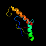
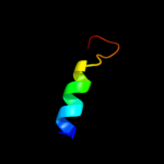
Top model
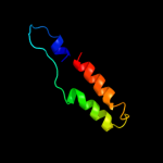
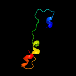
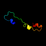
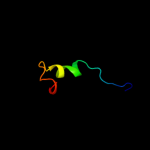
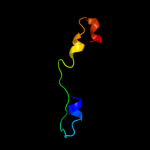
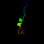
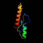
Image coloured by rainbow N → C terminus
Model (left) based on template c3mbfA_
Top template information
PDB header:lyase
Chain: A: PDB Molecule:fructose-bisphosphate aldolase;
PDBTitle: crystal structure of fructose bisphosphate aldolase from2 encephalitozoon cuniculi, bound to fructose 1,6-bisphosphate
Confidence and coverage
Confidence: 79.9% Coverage: 55%
52 residues ( 55% of your sequence) have been modelled with 79.9% confidence by the single highest scoring template.
You may wish to submit your sequence to Phyrealarm. This will automatically scan your sequence every week for new potential templates as they appear in the Phyre2 library.
Please note: You must be registered and logged in to use Phyrealarm.
3D viewing
Interactive 3D view in Jmol

Sequence analysis 

Secondary structure and disorder prediction 

   1........10.........20.........30.........40.........50.........60
Sequence  MSTRNIHVNTASYTLLVAGKKKNTGEEWDVLEFSSLTELKKYRKSHPEKMAFSYSYALSR
Secondary structure 


SS confidence 


Disorder  ??????


?
???????


Disorder confidence 


   .........70.........80.........90.....
Sequence  GVDTQFRHINIAEADHFKQFLRQIKRAGLDIRAIC
Secondary structure 


SS confidence 


Disorder 


??????
Disorder confidence 


Confidence Key
High(9)                    Low (0)
?Disordered
Alpha helix
Beta strand

Domain analysis 

Hover over an aligned region to see model and summary info

Please note, only up to the top 20 hits are modelled to reduce computer load

RankAligned region

PDB 3mbf chain A

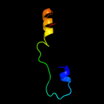
3D model

Region: 36 - 89
Aligned: 52
Modelled: 54
Confidence: 79.9%
Identity: 31%
PDB header:lyase
Chain: A: PDB Molecule:fructose-bisphosphate aldolase;
PDBTitle: crystal structure of fructose bisphosphate aldolase from2 encephalitozoon cuniculi, bound to fructose 1,6-bisphosphate

Phyre2

PDB 3kx6 chain C

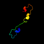
3D model

Region: 36 - 89
Aligned: 51
Modelled: 54
Confidence: 58.1%
Identity: 27%
PDB header:lyase
Chain: C: PDB Molecule:fructose-bisphosphate aldolase;
PDBTitle: crystal structure of fructose-1,6-bisphosphate aldolase from babesia2 bovis at 2.1a resolution

Phyre2

PDB 1zai chain A domain 1

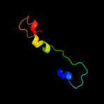
3D model

Region: 36 - 75
Aligned: 40
Modelled: 40
Confidence: 47.2%
Identity: 25%
Fold: TIM beta/alpha-barrel
Superfamily: Aldolase
Family: Class I aldolase

Phyre2

PDB 2itb chain A domain 1

3D model

Region: 43 - 94
Aligned: 52
Modelled: 52
Confidence: 43.3%
Identity: 21%
Fold: Ferritin-like
Superfamily: Ferritin-like
Family: MiaE-like

Phyre2

PDB 1tz9 chain A

3D model

Region: 15 - 91
Aligned: 69
Modelled: 72
Confidence: 39.6%
Identity: 25%
Fold: TIM beta/alpha-barrel
Superfamily: Xylose isomerase-like
Family: UxuA-like

Phyre2

PDB 1qo5 chain B

3D model

Region: 36 - 75
Aligned: 40
Modelled: 40
Confidence: 39.1%
Identity: 23%
Fold: TIM beta/alpha-barrel
Superfamily: Aldolase
Family: Class I aldolase

Phyre2

PDB 1fdj chain A

3D model

Region: 36 - 75
Aligned: 40
Modelled: 40
Confidence: 38.6%
Identity: 23%
Fold: TIM beta/alpha-barrel
Superfamily: Aldolase
Family: Class I aldolase

Phyre2

PDB 1f2j chain A

3D model

Region: 36 - 75
Aligned: 40
Modelled: 40
Confidence: 31.8%
Identity: 20%
Fold: TIM beta/alpha-barrel
Superfamily: Aldolase
Family: Class I aldolase

Phyre2

PDB 2qap chain C

3D model

Region: 36 - 75
Aligned: 40
Modelled: 40
Confidence: 30.5%
Identity: 20%
PDB header:lyase
Chain: C: PDB Molecule:fructose-1,6-bisphosphate aldolase;
PDBTitle: fructose-1,6-bisphosphate aldolase from leishmania mexicana

Phyre2

PDB 2qap chain A domain 1

3D model

Region: 36 - 75
Aligned: 40
Modelled: 40
Confidence: 30.2%
Identity: 20%
Fold: TIM beta/alpha-barrel
Superfamily: Aldolase
Family: Class I aldolase

Phyre2

PDB 3mmt chain C

3D model

Region: 46 - 74
Aligned: 29
Modelled: 29
Confidence: 27.3%
Identity: 34%
PDB header:hydrolase
Chain: C: PDB Molecule:fructose-bisphosphate aldolase;
PDBTitle: crystal structure of fructose bisphosphate aldolase from bartonella2 henselae, bound to fructose bisphosphate

Phyre2

PDB 1xfb chain A domain 1

3D model

Region: 36 - 74
Aligned: 39
Modelled: 39
Confidence: 24.2%
Identity: 26%
Fold: TIM beta/alpha-barrel
Superfamily: Aldolase
Family: Class I aldolase

Phyre2

PDB 2pc4 chain B

3D model

Region: 46 - 74
Aligned: 29
Modelled: 29
Confidence: 22.8%
Identity: 38%
PDB header:lyase
Chain: B: PDB Molecule:fructose-bisphosphate aldolase;
PDBTitle: crystal structure of fructose-bisphosphate aldolase from plasmodium2 falciparum in complex with trap-tail determined at 2.4 angstrom3 resolution

Phyre2

PDB 3bdk chain B

3D model

Region: 15 - 91
Aligned: 69
Modelled: 72
Confidence: 22.2%
Identity: 23%
PDB header:lyase
Chain: B: PDB Molecule:d-mannonate dehydratase;
PDBTitle: crystal structure of streptococcus suis mannonate2 dehydratase complexed with substrate analogue

Phyre2

PDB 1a5c chain A

3D model

Region: 46 - 74
Aligned: 29
Modelled: 29
Confidence: 21.8%
Identity: 38%
Fold: TIM beta/alpha-barrel
Superfamily: Aldolase
Family: Class I aldolase

Phyre2

PDB 1ro5 chain A

3D model

Region: 11 - 94
Aligned: 77
Modelled: 84
Confidence: 21.3%
Identity: 19%
Fold: Acyl-CoA N-acyltransferases (Nat)
Superfamily: Acyl-CoA N-acyltransferases (Nat)
Family: Autoinducer synthetase

Phyre2

PDB 2dt7 chain A

3D model

Region: 33 - 51
Aligned: 19
Modelled: 19
Confidence: 17.7%
Identity: 21%
PDB header:rna binding protein
Chain: A: PDB Molecule:splicing factor 3a subunit 3;
PDBTitle: solution structure of the second surp domain of human2 splicing factor sf3a120 in complex with a fragment of3 human splicing factor sf3a60

Phyre2

PDB 1fba chain A

3D model

Region: 36 - 89
Aligned: 54
Modelled: 54
Confidence: 16.3%
Identity: 22%
Fold: TIM beta/alpha-barrel
Superfamily: Aldolase
Family: Class I aldolase

Phyre2

PDB 2ekc chain A

3D model

Region: 34 - 90
Aligned: 45
Modelled: 57
Confidence: 14.7%
Identity: 22%
PDB header:lyase
Chain: A: PDB Molecule:tryptophan synthase alpha chain;
PDBTitle: structural study of project id aq_1548 from aquifex aeolicus vf5

Phyre2

PDB 2iqt chain A

3D model

Region: 49 - 65
Aligned: 17
Modelled: 17
Confidence: 13.8%
Identity: 29%
PDB header:lyase
Chain: A: PDB Molecule:fructose-bisphosphate aldolase class 1;
PDBTitle: crystal structure of fructose-bisphosphate aldolase, class i from2 porphyromonas gingivalis

Phyre2
1

c3mbfA_
2

c3kx6C_
3

d1zaia1
4

d2itba1
5

d1tz9a_
6

d1qo5b_
7

d1fdja_
8

d1f2ja_
9

c2qapC_
10

d2qapa1
11

c3mmtC_
12

d1xfba1
13

c2pc4B_
14

c3bdkB_
15

d1a5ca_
16

d1ro5a_
17

c2dt7A_
18

d1fbaa_
19

c2ekcA_
20

c2iqtA_
21


22


23


24


25


26


27


28


29


30


31


32


33


34


35


36


37


38


39


40


41


42


43


44


Detailed template information 

#
Template Alignment Coverage3D Model Confidence
% i.d. Template Information
1c3mbfA_


79.9 31 PDB header:lyase
Chain: A: PDB Molecule:fructose-bisphosphate aldolase;
PDBTitle: crystal structure of fructose bisphosphate aldolase from2 encephalitozoon cuniculi, bound to fructose 1,6-bisphosphate
2c3kx6C_


58.1 27 PDB header:lyase
Chain: C: PDB Molecule:fructose-bisphosphate aldolase;
PDBTitle: crystal structure of fructose-1,6-bisphosphate aldolase from babesia2 bovis at 2.1a resolution
3d1zaia1


47.2 25 Fold:TIM beta/alpha-barrel
Superfamily:Aldolase
Family:Class I aldolase
4d2itba1


43.3 21 Fold:Ferritin-like
Superfamily:Ferritin-like
Family:MiaE-like
5d1tz9a_


39.6 25 Fold:TIM beta/alpha-barrel
Superfamily:Xylose isomerase-like
Family:UxuA-like
6d1qo5b_


39.1 23 Fold:TIM beta/alpha-barrel
Superfamily:Aldolase
Family:Class I aldolase
7d1fdja_


38.6 23 Fold:TIM beta/alpha-barrel
Superfamily:Aldolase
Family:Class I aldolase
8d1f2ja_


31.8 20 Fold:TIM beta/alpha-barrel
Superfamily:Aldolase
Family:Class I aldolase
9c2qapC_


30.5 20 PDB header:lyase
Chain: C: PDB Molecule:fructose-1,6-bisphosphate aldolase;
PDBTitle: fructose-1,6-bisphosphate aldolase from leishmania mexicana
10d2qapa1


30.2 20 Fold:TIM beta/alpha-barrel
Superfamily:Aldolase
Family:Class I aldolase
11c3mmtC_


27.3 34 PDB header:hydrolase
Chain: C: PDB Molecule:fructose-bisphosphate aldolase;
PDBTitle: crystal structure of fructose bisphosphate aldolase from bartonella2 henselae, bound to fructose bisphosphate
12d1xfba1


24.2 26 Fold:TIM beta/alpha-barrel
Superfamily:Aldolase
Family:Class I aldolase
13c2pc4B_


22.8 38 PDB header:lyase
Chain: B: PDB Molecule:fructose-bisphosphate aldolase;
PDBTitle: crystal structure of fructose-bisphosphate aldolase from plasmodium2 falciparum in complex with trap-tail determined at 2.4 angstrom3 resolution
14c3bdkB_


22.2 23 PDB header:lyase
Chain: B: PDB Molecule:d-mannonate dehydratase;
PDBTitle: crystal structure of streptococcus suis mannonate2 dehydratase complexed with substrate analogue
15d1a5ca_


21.8 38 Fold:TIM beta/alpha-barrel
Superfamily:Aldolase
Family:Class I aldolase
16d1ro5a_


21.3 19 Fold:Acyl-CoA N-acyltransferases (Nat)
Superfamily:Acyl-CoA N-acyltransferases (Nat)
Family:Autoinducer synthetase
17c2dt7A_


17.7 21 PDB header:rna binding protein
Chain: A: PDB Molecule:splicing factor 3a subunit 3;
PDBTitle: solution structure of the second surp domain of human2 splicing factor sf3a120 in complex with a fragment of3 human splicing factor sf3a60
18d1fbaa_


16.3 22 Fold:TIM beta/alpha-barrel
Superfamily:Aldolase
Family:Class I aldolase
19c2ekcA_


14.7 22 PDB header:lyase
Chain: A: PDB Molecule:tryptophan synthase alpha chain;
PDBTitle: structural study of project id aq_1548 from aquifex aeolicus vf5
20c2iqtA_


13.8 29 PDB header:lyase
Chain: A: PDB Molecule:fructose-bisphosphate aldolase class 1;
PDBTitle: crystal structure of fructose-bisphosphate aldolase, class i from2 porphyromonas gingivalis
21c3q4nA_


not modelled 13.5 16 PDB header:unknown function
Chain: A: PDB Molecule:uncharacterized protein mj0754;
PDBTitle: crystal structure of hypothetical protein mj0754 from methanococcus2 jannaschii dsm 2661
22c2kp7A_


not modelled 12.6 28 PDB header:hydrolase
Chain: A: PDB Molecule:crossover junction endonuclease mus81;
PDBTitle: solution nmr structure of the mus81 n-terminal hhh.2 northeast structural genomics consortium target mmt1a
23c3p2fA_


not modelled 12.2 17 PDB header:signaling protein
Chain: A: PDB Molecule:ahl synthase;
PDBTitle: crystal structure of tofi in an apo form
24d1ofcx1


not modelled 12.1 37 Fold:DNA/RNA-binding 3-helical bundle
Superfamily:Homeodomain-like
Family:Myb/SANT domain
25c3lo3E_


not modelled 10.6 25 PDB header:structure genomics, unknown function
Chain: E: PDB Molecule:uncharacterized conserved protein;
PDBTitle: the crystal structure of a conserved functionally unknown2 protein from colwellia psychrerythraea 34h.
26d1kzfa_


not modelled 10.4 20 Fold:Acyl-CoA N-acyltransferases (Nat)
Superfamily:Acyl-CoA N-acyltransferases (Nat)
Family:Autoinducer synthetase
27c2ktaA_


not modelled 8.6 22 PDB header:hydrolase
Chain: A: PDB Molecule:putative helicase;
PDBTitle: solution nmr structure of a domain of protein a6ky75 from bacteroides2 vulgatus, northeast structural genomics target bvr106a
28d1udxa3


not modelled 8.1 13 Fold:Obg GTP-binding protein C-terminal domain
Superfamily:Obg GTP-binding protein C-terminal domain
Family:Obg GTP-binding protein C-terminal domain
29d2cpya1


not modelled 8.0 27 Fold:Ferredoxin-like
Superfamily:RNA-binding domain, RBD
Family:Canonical RBD
30c3cz8A_


not modelled 7.9 19 PDB header:hydrolase
Chain: A: PDB Molecule:putative sporulation-specific glycosylase ydhd;
PDBTitle: crystal structure of putative sporulation-specific glycosylase ydhd2 from bacillus subtilis
31d1wapa_


not modelled 7.7 64 Fold:Double-stranded beta-helix
Superfamily:TRAP-like
Family:Trp RNA-binding attenuation protein (TRAP)
32d1gtfa_


not modelled 7.5 64 Fold:Double-stranded beta-helix
Superfamily:TRAP-like
Family:Trp RNA-binding attenuation protein (TRAP)
33d1e0ba_


not modelled 7.5 45 Fold:SH3-like barrel
Superfamily:Chromo domain-like
Family:Chromo domain
34d1iapa_


not modelled 6.9 21 Fold:Regulator of G-protein signaling, RGS
Superfamily:Regulator of G-protein signaling, RGS
Family:Regulator of G-protein signaling, RGS
35c3qpiA_


not modelled 6.7 21 PDB header:oxidoreductase
Chain: A: PDB Molecule:chlorite dismutase;
PDBTitle: crystal structure of dimeric chlorite dismutases from nitrobacter2 winogradskyi
36d1q4ra_


not modelled 6.7 26 Fold:Ferredoxin-like
Superfamily:Dimeric alpha+beta barrel
Family:Plant stress-induced protein
37c1y7xA_


not modelled 6.5 57 PDB header:structural protein, protein binding
Chain: A: PDB Molecule:major vault protein;
PDBTitle: solution structure of a two-repeat fragment of major vault2 protein
38d1usta_


not modelled 6.3 29 Fold:DNA/RNA-binding 3-helical bundle
Superfamily:"Winged helix" DNA-binding domain
Family:Linker histone H1/H5
39c2rh0B_


not modelled 6.3 24 PDB header:nuclear protein
Chain: B: PDB Molecule:nudc domain-containing protein 2;
PDBTitle: crystal structure of nudc domain-containing protein 22 (13542905) from mus musculus at 1.95 a resolution
40c3cxnB_


not modelled 5.9 21 PDB header:chaperone
Chain: B: PDB Molecule:urease accessory protein uref;
PDBTitle: structure of the urease accessory protein uref from helicobacter2 pylori
41d2nt0a2


not modelled 5.9 29 Fold:TIM beta/alpha-barrel
Superfamily:(Trans)glycosidases
Family:beta-glycanases
42c2rqpA_


not modelled 5.5 23 PDB header:gene regulation
Chain: A: PDB Molecule:heterochromatin protein 1-binding protein 3;
PDBTitle: the solution structure of heterochromatin protein 1-binding2 protein 74 histone h1 like domain
43c1u6tA_


not modelled 5.4 14 PDB header:protein binding, signaling protein
Chain: A: PDB Molecule:sh3 domain-binding glutamic acid-rich-like
PDBTitle: crystal structure of the human sh3 binding glutamic-rich2 protein like
44c2qr4B_


not modelled 5.3 14 PDB header:hydrolase
Chain: B: PDB Molecule:peptidase m3b, oligoendopeptidase f;
PDBTitle: crystal structure of oligoendopeptidase-f from enterococcus faecium

Binding site prediction 

Due to computational demand, binding site predictions are not run for batch jobs

If you want to predict binding sites, please manually submit your model of choice to 3DLigandSite


Phyre is for academic use only

Please cite: Protein structure prediction on the web: a case study using the Phyre server
Kelley LA and Sternberg MJE. Nature Protocols 4, 363 - 371 (2009) [pdf] [Import into BibTeX]
 
If you use the binding site predictions from 3DLigandSite, please also cite:
3DLigandSite: predicting ligand-binding sites using similar structures.
Wass MN, Kelley LA and Sternberg MJ Nucleic Acids Research 38, W469-73 (2010) [PubMed]
 
© Structural Bioinformatics Group
Imperial College London
Lawrence Kelley, Benjamin Jefferys
Disclaimer
Terms and Conditions
Component software
Template detection: HHpred 1.51
Secondary structure prediction: Psi-pred 2.5
Disorder prediction: Disopred 2.4
Transmembrane prediction: Memsat_SVM
Multi-template modelling and ab initio: Poing 1.0