Phyre Home Retrieve Phyre Job Id
Emaill.a.kelley@imperial.ac.uk
DescriptionQ9JMS6
DateThu Jan 5 12:38:02 GMT 2012
Unique Job IDfacdcc9597abd74e

Summary 

Top model
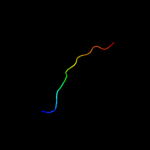
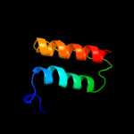
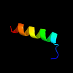
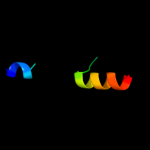
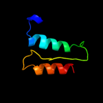
Image coloured by rainbow N → C terminus
Model (left) based on template c3nm7C_
Top template information
PDB header:nucleic acid binding protein
Chain: C: PDB Molecule:uncharacterized protein;
PDBTitle: crystal structure of borrelia burgdorferi pur-alpha
Confidence and coverage
Confidence: 12.5% Coverage: 17%
13 residues ( 17% of your sequence) have been modelled with 12.5% confidence by the single highest scoring template.
You may wish to submit your sequence to Phyrealarm. This will automatically scan your sequence every week for new potential templates as they appear in the Phyre2 library.
Please note: You must be registered and logged in to use Phyrealarm.
3D viewing
Interactive 3D view in Jmol

Sequence analysis 

Secondary structure and disorder prediction 

   1........10.........20.........30.........40.........50.........60
Sequence  MVRYHGEFTYYVYQQSGRYFFCKKLTKKRDTSNCNHLHIIRELSFNEDELELIDFSTDGL
Secondary structure 


SS confidence 


Disorder  ??????


???????


Disorder confidence 


   .........70.......
Sequence  DANDKEIIRGMIDELKK
Secondary structure 

SS confidence 


Disorder 


??
Disorder confidence 


Confidence Key
High(9)                    Low (0)
?Disordered
Alpha helix
Beta strand

Domain analysis 

Hover over an aligned region to see model and summary info

Please note, only up to the top 20 hits are modelled to reduce computer load

RankAligned region

PDB 3nm7 chain C

3D model

Region: 6 - 18
Aligned: 13
Modelled: 13
Confidence: 12.5%
Identity: 31%
PDB header:nucleic acid binding protein
Chain: C: PDB Molecule:uncharacterized protein;
PDBTitle: crystal structure of borrelia burgdorferi pur-alpha

Phyre2

PDB 3nm7 chain D

3D model

Region: 6 - 18
Aligned: 13
Modelled: 13
Confidence: 12.0%
Identity: 31%
PDB header:nucleic acid binding protein
Chain: D: PDB Molecule:uncharacterized protein;
PDBTitle: crystal structure of borrelia burgdorferi pur-alpha

Phyre2

PDB 3n8b chain A

3D model

Region: 6 - 18
Aligned: 13
Modelled: 13
Confidence: 12.0%
Identity: 31%
PDB header:nucleic acid binding protein
Chain: A: PDB Molecule:uncharacterized protein;
PDBTitle: crystal structure of borrelia burgdorferi pur-alpha

Phyre2

PDB 3n8b chain B

3D model

Region: 6 - 18
Aligned: 13
Modelled: 13
Confidence: 11.8%
Identity: 31%
PDB header:nucleic acid binding protein
Chain: B: PDB Molecule:uncharacterized protein;
PDBTitle: crystal structure of borrelia burgdorferi pur-alpha

Phyre2

PDB 2pcj chain B

3D model

Region: 23 - 77
Aligned: 53
Modelled: 55
Confidence: 11.4%
Identity: 17%
PDB header:hydrolase
Chain: B: PDB Molecule:lipoprotein-releasing system atp-binding protein lold;
PDBTitle: crystal structure of abc transporter (aq_297) from aquifex aeolicus2 vf5

Phyre2

PDB 1bjt chain A

3D model

Region: 51 - 73
Aligned: 23
Modelled: 23
Confidence: 11.2%
Identity: 35%
PDB header:topoisomerase
Chain: A: PDB Molecule:topoisomerase ii;
PDBTitle: topoisomerase ii residues 409-1201

Phyre2

PDB 1bjt chain A

3D model

Region: 51 - 73
Aligned: 23
Modelled: 23
Confidence: 11.2%
Identity: 35%
Fold: Type II DNA topoisomerase
Superfamily: Type II DNA topoisomerase
Family: Type II DNA topoisomerase

Phyre2

PDB 1l4d chain B

3D model

Region: 38 - 58
Aligned: 21
Modelled: 21
Confidence: 11.1%
Identity: 38%
Fold: beta-Grasp (ubiquitin-like)
Superfamily: Staphylokinase/streptokinase
Family: Staphylokinase/streptokinase

Phyre2

PDB 1uw0 chain A

3D model

Region: 58 - 77
Aligned: 20
Modelled: 20
Confidence: 11.0%
Identity: 35%
Fold: Glucocorticoid receptor-like (DNA-binding domain)
Superfamily: Glucocorticoid receptor-like (DNA-binding domain)
Family: PARP-type zinc finger

Phyre2

PDB 1l2t chain A

3D model

Region: 39 - 77
Aligned: 39
Modelled: 39
Confidence: 10.1%
Identity: 26%
Fold: P-loop containing nucleoside triphosphate hydrolases
Superfamily: P-loop containing nucleoside triphosphate hydrolases
Family: ABC transporter ATPase domain-like

Phyre2

PDB 1bml chain C domain 1

3D model

Region: 44 - 58
Aligned: 15
Modelled: 15
Confidence: 10.0%
Identity: 33%
Fold: beta-Grasp (ubiquitin-like)
Superfamily: Staphylokinase/streptokinase
Family: Staphylokinase/streptokinase

Phyre2

PDB 3gkx chain B

3D model

Region: 32 - 71
Aligned: 40
Modelled: 40
Confidence: 10.0%
Identity: 13%
PDB header:structural genomics, unknown function
Chain: B: PDB Molecule:putative arsc family related protein;
PDBTitle: crystal structure of putative arsc family related protein from2 bacteroides fragilis

Phyre2

PDB 3qx3 chain B

3D model

Region: 51 - 73
Aligned: 23
Modelled: 23
Confidence: 10.0%
Identity: 30%
PDB header:isomerase/dna/isomerase inhibitor
Chain: B: PDB Molecule:dna topoisomerase 2-beta;
PDBTitle: human topoisomerase iibeta in complex with dna and etoposide

Phyre2

PDB 1v43 chain A domain 3

3D model

Region: 23 - 76
Aligned: 52
Modelled: 54
Confidence: 9.6%
Identity: 19%
Fold: P-loop containing nucleoside triphosphate hydrolases
Superfamily: P-loop containing nucleoside triphosphate hydrolases
Family: ABC transporter ATPase domain-like

Phyre2

PDB 1l4z chain B

3D model

Region: 38 - 58
Aligned: 21
Modelled: 21
Confidence: 9.5%
Identity: 38%
Fold: beta-Grasp (ubiquitin-like)
Superfamily: Staphylokinase/streptokinase
Family: Staphylokinase/streptokinase

Phyre2

PDB 2fok chain A domain 4

3D model

Region: 50 - 70
Aligned: 21
Modelled: 21
Confidence: 8.7%
Identity: 19%
Fold: Restriction endonuclease-like
Superfamily: Restriction endonuclease-like
Family: Restriction endonuclease FokI, C-terminal (catalytic) domain

Phyre2

PDB 1sgw chain A

3D model

Region: 23 - 77
Aligned: 53
Modelled: 55
Confidence: 8.6%
Identity: 21%
Fold: P-loop containing nucleoside triphosphate hydrolases
Superfamily: P-loop containing nucleoside triphosphate hydrolases
Family: ABC transporter ATPase domain-like

Phyre2

PDB 3nym chain A

3D model

Region: 38 - 77
Aligned: 40
Modelled: 37
Confidence: 8.2%
Identity: 20%
PDB header:structural genomics, unknown function
Chain: A: PDB Molecule:uncharacterized protein;
PDBTitle: the crystal structure of functionally unknown protein from neisseria2 meningitidis mc58

Phyre2

PDB 2wsh chain C

3D model

Region: 10 - 53
Aligned: 44
Modelled: 44
Confidence: 8.0%
Identity: 14%
PDB header:hydrolase
Chain: C: PDB Molecule:endonuclease ii;
PDBTitle: structure of bacteriophage t4 endoii e118a mutant

Phyre2

PDB 3d31 chain A domain 2

3D model

Region: 40 - 77
Aligned: 38
Modelled: 38
Confidence: 7.8%
Identity: 24%
Fold: P-loop containing nucleoside triphosphate hydrolases
Superfamily: P-loop containing nucleoside triphosphate hydrolases
Family: ABC transporter ATPase domain-like

Phyre2
1

c3nm7C_
2

c3nm7D_
3

c3n8bA_
4

c3n8bB_
5

c2pcjB_
6

c1bjtA_
7

d1bjta_
8

d1l4db_
9

d1uw0a_
10

d1l2ta_
11

d1bmlc1
12

c3gkxB_
13

c3qx3B_
14

d1v43a3
15

d1l4zb_
16

d2foka4
17

d1sgwa_
18

c3nymA_
19

c2wshC_
20

d3d31a2
21


22


23


24


25


26


27


28


Detailed template information 

#
Template Alignment Coverage3D Model Confidence
% i.d. Template Information
1c3nm7C_


12.5 31 PDB header:nucleic acid binding protein
Chain: C: PDB Molecule:uncharacterized protein;
PDBTitle: crystal structure of borrelia burgdorferi pur-alpha
2c3nm7D_


12.0 31 PDB header:nucleic acid binding protein
Chain: D: PDB Molecule:uncharacterized protein;
PDBTitle: crystal structure of borrelia burgdorferi pur-alpha
3c3n8bA_


12.0 31 PDB header:nucleic acid binding protein
Chain: A: PDB Molecule:uncharacterized protein;
PDBTitle: crystal structure of borrelia burgdorferi pur-alpha
4c3n8bB_


11.8 31 PDB header:nucleic acid binding protein
Chain: B: PDB Molecule:uncharacterized protein;
PDBTitle: crystal structure of borrelia burgdorferi pur-alpha
5c2pcjB_


11.4 17 PDB header:hydrolase
Chain: B: PDB Molecule:lipoprotein-releasing system atp-binding protein lold;
PDBTitle: crystal structure of abc transporter (aq_297) from aquifex aeolicus2 vf5
6c1bjtA_


11.2 35 PDB header:topoisomerase
Chain: A: PDB Molecule:topoisomerase ii;
PDBTitle: topoisomerase ii residues 409-1201
7d1bjta_


11.2 35 Fold:Type II DNA topoisomerase
Superfamily:Type II DNA topoisomerase
Family:Type II DNA topoisomerase
8d1l4db_


11.1 38 Fold:beta-Grasp (ubiquitin-like)
Superfamily:Staphylokinase/streptokinase
Family:Staphylokinase/streptokinase
9d1uw0a_


11.0 35 Fold:Glucocorticoid receptor-like (DNA-binding domain)
Superfamily:Glucocorticoid receptor-like (DNA-binding domain)
Family:PARP-type zinc finger
10d1l2ta_


10.1 26 Fold:P-loop containing nucleoside triphosphate hydrolases
Superfamily:P-loop containing nucleoside triphosphate hydrolases
Family:ABC transporter ATPase domain-like
11d1bmlc1


10.0 33 Fold:beta-Grasp (ubiquitin-like)
Superfamily:Staphylokinase/streptokinase
Family:Staphylokinase/streptokinase
12c3gkxB_


10.0 13 PDB header:structural genomics, unknown function
Chain: B: PDB Molecule:putative arsc family related protein;
PDBTitle: crystal structure of putative arsc family related protein from2 bacteroides fragilis
13c3qx3B_


10.0 30 PDB header:isomerase/dna/isomerase inhibitor
Chain: B: PDB Molecule:dna topoisomerase 2-beta;
PDBTitle: human topoisomerase iibeta in complex with dna and etoposide
14d1v43a3


9.6 19 Fold:P-loop containing nucleoside triphosphate hydrolases
Superfamily:P-loop containing nucleoside triphosphate hydrolases
Family:ABC transporter ATPase domain-like
15d1l4zb_


9.5 38 Fold:beta-Grasp (ubiquitin-like)
Superfamily:Staphylokinase/streptokinase
Family:Staphylokinase/streptokinase
16d2foka4


8.7 19 Fold:Restriction endonuclease-like
Superfamily:Restriction endonuclease-like
Family:Restriction endonuclease FokI, C-terminal (catalytic) domain
17d1sgwa_


8.6 21 Fold:P-loop containing nucleoside triphosphate hydrolases
Superfamily:P-loop containing nucleoside triphosphate hydrolases
Family:ABC transporter ATPase domain-like
18c3nymA_


8.2 20 PDB header:structural genomics, unknown function
Chain: A: PDB Molecule:uncharacterized protein;
PDBTitle: the crystal structure of functionally unknown protein from neisseria2 meningitidis mc58
19c2wshC_


8.0 14 PDB header:hydrolase
Chain: C: PDB Molecule:endonuclease ii;
PDBTitle: structure of bacteriophage t4 endoii e118a mutant
20d3d31a2


7.8 24 Fold:P-loop containing nucleoside triphosphate hydrolases
Superfamily:P-loop containing nucleoside triphosphate hydrolases
Family:ABC transporter ATPase domain-like
21c2cs2A_


not modelled 6.8 25 PDB header:transferase
Chain: A: PDB Molecule:poly [adp-ribose] polymerase-1;
PDBTitle: solution structure of the second zn-finger domain of2 poly(adp-ribose) polymerase-1
22c1s2xA_


not modelled 6.5 35 PDB header:unknown function
Chain: A: PDB Molecule:cag-z;
PDBTitle: crystal structure of cag-z from helicobacter pylori
23d1s2xa_


not modelled 6.5 35 Fold:STAT-like
Superfamily:Cag-Z
Family:Cag-Z
24d1vpla_


not modelled 6.3 21 Fold:P-loop containing nucleoside triphosphate hydrolases
Superfamily:P-loop containing nucleoside triphosphate hydrolases
Family:ABC transporter ATPase domain-like
25d1o8ba1


not modelled 6.3 21 Fold:NagB/RpiA/CoA transferase-like
Superfamily:NagB/RpiA/CoA transferase-like
Family:D-ribose-5-phosphate isomerase (RpiA), catalytic domain
26d1ueba2


not modelled 6.0 27 Fold:OB-fold
Superfamily:Nucleic acid-binding proteins
Family:Cold shock DNA-binding domain-like
27c3rnvA_


not modelled 5.6 36 PDB header:hydrolase
Chain: A: PDB Molecule:helper component proteinase;
PDBTitle: structure of the autocatalytic cysteine protease domain of potyvirus2 helper-component proteinase
28d1uura1


not modelled 5.5 17 Fold:STAT-like
Superfamily:STAT
Family:STAT

Binding site prediction 

Due to computational demand, binding site predictions are not run for batch jobs

If you want to predict binding sites, please manually submit your model of choice to 3DLigandSite


Phyre is for academic use only

Please cite: Protein structure prediction on the web: a case study using the Phyre server
Kelley LA and Sternberg MJE. Nature Protocols 4, 363 - 371 (2009) [pdf] [Import into BibTeX]
 
If you use the binding site predictions from 3DLigandSite, please also cite:
3DLigandSite: predicting ligand-binding sites using similar structures.
Wass MN, Kelley LA and Sternberg MJ Nucleic Acids Research 38, W469-73 (2010) [PubMed]
 
© Structural Bioinformatics Group
Imperial College London
Lawrence Kelley, Benjamin Jefferys
Disclaimer
Terms and Conditions
Component software
Template detection: HHpred 1.51
Secondary structure prediction: Psi-pred 2.5
Disorder prediction: Disopred 2.4
Transmembrane prediction: Memsat_SVM
Multi-template modelling and ab initio: Poing 1.0