Phyre Home Retrieve Phyre Job Id
Emaill.a.kelley@imperial.ac.uk
DescriptionQ47152
DateThu Jan 5 12:36:27 GMT 2012
Unique Job IDf9e8c21fa367e305

Summary 

Top model
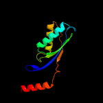
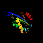
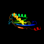
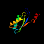
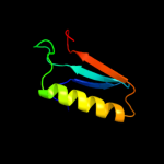
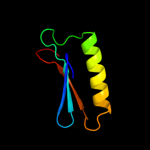
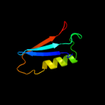
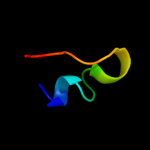
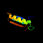
Image coloured by rainbow N → C terminus
Model (left) based on template d2f5ga1
Top template information
Fold:Ferredoxin-like
Superfamily:Transposase IS200-like
Family:Transposase IS200-like
Confidence and coverage
Confidence: 99.9% Coverage: 73%
120 residues ( 73% of your sequence) have been modelled with 99.9% confidence by the single highest scoring template.
3D viewing
Interactive 3D view in Jmol

Sequence analysis 

Secondary structure and disorder prediction 

   1........10.........20.........30.........40.........50.........60
Sequence  MSEYRRYYIKGGTWFFTVNLRNRRSQLLTTQYQMLRHAIIKVKRDRPFEINAWVVLPEHM
Secondary structure 


SS confidence 


Disorder  ???????


Disorder confidence 


   .........70.........80.........90.........100.........110.........120
Sequence  HCIWTLPEGDDDFSSRWREIKKQFTHACGLKNIWQPRFWEHAIRNTKDYRHHVDYIYINP
Secondary structure 


SS confidence 


Disorder 


????


Disorder confidence 


   .........130.........140.........150.........160.....
Sequence  VKHGWVKQVSDWPFSTFHRDVARGLYPIDWAGDVTDFSAGERIIS
Secondary structure 


SS confidence 


Disorder 


??????????
Disorder confidence 


Confidence Key
High(9)                    Low (0)
?Disordered
Alpha helix
Beta strand

Domain analysis 

Hover over an aligned region to see model and summary info

Please note, only up to the top 20 hits are modelled to reduce computer load

RankAligned region

PDB 2f5g chain A domain 1

3D model

Region: 3 - 124
Aligned: 120
Modelled: 122
Confidence: 99.9%
Identity: 18%
Fold: Ferredoxin-like
Superfamily: Transposase IS200-like
Family: Transposase IS200-like

Phyre2

PDB 2fyx chain A domain 1

3D model

Region: 3 - 118
Aligned: 114
Modelled: 116
Confidence: 99.9%
Identity: 15%
Fold: Ferredoxin-like
Superfamily: Transposase IS200-like
Family: Transposase IS200-like

Phyre2

PDB 2vhg chain B

3D model

Region: 1 - 118
Aligned: 116
Modelled: 118
Confidence: 99.9%
Identity: 14%
PDB header:dna-binding protein
Chain: B: PDB Molecule:transposase orfa;
PDBTitle: crystal structure of the ishp608 transposase in complex2 with right end 31-mer dna

Phyre2

PDB 2vjv chain A domain 1

3D model

Region: 1 - 118
Aligned: 116
Modelled: 118
Confidence: 99.9%
Identity: 14%
Fold: Ferredoxin-like
Superfamily: Transposase IS200-like
Family: Transposase IS200-like

Phyre2

PDB 1nzy chain A

3D model

Region: 1 - 60
Aligned: 60
Modelled: 60
Confidence: 19.2%
Identity: 17%
Fold: ClpP/crotonase
Superfamily: ClpP/crotonase
Family: Crotonase-like

Phyre2

PDB 2ej5 chain B

3D model

Region: 1 - 60
Aligned: 60
Modelled: 60
Confidence: 18.3%
Identity: 17%
PDB header:lyase
Chain: B: PDB Molecule:enoyl-coa hydratase subunit ii;
PDBTitle: crystal structure of gk2038 protein (enoyl-coa hydratase subunit ii)2 from geobacillus kaustophilus

Phyre2

PDB 1qgv chain A

3D model

Region: 103 - 132
Aligned: 30
Modelled: 30
Confidence: 17.3%
Identity: 17%
Fold: Thioredoxin fold
Superfamily: Thioredoxin-like
Family: spliceosomal protein U5-15Kd

Phyre2

PDB 2hld chain H

3D model

Region: 52 - 69
Aligned: 18
Modelled: 18
Confidence: 15.6%
Identity: 28%
PDB header:hydrolase
Chain: H: PDB Molecule:atp synthase delta chain, mitochondrial;
PDBTitle: crystal structure of yeast mitochondrial f1-atpase

Phyre2

PDB 2vx2 chain D

3D model

Region: 2 - 60
Aligned: 59
Modelled: 59
Confidence: 13.7%
Identity: 8%
PDB header:isomerase
Chain: D: PDB Molecule:enoyl-coa hydratase domain-containing protein 3;
PDBTitle: crystal structure of human enoyl coenzyme a hydratase2 domain-containing protein 3 (echdc3)

Phyre2

PDB 2al3 chain A domain 1

3D model

Region: 16 - 56
Aligned: 41
Modelled: 41
Confidence: 13.4%
Identity: 15%
Fold: beta-Grasp (ubiquitin-like)
Superfamily: Ubiquitin-like
Family: UBX domain

Phyre2

PDB 2rq7 chain A

3D model

Region: 53 - 69
Aligned: 17
Modelled: 17
Confidence: 12.9%
Identity: 24%
PDB header:hydrolase
Chain: A: PDB Molecule:atp synthase epsilon chain;
PDBTitle: solution structure of the epsilon subunit chimera combining2 the n-terminal beta-sandwich domain from t. elongatus bp-13 f1 and the c-terminal alpha-helical domain from spinach4 chloroplast f1

Phyre2

PDB 1fs0 chain E

3D model

Region: 52 - 69
Aligned: 18
Modelled: 18
Confidence: 11.8%
Identity: 17%
PDB header:hydrolase
Chain: E: PDB Molecule:atp synthase epsilon subunit;
PDBTitle: complex of gamma/epsilon atp synthase from e.coli

Phyre2

PDB 1y0u chain A

3D model

Region: 107 - 131
Aligned: 25
Modelled: 25
Confidence: 11.5%
Identity: 20%
Fold: DNA/RNA-binding 3-helical bundle
Superfamily: "Winged helix" DNA-binding domain
Family: ArsR-like transcriptional regulators

Phyre2

PDB 3eug chain A

3D model

Region: 134 - 158
Aligned: 25
Modelled: 25
Confidence: 9.7%
Identity: 28%
Fold: Uracil-DNA glycosylase-like
Superfamily: Uracil-DNA glycosylase-like
Family: Uracil-DNA glycosylase

Phyre2

PDB 3i0u chain A

3D model

Region: 109 - 138
Aligned: 30
Modelled: 30
Confidence: 9.5%
Identity: 13%
PDB header:lyase
Chain: A: PDB Molecule:phosphothreonine lyase ospf;
PDBTitle: structure of the type iii effector/phosphothreonine lyase ospf from2 shigella flexneri

Phyre2

PDB 2x3g chain A

3D model

Region: 12 - 76
Aligned: 63
Modelled: 65
Confidence: 9.4%
Identity: 14%
PDB header:viral protein
Chain: A: PDB Molecule:sirv1 hypothetical protein orf119;
PDBTitle: crystal structure of the hypothetical protein orf119 from2 sulfolobus islandicus rod-shaped virus 1

Phyre2

PDB 1nvm chain A domain 1

3D model

Region: 134 - 149
Aligned: 16
Modelled: 16
Confidence: 9.0%
Identity: 31%
Fold: RuvA C-terminal domain-like
Superfamily: post-HMGL domain-like
Family: DmpG/LeuA communication domain-like

Phyre2

PDB 3tr7 chain A

3D model

Region: 134 - 151
Aligned: 18
Modelled: 18
Confidence: 8.9%
Identity: 39%
PDB header:hydrolase
Chain: A: PDB Molecule:uracil-dna glycosylase;
PDBTitle: structure of a uracil-dna glycosylase (ung) from coxiella burnetii

Phyre2

PDB 3trr chain A

3D model

Region: 1 - 60
Aligned: 60
Modelled: 60
Confidence: 7.9%
Identity: 12%
PDB header:isomerase
Chain: A: PDB Molecule:probable enoyl-coa hydratase/isomerase;
PDBTitle: crystal structure of a probable enoyl-coa hydratase/isomerase from2 mycobacterium abscessus

Phyre2

PDB 1aqt chain A domain 2

3D model

Region: 52 - 69
Aligned: 18
Modelled: 18
Confidence: 7.6%
Identity: 17%
Fold: Epsilon subunit of F1F0-ATP synthase N-terminal domain
Superfamily: Epsilon subunit of F1F0-ATP synthase N-terminal domain
Family: Epsilon subunit of F1F0-ATP synthase N-terminal domain

Phyre2
1

d2f5ga1
2

d2fyxa1
3

c2vhgB_
4

d2vjva1
5

d1nzya_
6

c2ej5B_
7

d1qgva_
8

c2hldH_
9

c2vx2D_
10

d2al3a1
11

c2rq7A_
12

c1fs0E_
13

d1y0ua_
14

d3euga_
15

c3i0uA_
16

c2x3gA_
17

d1nvma1
18

c3tr7A_
19

c3trrA_
20

d1aqta2
21


22


23


24


25


26


27


28


29


Detailed template information 

#
Template Alignment Coverage3D Model Confidence
% i.d. Template Information
1d2f5ga1


99.9 18 Fold:Ferredoxin-like
Superfamily:Transposase IS200-like
Family:Transposase IS200-like
2d2fyxa1


99.9 15 Fold:Ferredoxin-like
Superfamily:Transposase IS200-like
Family:Transposase IS200-like
3c2vhgB_


99.9 14 PDB header:dna-binding protein
Chain: B: PDB Molecule:transposase orfa;
PDBTitle: crystal structure of the ishp608 transposase in complex2 with right end 31-mer dna
4d2vjva1


99.9 14 Fold:Ferredoxin-like
Superfamily:Transposase IS200-like
Family:Transposase IS200-like
5d1nzya_


19.2 17 Fold:ClpP/crotonase
Superfamily:ClpP/crotonase
Family:Crotonase-like
6c2ej5B_


18.3 17 PDB header:lyase
Chain: B: PDB Molecule:enoyl-coa hydratase subunit ii;
PDBTitle: crystal structure of gk2038 protein (enoyl-coa hydratase subunit ii)2 from geobacillus kaustophilus
7d1qgva_


17.3 17 Fold:Thioredoxin fold
Superfamily:Thioredoxin-like
Family:spliceosomal protein U5-15Kd
8c2hldH_


15.6 28 PDB header:hydrolase
Chain: H: PDB Molecule:atp synthase delta chain, mitochondrial;
PDBTitle: crystal structure of yeast mitochondrial f1-atpase
9c2vx2D_


13.7 8 PDB header:isomerase
Chain: D: PDB Molecule:enoyl-coa hydratase domain-containing protein 3;
PDBTitle: crystal structure of human enoyl coenzyme a hydratase2 domain-containing protein 3 (echdc3)
10d2al3a1


13.4 15 Fold:beta-Grasp (ubiquitin-like)
Superfamily:Ubiquitin-like
Family:UBX domain
11c2rq7A_


12.9 24 PDB header:hydrolase
Chain: A: PDB Molecule:atp synthase epsilon chain;
PDBTitle: solution structure of the epsilon subunit chimera combining2 the n-terminal beta-sandwich domain from t. elongatus bp-13 f1 and the c-terminal alpha-helical domain from spinach4 chloroplast f1
12c1fs0E_


11.8 17 PDB header:hydrolase
Chain: E: PDB Molecule:atp synthase epsilon subunit;
PDBTitle: complex of gamma/epsilon atp synthase from e.coli
13d1y0ua_


11.5 20 Fold:DNA/RNA-binding 3-helical bundle
Superfamily:"Winged helix" DNA-binding domain
Family:ArsR-like transcriptional regulators
14d3euga_


9.7 28 Fold:Uracil-DNA glycosylase-like
Superfamily:Uracil-DNA glycosylase-like
Family:Uracil-DNA glycosylase
15c3i0uA_


9.5 13 PDB header:lyase
Chain: A: PDB Molecule:phosphothreonine lyase ospf;
PDBTitle: structure of the type iii effector/phosphothreonine lyase ospf from2 shigella flexneri
16c2x3gA_


9.4 14 PDB header:viral protein
Chain: A: PDB Molecule:sirv1 hypothetical protein orf119;
PDBTitle: crystal structure of the hypothetical protein orf119 from2 sulfolobus islandicus rod-shaped virus 1
17d1nvma1


9.0 31 Fold:RuvA C-terminal domain-like
Superfamily:post-HMGL domain-like
Family:DmpG/LeuA communication domain-like
18c3tr7A_


8.9 39 PDB header:hydrolase
Chain: A: PDB Molecule:uracil-dna glycosylase;
PDBTitle: structure of a uracil-dna glycosylase (ung) from coxiella burnetii
19c3trrA_


7.9 12 PDB header:isomerase
Chain: A: PDB Molecule:probable enoyl-coa hydratase/isomerase;
PDBTitle: crystal structure of a probable enoyl-coa hydratase/isomerase from2 mycobacterium abscessus
20d1aqta2


7.6 17 Fold:Epsilon subunit of F1F0-ATP synthase N-terminal domain
Superfamily:Epsilon subunit of F1F0-ATP synthase N-terminal domain
Family:Epsilon subunit of F1F0-ATP synthase N-terminal domain
21d1laue_


not modelled 7.4 37 Fold:Uracil-DNA glycosylase-like
Superfamily:Uracil-DNA glycosylase-like
Family:Uracil-DNA glycosylase
22c2qe7H_


not modelled 7.2 17 PDB header:hydrolase
Chain: H: PDB Molecule:atp synthase subunit epsilon;
PDBTitle: crystal structure of the f1-atpase from the thermoalkaliphilic2 bacterium bacillus sp. ta2.a1
23c2owrD_


not modelled 6.9 20 PDB header:hydrolase
Chain: D: PDB Molecule:uracil-dna glycosylase;
PDBTitle: crystal structure of vaccinia virus uracil-dna glycosylase
24c2e5yA_


not modelled 6.5 22 PDB header:hydrolase
Chain: A: PDB Molecule:atp synthase epsilon chain;
PDBTitle: epsilon subunit and atp complex of f1f0-atp synthase from2 the thermophilic bacillus ps3
25c3swxB_


not modelled 6.2 15 PDB header:isomerase
Chain: B: PDB Molecule:probable enoyl-coa hydratase/isomerase;
PDBTitle: crystal structure of a probable enoyl-coa hydratase/isomerase from2 mycobacterium abscessus
26d2hxma1


not modelled 6.0 37 Fold:Uracil-DNA glycosylase-like
Superfamily:Uracil-DNA glycosylase-like
Family:Uracil-DNA glycosylase
27d1ysja1


not modelled 5.7 7 Fold:Phosphorylase/hydrolase-like
Superfamily:Zn-dependent exopeptidases
Family:Bacterial dinuclear zinc exopeptidases
28c1h8eH_


not modelled 5.6 28 PDB header:hydrolase
Chain: H: PDB Molecule:bovine mitochondrial f1-atpase;
PDBTitle: (adp.alf4)2(adp.so4) bovine f1-atpase2 (all three catalytic sites occupied)
29d2aqaa1


not modelled 5.2 32 Fold:Rubredoxin-like
Superfamily:Nop10-like SnoRNP
Family:Nucleolar RNA-binding protein Nop10-like

Binding site prediction 

Due to computational demand, binding site predictions are not run for batch jobs

If you want to predict binding sites, please manually submit your model of choice to 3DLigandSite


Phyre is for academic use only

Please cite: Protein structure prediction on the web: a case study using the Phyre server
Kelley LA and Sternberg MJE. Nature Protocols 4, 363 - 371 (2009) [pdf] [Import into BibTeX]
 
If you use the binding site predictions from 3DLigandSite, please also cite:
3DLigandSite: predicting ligand-binding sites using similar structures.
Wass MN, Kelley LA and Sternberg MJ Nucleic Acids Research 38, W469-73 (2010) [PubMed]
 
© Structural Bioinformatics Group
Imperial College London
Lawrence Kelley, Benjamin Jefferys
Disclaimer
Terms and Conditions
Component software
Template detection: HHpred 1.51
Secondary structure prediction: Psi-pred 2.5
Disorder prediction: Disopred 2.4
Transmembrane prediction: Memsat_SVM
Multi-template modelling and ab initio: Poing 1.0