| 1 |
|
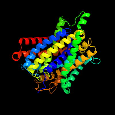
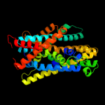
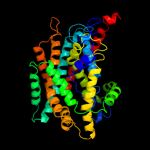
PDB 3gia chain A
Region: 16 - 427
Aligned: 393
Modelled: 411
Confidence: 100.0%
Identity: 11%
PDB header:transport protein
Chain: A: PDB Molecule:uncharacterized protein mj0609;
PDBTitle: crystal structure of apct transporter
Phyre2
| 2 |
|
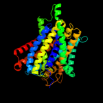
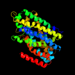
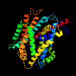
PDB 3lrc chain C
Region: 17 - 425
Aligned: 372
Modelled: 372
Confidence: 100.0%
Identity: 13%
PDB header:transport protein
Chain: C: PDB Molecule:arginine/agmatine antiporter;
PDBTitle: structure of e. coli adic (p1)
Phyre2
| 3 |
|
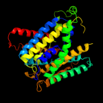
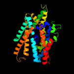
PDB 2jln chain A
Region: 5 - 424
Aligned: 399
Modelled: 399
Confidence: 99.9%
Identity: 9%
PDB header:membrane protein
Chain: A: PDB Molecule:mhp1;
PDBTitle: structure of mhp1, a nucleobase-cation-symport-1 family2 transporter
Phyre2
| 4 |
|
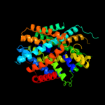
PDB 3dh4 chain A
Region: 2 - 425
Aligned: 411
Modelled: 397
Confidence: 99.3%
Identity: 11%
PDB header:transport protein
Chain: A: PDB Molecule:sodium/glucose cotransporter;
PDBTitle: crystal structure of sodium/sugar symporter with bound galactose from2 vibrio parahaemolyticus
Phyre2
| 5 |
|
PDB 2xq2 chain A
Region: 18 - 425
Aligned: 396
Modelled: 396
Confidence: 99.1%
Identity: 11%
PDB header:transport protein
Chain: A: PDB Molecule:sodium/glucose cotransporter;
PDBTitle: structure of the k294a mutant of vsglt
Phyre2
| 6 |
|
PDB 2a65 chain A domain 1
Region: 20 - 426
Aligned: 395
Modelled: 395
Confidence: 98.6%
Identity: 14%
Fold: SNF-like
Superfamily: SNF-like
Family: SNF-like
Phyre2
| 7 |
|
PDB 2w8a chain C
Region: 8 - 406
Aligned: 389
Modelled: 399
Confidence: 98.3%
Identity: 11%
PDB header:membrane protein
Chain: C: PDB Molecule:glycine betaine transporter betp;
PDBTitle: crystal structure of the sodium-coupled glycine betaine2 symporter betp from corynebacterium glutamicum with bound3 substrate
Phyre2
| 8 |
|
PDB 3hfx chain A
Region: 4 - 401
Aligned: 396
Modelled: 398
Confidence: 97.2%
Identity: 8%
PDB header:transport protein
Chain: A: PDB Molecule:l-carnitine/gamma-butyrobetaine antiporter;
PDBTitle: crystal structure of carnitine transporter
Phyre2
| 9 |
|
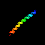
PDB 3hd7 chain A
Region: 396 - 429
Aligned: 34
Modelled: 34
Confidence: 10.4%
Identity: 12%
PDB header:exocytosis
Chain: A: PDB Molecule:vesicle-associated membrane protein 2;
PDBTitle: helical extension of the neuronal snare complex into the membrane,2 spacegroup c 1 2 1
Phyre2
| 10 |
|
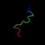
PDB 2zkr chain 3
Region: 6 - 24
Aligned: 19
Modelled: 19
Confidence: 7.7%
Identity: 26%
PDB header:ribosomal protein/rna
Chain: 3: PDB Molecule:60s ribosomal protein l39e;
PDBTitle: structure of a mammalian ribosomal 60s subunit within an2 80s complex obtained by docking homology models of the rna3 and proteins into an 8.7 a cryo-em map
Phyre2
| 11 |
|
PDB 1vqo chain 2 domain 1
Region: 8 - 22
Aligned: 15
Modelled: 15
Confidence: 7.3%
Identity: 33%
Fold: Non-globular all-alpha subunits of globular proteins
Superfamily: Ribosomal protein L39e
Family: Ribosomal protein L39e
Phyre2
| 12 |
|
PDB 4a1b chain B
Region: 1 - 24
Aligned: 24
Modelled: 24
Confidence: 6.9%
Identity: 21%
PDB header:ribosome
Chain: B: PDB Molecule:rpl39;
PDBTitle: t.thermophila 60s ribosomal subunit in complex with2 initiation factor 6. this file contains 26s rrna and3 proteins of molecule 3.
Phyre2