Phyre Home Retrieve Phyre Job Id
Emaill.a.kelley@imperial.ac.uk
DescriptionP0AB35
DateThu Jan 5 11:14:31 GMT 2012
Unique Job IDea7a9c13850ef6b7

Summary 

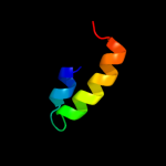
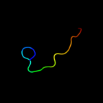
Top model
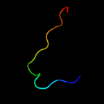
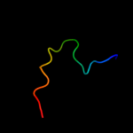
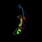
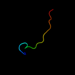
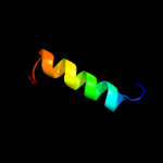
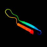
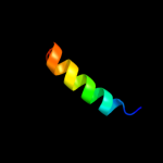
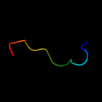
Image coloured by rainbow N → C terminus
Model (left) based on template d1i4ma_
Top template information
Fold:Prion-like
Superfamily:Prion-like
Family:Prion-like
Confidence and coverage
Confidence: 39.7% Coverage: 10%
18 residues ( 10% of your sequence) have been modelled with 39.7% confidence by the single highest scoring template.
You may wish to submit your sequence to Phyrealarm. This will automatically scan your sequence every week for new potential templates as they appear in the Phyre2 library.
Please note: You must be registered and logged in to use Phyrealarm.
3D viewing
Interactive 3D view in Jmol

Sequence analysis 

Secondary structure and disorder prediction 

   1........10.........20.........30.........40.........50.........60
Sequence  MNKSMLAGIGIGVAAALGVAAVASLNVFERGPQYAQVVSATPIKETVKTPRQECRNVTVT
Secondary structure 


SS confidence 


Disorder  ????


?
?


??


Disorder confidence 


   .........70.........80.........90.........100.........110.........120
Sequence  HRRPVQDENRITGSVLGAVAGGVIGHQFGGGRGKDVATVVGALGGGYAGNQIQGSLQESD
Secondary structure 


SS confidence 


Disorder  ???????????


?????????


????????????????
Disorder confidence 


   .........130.........140.........150.........160.........170.........
Sequence  TYTTTQQRCKTVYDKSEKMLGYDVTYKIGDQQGKIRMDRDPGTQIPLDSNGQLILNNKV
Secondary structure 


SS confidence 


Disorder  ???


??
?


?????
Disorder confidence 


Confidence Key
High(9)                    Low (0)
?Disordered
Alpha helix
Beta strand

Domain analysis 

Hover over an aligned region to see model and summary info

Please note, only up to the top 20 hits are modelled to reduce computer load

RankAligned region

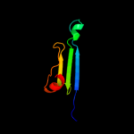
PDB 1i4m chain A

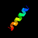
3D model

Region: 73 - 90
Aligned: 18
Modelled: 18
Confidence: 39.7%
Identity: 28%
Fold: Prion-like
Superfamily: Prion-like
Family: Prion-like

Phyre2

PDB 2lbg chain A

3D model

Region: 71 - 89
Aligned: 19
Modelled: 19
Confidence: 23.2%
Identity: 37%
PDB header:membrane protein
Chain: A: PDB Molecule:major prion protein;
PDBTitle: structure of the chr of the prion protein in dpc micelles

Phyre2

PDB 1v7w chain A

3D model

Region: 141 - 174
Aligned: 34
Modelled: 34
Confidence: 22.5%
Identity: 21%
PDB header:transferase
Chain: A: PDB Molecule:chitobiose phosphorylase;
PDBTitle: crystal structure of vibrio proteolyticus chitobiose phosphorylase in2 complex with glcnac

Phyre2

PDB 2cqt chain A

3D model

Region: 141 - 179
Aligned: 39
Modelled: 39
Confidence: 22.0%
Identity: 23%
PDB header:transferase
Chain: A: PDB Molecule:cellobiose phosphorylase;
PDBTitle: crystal structure of cellvibrio gilvus cellobiose phosphorylase2 crystallized from sodium/potassium phosphate

Phyre2

PDB 1u5l chain A

3D model

Region: 73 - 90
Aligned: 18
Modelled: 18
Confidence: 21.8%
Identity: 39%
Fold: Prion-like
Superfamily: Prion-like
Family: Prion-like

Phyre2

PDB 3hfw chain A

3D model

Region: 90 - 109
Aligned: 20
Modelled: 20
Confidence: 21.2%
Identity: 30%
PDB header:hydrolase
Chain: A: PDB Molecule:protein adp-ribosylarginine hydrolase;
PDBTitle: crystal structure of human adp-ribosylhydrolase 1 (harh1)

Phyre2

PDB 1ktd chain B domain 2

3D model

Region: 104 - 173
Aligned: 67
Modelled: 70
Confidence: 20.7%
Identity: 18%
Fold: MHC antigen-recognition domain
Superfamily: MHC antigen-recognition domain
Family: MHC antigen-recognition domain

Phyre2

PDB 1i3r chain B domain 2

3D model

Region: 104 - 173
Aligned: 67
Modelled: 70
Confidence: 17.4%
Identity: 18%
Fold: MHC antigen-recognition domain
Superfamily: MHC antigen-recognition domain
Family: MHC antigen-recognition domain

Phyre2

PDB 2qty chain B

3D model

Region: 90 - 111
Aligned: 22
Modelled: 22
Confidence: 16.0%
Identity: 41%
PDB header:hydrolase
Chain: B: PDB Molecule:poly(adp-ribose) glycohydrolase arh3;
PDBTitle: crystal structure of mouse adp-ribosylhydrolase 3 (marh3)

Phyre2

PDB 1t5j chain A

3D model

Region: 90 - 111
Aligned: 22
Modelled: 22
Confidence: 16.0%
Identity: 36%
Fold: ADP-ribosylglycohydrolase
Superfamily: ADP-ribosylglycohydrolase
Family: ADP-ribosylglycohydrolase

Phyre2

PDB 1u3m chain A

3D model

Region: 73 - 90
Aligned: 18
Modelled: 18
Confidence: 15.9%
Identity: 39%
Fold: Prion-like
Superfamily: Prion-like
Family: Prion-like

Phyre2

PDB 3g9d chain B

3D model

Region: 90 - 111
Aligned: 22
Modelled: 22
Confidence: 13.6%
Identity: 27%
PDB header:hydrolase
Chain: B: PDB Molecule:dinitrogenase reductase activacting
PDBTitle: crystal structure glycohydrolase

Phyre2

PDB 1i7o chain C

3D model

Region: 160 - 179
Aligned: 20
Modelled: 20
Confidence: 11.5%
Identity: 30%
PDB header:isomerase, lyase
Chain: C: PDB Molecule:4-hydroxyphenylacetate degradation bifunctional
PDBTitle: crystal structure of hpce

Phyre2

PDB 2woc chain A

3D model

Region: 90 - 111
Aligned: 22
Modelled: 22
Confidence: 11.5%
Identity: 27%
PDB header:hydrolase
Chain: A: PDB Molecule:adp-ribosyl-[dinitrogen reductase] glycohydrolase;
PDBTitle: crystal structure of the dinitrogenase reductase-activating2 glycohydrolase (drag) from rhodospirillum rubrum

Phyre2

PDB 1xyk chain A

3D model

Region: 75 - 90
Aligned: 16
Modelled: 16
Confidence: 10.3%
Identity: 25%
Fold: Prion-like
Superfamily: Prion-like
Family: Prion-like

Phyre2

PDB 1bx2 chain B domain 2

3D model

Region: 117 - 176
Aligned: 57
Modelled: 60
Confidence: 9.8%
Identity: 12%
Fold: MHC antigen-recognition domain
Superfamily: MHC antigen-recognition domain
Family: MHC antigen-recognition domain

Phyre2

PDB 2vv5 chain A domain 1

3D model

Region: 158 - 168
Aligned: 11
Modelled: 11
Confidence: 9.4%
Identity: 18%
Fold: Sm-like fold
Superfamily: Sm-like ribonucleoproteins
Family: Mechanosensitive channel protein MscS (YggB), middle domain

Phyre2

PDB 1aml chain A

3D model

Region: 69 - 84
Aligned: 16
Modelled: 16
Confidence: 9.2%
Identity: 31%
PDB header:serine protease inhibitor
Chain: A: PDB Molecule:amyloid a4;
PDBTitle: the alzheimer`s disease amyloid a4 peptide (residues 1-40)

Phyre2

PDB 2yzw chain A

3D model

Region: 82 - 111
Aligned: 30
Modelled: 30
Confidence: 8.9%
Identity: 33%
PDB header:hydrolase
Chain: A: PDB Molecule:adp-ribosylglycohydrolase;
PDBTitle: adp-ribosylglycohydrolase-related protein complex

Phyre2

PDB 1xyx chain A

3D model

Region: 75 - 90
Aligned: 16
Modelled: 16
Confidence: 8.9%
Identity: 25%
Fold: Prion-like
Superfamily: Prion-like
Family: Prion-like

Phyre2
1

d1i4ma_
2

c2lbgA_
3

c1v7wA_
4

c2cqtA_
5

d1u5la_
6

c3hfwA_
7

d1ktdb2
8

d1i3rb2
9

c2qtyB_
10

d1t5ja_
11

d1u3ma_
12

c3g9dB_
13

c1i7oC_
14

c2wocA_
15

d1xyka_
16

d1bx2b2
17

d2vv5a1
18

c1amlA_
19

c2yzwA_
20

d1xyxa_
21


22


23


24


25


26


27


28


29


30


31


32


33


34


35


36


Detailed template information 

#
Template Alignment Coverage3D Model Confidence
% i.d. Template Information
1d1i4ma_


39.7 28 Fold:Prion-like
Superfamily:Prion-like
Family:Prion-like
2c2lbgA_


23.2 37 PDB header:membrane protein
Chain: A: PDB Molecule:major prion protein;
PDBTitle: structure of the chr of the prion protein in dpc micelles
3c1v7wA_


22.5 21 PDB header:transferase
Chain: A: PDB Molecule:chitobiose phosphorylase;
PDBTitle: crystal structure of vibrio proteolyticus chitobiose phosphorylase in2 complex with glcnac
4c2cqtA_


22.0 23 PDB header:transferase
Chain: A: PDB Molecule:cellobiose phosphorylase;
PDBTitle: crystal structure of cellvibrio gilvus cellobiose phosphorylase2 crystallized from sodium/potassium phosphate
5d1u5la_


21.8 39 Fold:Prion-like
Superfamily:Prion-like
Family:Prion-like
6c3hfwA_


21.2 30 PDB header:hydrolase
Chain: A: PDB Molecule:protein adp-ribosylarginine hydrolase;
PDBTitle: crystal structure of human adp-ribosylhydrolase 1 (harh1)
7d1ktdb2


20.7 18 Fold:MHC antigen-recognition domain
Superfamily:MHC antigen-recognition domain
Family:MHC antigen-recognition domain
8d1i3rb2


17.4 18 Fold:MHC antigen-recognition domain
Superfamily:MHC antigen-recognition domain
Family:MHC antigen-recognition domain
9c2qtyB_


16.0 41 PDB header:hydrolase
Chain: B: PDB Molecule:poly(adp-ribose) glycohydrolase arh3;
PDBTitle: crystal structure of mouse adp-ribosylhydrolase 3 (marh3)
10d1t5ja_


16.0 36 Fold:ADP-ribosylglycohydrolase
Superfamily:ADP-ribosylglycohydrolase
Family:ADP-ribosylglycohydrolase
11d1u3ma_


15.9 39 Fold:Prion-like
Superfamily:Prion-like
Family:Prion-like
12c3g9dB_


13.6 27 PDB header:hydrolase
Chain: B: PDB Molecule:dinitrogenase reductase activacting
PDBTitle: crystal structure glycohydrolase
13c1i7oC_


11.5 30 PDB header:isomerase, lyase
Chain: C: PDB Molecule:4-hydroxyphenylacetate degradation bifunctional
PDBTitle: crystal structure of hpce
14c2wocA_


11.5 27 PDB header:hydrolase
Chain: A: PDB Molecule:adp-ribosyl-[dinitrogen reductase] glycohydrolase;
PDBTitle: crystal structure of the dinitrogenase reductase-activating2 glycohydrolase (drag) from rhodospirillum rubrum
15d1xyka_


10.3 25 Fold:Prion-like
Superfamily:Prion-like
Family:Prion-like
16d1bx2b2


9.8 12 Fold:MHC antigen-recognition domain
Superfamily:MHC antigen-recognition domain
Family:MHC antigen-recognition domain
17d2vv5a1


9.4 18 Fold:Sm-like fold
Superfamily:Sm-like ribonucleoproteins
Family:Mechanosensitive channel protein MscS (YggB), middle domain
18c1amlA_


9.2 31 PDB header:serine protease inhibitor
Chain: A: PDB Molecule:amyloid a4;
PDBTitle: the alzheimer`s disease amyloid a4 peptide (residues 1-40)
19c2yzwA_


8.9 33 PDB header:hydrolase
Chain: A: PDB Molecule:adp-ribosylglycohydrolase;
PDBTitle: adp-ribosylglycohydrolase-related protein complex
20d1xyxa_


8.9 25 Fold:Prion-like
Superfamily:Prion-like
Family:Prion-like
21d1xyqa_


not modelled 8.9 25 Fold:Prion-like
Superfamily:Prion-like
Family:Prion-like
22d1nppa2


not modelled 8.8 15 Fold:SH3-like barrel
Superfamily:Translation proteins SH3-like domain
Family:N-utilization substance G protein NusG, C-terminal domain
23d1d5mb2


not modelled 7.6 13 Fold:MHC antigen-recognition domain
Superfamily:MHC antigen-recognition domain
Family:MHC antigen-recognition domain
24d1xyua_


not modelled 7.5 25 Fold:Prion-like
Superfamily:Prion-like
Family:Prion-like
25d2gu3a1


not modelled 7.4 24 Fold:Cystatin-like
Superfamily:Cystatin/monellin
Family:PepSY-like
26d1v7wa1


not modelled 6.6 21 Fold:alpha/alpha toroid
Superfamily:Six-hairpin glycosidases
Family:Glycosyltransferase family 36 C-terminal domain
27d1q5xa_


not modelled 6.2 15 Fold:The "swivelling" beta/beta/alpha domain
Superfamily:RraA-like
Family:RraA-like
28d2ifra1


not modelled 5.8 18 Fold:Concanavalin A-like lectins/glucanases
Superfamily:Concanavalin A-like lectins/glucanases
Family:Peptidase A4
29c3ge2A_


not modelled 5.5 27 PDB header:lipoprotein
Chain: A: PDB Molecule:lipoprotein, putative;
PDBTitle: crystal structure of putative lipoprotein sp_0198 from streptococcus2 pneumoniae
30d1fngb2


not modelled 5.4 15 Fold:MHC antigen-recognition domain
Superfamily:MHC antigen-recognition domain
Family:MHC antigen-recognition domain
31c1iclA_


not modelled 5.4 33 PDB header:de novo protein
Chain: A: PDB Molecule:th1ox;
PDBTitle: solution structure of designed beta-sheet mini-protein th1ox
32d1nfia2


not modelled 5.4 13 Fold:Common fold of diphtheria toxin/transcription factors/cytochrome f
Superfamily:p53-like transcription factors
Family:Rel/Dorsal transcription factors, DNA-binding domain
33d1mj4a_


not modelled 5.3 42 Fold:Cytochrome b5-like heme/steroid binding domain
Superfamily:Cytochrome b5-like heme/steroid binding domain
Family:Cytochrome b5
34c2jcbA_


not modelled 5.2 19 PDB header:ligase
Chain: A: PDB Molecule:5-formyltetrahydrofolate cyclo-ligase family protein;
PDBTitle: the crystal structure of 5-formyl-tetrahydrofolate2 cycloligase from bacillus anthracis (ba4489)
35c2rfuA_


not modelled 5.1 19 PDB header:viral protein
Chain: A: PDB Molecule:influenza b hemagglutinin (ha);
PDBTitle: crystal structure of influenza b virus hemagglutinin in complex with2 lstc receptor analog
36c3c8oB_


not modelled 5.1 25 PDB header:hydrolase regulator
Chain: B: PDB Molecule:regulator of ribonuclease activity a;
PDBTitle: the crystal structure of rraa from pao1

Binding site prediction 

Due to computational demand, binding site predictions are not run for batch jobs

If you want to predict binding sites, please manually submit your model of choice to 3DLigandSite


Phyre is for academic use only

Please cite: Protein structure prediction on the web: a case study using the Phyre server
Kelley LA and Sternberg MJE. Nature Protocols 4, 363 - 371 (2009) [pdf] [Import into BibTeX]
 
If you use the binding site predictions from 3DLigandSite, please also cite:
3DLigandSite: predicting ligand-binding sites using similar structures.
Wass MN, Kelley LA and Sternberg MJ Nucleic Acids Research 38, W469-73 (2010) [PubMed]
 
© Structural Bioinformatics Group
Imperial College London
Lawrence Kelley, Benjamin Jefferys
Disclaimer
Terms and Conditions
Component software
Template detection: HHpred 1.51
Secondary structure prediction: Psi-pred 2.5
Disorder prediction: Disopred 2.4
Transmembrane prediction: Memsat_SVM
Multi-template modelling and ab initio: Poing 1.0