Phyre Home Retrieve Phyre Job Id
Emaill.a.kelley@imperial.ac.uk
DescriptionP60720
DateThu Jan 5 12:07:03 GMT 2012
Unique Job IDe76ed21bf11c66d4

Summary 

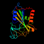
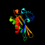
Top model
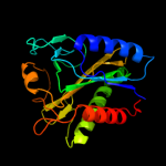
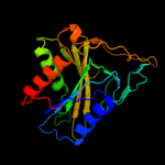
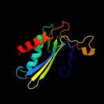
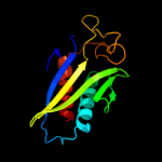
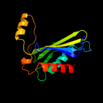
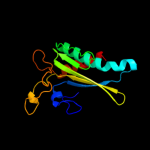
Image coloured by rainbow N → C terminus
Model (left) based on template d1w66a1
Top template information
Fold:Class II aaRS and biotin synthetases
Superfamily:Class II aaRS and biotin synthetases
Family:LplA-like
Confidence and coverage
Confidence:100.0% Coverage: 95%
203 residues ( 95% of your sequence) have been modelled with 100.0% confidence by the single highest scoring template.
3D viewing
Interactive 3D view in Jmol

Sequence analysis 

Secondary structure and disorder prediction 

   1........10.........20.........30.........40.........50.........60
Sequence  MYQDKILVRQLGLQPYEPISQAMHEFTDTRDDSTLDEIWLVEHYPVFTQGQAGKAEHILM
Secondary structure 


SS confidence 


Disorder  ?????


??


?
?

Disorder confidence 


   .........70.........80.........90.........100.........110.........120
Sequence  PGDIPVIQSDRGGQVTYHGPGQQVMYVLLNLKRRKLGVRELVTLLEQTVVNTLAELGIEA
Secondary structure 


SS confidence 


Disorder 


Disorder confidence 


   .........130.........140.........150.........160.........170.........180
Sequence  HPRADAPGVYVGEKKICSLGLRIRRGCSFHGLALNVNMDLSPFLRINPCGYAGMEMAKIS
Secondary structure 


SS confidence 


Disorder 


Disorder confidence 


   .........190.........200.........210...
Sequence  QWKPEATTNNIAPRLLENILALLNNPDFEYITA
Secondary structure 


SS confidence 


Disorder 


????
Disorder confidence 


Confidence Key
High(9)                    Low (0)
?Disordered
Alpha helix
Beta strand

Domain analysis 

Hover over an aligned region to see model and summary info

Please note, only up to the top 20 hits are modelled to reduce computer load

RankAligned region

PDB 1w66 chain A domain 1

3D model

Region: 2 - 206
Aligned: 203
Modelled: 205
Confidence: 100.0%
Identity: 31%
Fold: Class II aaRS and biotin synthetases
Superfamily: Class II aaRS and biotin synthetases
Family: LplA-like

Phyre2

PDB 2qhv chain A

3D model

Region: 5 - 208
Aligned: 201
Modelled: 204
Confidence: 100.0%
Identity: 32%
PDB header:transferase
Chain: A: PDB Molecule:lipoyltransferase;
PDBTitle: structural basis of octanoic acid recognition by lipoate-protein2 ligase b

Phyre2

PDB 2qhs chain A

3D model

Region: 5 - 208
Aligned: 201
Modelled: 204
Confidence: 100.0%
Identity: 32%
PDB header:transferase
Chain: A: PDB Molecule:lipoyltransferase;
PDBTitle: structural basis of octanoic acid recognition by lipoate-protein2 ligase b

Phyre2

PDB 1x2g chain B

3D model

Region: 4 - 206
Aligned: 195
Modelled: 197
Confidence: 100.0%
Identity: 16%
PDB header:ligase
Chain: B: PDB Molecule:lipoate-protein ligase a;
PDBTitle: crystal structure of lipate-protein ligase a from2 escherichia coli

Phyre2

PDB 1vqz chain A

3D model

Region: 6 - 212
Aligned: 202
Modelled: 206
Confidence: 100.0%
Identity: 17%
PDB header:ligase
Chain: A: PDB Molecule:lipoate-protein ligase, putative;
PDBTitle: crystal structure of a putative lipoate-protein ligase a (sp_1160)2 from streptococcus pneumoniae tigr4 at 1.99 a resolution

Phyre2

PDB 2e5a chain A

3D model

Region: 7 - 211
Aligned: 200
Modelled: 205
Confidence: 100.0%
Identity: 15%
PDB header:ligase
Chain: A: PDB Molecule:lipoyltransferase 1;
PDBTitle: crystal structure of bovine lipoyltransferase in complex2 with lipoyl-amp

Phyre2

PDB 1x2g chain A domain 2

3D model

Region: 4 - 213
Aligned: 202
Modelled: 204
Confidence: 100.0%
Identity: 17%
Fold: Class II aaRS and biotin synthetases
Superfamily: Class II aaRS and biotin synthetases
Family: LplA-like

Phyre2

PDB 2c8m chain A domain 1

3D model

Region: 4 - 206
Aligned: 195
Modelled: 203
Confidence: 100.0%
Identity: 18%
Fold: Class II aaRS and biotin synthetases
Superfamily: Class II aaRS and biotin synthetases
Family: LplA-like

Phyre2

PDB 1vqz chain A domain 2

3D model

Region: 7 - 212
Aligned: 201
Modelled: 203
Confidence: 100.0%
Identity: 17%
Fold: Class II aaRS and biotin synthetases
Superfamily: Class II aaRS and biotin synthetases
Family: LplA-like

Phyre2

PDB 2p5i chain A domain 1

3D model

Region: 7 - 212
Aligned: 205
Modelled: 206
Confidence: 100.0%
Identity: 13%
Fold: Class II aaRS and biotin synthetases
Superfamily: Class II aaRS and biotin synthetases
Family: LplA-like

Phyre2

PDB 2p0l chain A domain 1

3D model

Region: 3 - 207
Aligned: 203
Modelled: 205
Confidence: 100.0%
Identity: 11%
Fold: Class II aaRS and biotin synthetases
Superfamily: Class II aaRS and biotin synthetases
Family: LplA-like

Phyre2

PDB 2zgw chain A domain 2

3D model

Region: 69 - 203
Aligned: 122
Modelled: 135
Confidence: 99.2%
Identity: 20%
Fold: Class II aaRS and biotin synthetases
Superfamily: Class II aaRS and biotin synthetases
Family: Biotin holoenzyme synthetase

Phyre2

PDB 2ewn chain A

3D model

Region: 69 - 203
Aligned: 129
Modelled: 135
Confidence: 99.0%
Identity: 15%
PDB header:ligase, transcription
Chain: A: PDB Molecule:bira bifunctional protein;
PDBTitle: ecoli biotin repressor with co-repressor analog

Phyre2

PDB 2ej9 chain A

3D model

Region: 69 - 203
Aligned: 123
Modelled: 135
Confidence: 99.0%
Identity: 18%
PDB header:ligase
Chain: A: PDB Molecule:putative biotin ligase;
PDBTitle: crystal structure of biotin protein ligase from2 methanococcus jannaschii

Phyre2

PDB 2eay chain B

3D model

Region: 66 - 203
Aligned: 122
Modelled: 132
Confidence: 98.9%
Identity: 21%
PDB header:ligase
Chain: B: PDB Molecule:biotin [acetyl-coa-carboxylase] ligase;
PDBTitle: crystal structure of biotin protein ligase from aquifex2 aeolicus

Phyre2

PDB 3bfm chain A

3D model

Region: 69 - 203
Aligned: 125
Modelled: 135
Confidence: 98.8%
Identity: 11%
PDB header:unknown function
Chain: A: PDB Molecule:biotin protein ligase-like protein of unknown function;
PDBTitle: crystal structure of a biotin protein ligase-like protein of unknown2 function (tm1040_0394) from silicibacter sp. tm1040 at 1.70 a3 resolution

Phyre2

PDB 2dzc chain A

3D model

Region: 66 - 203
Aligned: 118
Modelled: 138
Confidence: 98.8%
Identity: 23%
PDB header:ligase
Chain: A: PDB Molecule:biotin--[acetyl-coa-carboxylase] ligase;
PDBTitle: crystal structure of biotin protein ligase from pyrococcus2 horikoshii, mutation r48a

Phyre2

PDB 1bia chain A domain 3

3D model

Region: 44 - 203
Aligned: 122
Modelled: 141
Confidence: 98.6%
Identity: 16%
Fold: Class II aaRS and biotin synthetases
Superfamily: Class II aaRS and biotin synthetases
Family: Biotin holoenzyme synthetase

Phyre2

PDB 2cgh chain B

3D model

Region: 81 - 203
Aligned: 107
Modelled: 109
Confidence: 98.6%
Identity: 19%
PDB header:ligase
Chain: B: PDB Molecule:biotin ligase;
PDBTitle: crystal structure of biotin ligase from mycobacterium2 tuberculosis

Phyre2

PDB 2ddz chain A domain 1

3D model

Region: 103 - 204
Aligned: 88
Modelled: 102
Confidence: 97.5%
Identity: 20%
Fold: Class II aaRS and biotin synthetases
Superfamily: Class II aaRS and biotin synthetases
Family: PH0223-like

Phyre2
1

d1w66a1
2

c2qhvA_
3

c2qhsA_
4

c1x2gB_
5

c1vqzA_
6

c2e5aA_
7

d1x2ga2
8

d2c8ma1
9

d1vqza2
10

d2p5ia1
11

d2p0la1
12

d2zgwa2
13

c2ewnA_
14

c2ej9A_
15

c2eayB_
16

c3bfmA_
17

c2dzcA_
18

d1biaa3
19

c2cghB_
20

d2ddza1
21


22


23


24


25


26


27


28


29


30


31


32


33


34


35


36


37


38


39


Detailed template information 

#
Template Alignment Coverage3D Model Confidence
% i.d. Template Information
1d1w66a1


100.0 31 Fold:Class II aaRS and biotin synthetases
Superfamily:Class II aaRS and biotin synthetases
Family:LplA-like
2c2qhvA_


100.0 32 PDB header:transferase
Chain: A: PDB Molecule:lipoyltransferase;
PDBTitle: structural basis of octanoic acid recognition by lipoate-protein2 ligase b
3c2qhsA_


100.0 32 PDB header:transferase
Chain: A: PDB Molecule:lipoyltransferase;
PDBTitle: structural basis of octanoic acid recognition by lipoate-protein2 ligase b
4c1x2gB_


100.0 16 PDB header:ligase
Chain: B: PDB Molecule:lipoate-protein ligase a;
PDBTitle: crystal structure of lipate-protein ligase a from2 escherichia coli
5c1vqzA_


100.0 17 PDB header:ligase
Chain: A: PDB Molecule:lipoate-protein ligase, putative;
PDBTitle: crystal structure of a putative lipoate-protein ligase a (sp_1160)2 from streptococcus pneumoniae tigr4 at 1.99 a resolution
6c2e5aA_


100.0 15 PDB header:ligase
Chain: A: PDB Molecule:lipoyltransferase 1;
PDBTitle: crystal structure of bovine lipoyltransferase in complex2 with lipoyl-amp
7d1x2ga2


100.0 17 Fold:Class II aaRS and biotin synthetases
Superfamily:Class II aaRS and biotin synthetases
Family:LplA-like
8d2c8ma1


100.0 18 Fold:Class II aaRS and biotin synthetases
Superfamily:Class II aaRS and biotin synthetases
Family:LplA-like
9d1vqza2


100.0 17 Fold:Class II aaRS and biotin synthetases
Superfamily:Class II aaRS and biotin synthetases
Family:LplA-like
10d2p5ia1


100.0 13 Fold:Class II aaRS and biotin synthetases
Superfamily:Class II aaRS and biotin synthetases
Family:LplA-like
11d2p0la1


100.0 11 Fold:Class II aaRS and biotin synthetases
Superfamily:Class II aaRS and biotin synthetases
Family:LplA-like
12d2zgwa2


99.2 20 Fold:Class II aaRS and biotin synthetases
Superfamily:Class II aaRS and biotin synthetases
Family:Biotin holoenzyme synthetase
13c2ewnA_


99.0 15 PDB header:ligase, transcription
Chain: A: PDB Molecule:bira bifunctional protein;
PDBTitle: ecoli biotin repressor with co-repressor analog
14c2ej9A_


99.0 18 PDB header:ligase
Chain: A: PDB Molecule:putative biotin ligase;
PDBTitle: crystal structure of biotin protein ligase from2 methanococcus jannaschii
15c2eayB_


98.9 21 PDB header:ligase
Chain: B: PDB Molecule:biotin [acetyl-coa-carboxylase] ligase;
PDBTitle: crystal structure of biotin protein ligase from aquifex2 aeolicus
16c3bfmA_


98.8 11 PDB header:unknown function
Chain: A: PDB Molecule:biotin protein ligase-like protein of unknown function;
PDBTitle: crystal structure of a biotin protein ligase-like protein of unknown2 function (tm1040_0394) from silicibacter sp. tm1040 at 1.70 a3 resolution
17c2dzcA_


98.8 23 PDB header:ligase
Chain: A: PDB Molecule:biotin--[acetyl-coa-carboxylase] ligase;
PDBTitle: crystal structure of biotin protein ligase from pyrococcus2 horikoshii, mutation r48a
18d1biaa3


98.6 16 Fold:Class II aaRS and biotin synthetases
Superfamily:Class II aaRS and biotin synthetases
Family:Biotin holoenzyme synthetase
19c2cghB_


98.6 19 PDB header:ligase
Chain: B: PDB Molecule:biotin ligase;
PDBTitle: crystal structure of biotin ligase from mycobacterium2 tuberculosis
20d2ddza1


97.5 20 Fold:Class II aaRS and biotin synthetases
Superfamily:Class II aaRS and biotin synthetases
Family:PH0223-like
21d2qfra2


not modelled 22.8 16 Fold:Metallo-dependent phosphatases
Superfamily:Metallo-dependent phosphatases
Family:Purple acid phosphatase-like
22c3lr4A_


not modelled 19.7 19 PDB header:transferase
Chain: A: PDB Molecule:sensor protein;
PDBTitle: periplasmic domain of the riss sensor protein from burkholderia2 pseudomallei, barium phased at low ph
23d1knxa1


not modelled 17.9 11 Fold:MurF and HprK N-domain-like
Superfamily:HprK N-terminal domain-like
Family:HPr kinase/phoshatase HprK N-terminal domain
24c1xzwB_


not modelled 12.1 16 PDB header:hydrolase
Chain: B: PDB Molecule:purple acid phosphatase;
PDBTitle: sweet potato purple acid phosphatase/phosphate complex
25c1kbpB_


not modelled 11.9 16 PDB header:hydrolase (phosphoric monoester)
Chain: B: PDB Molecule:purple acid phosphatase;
PDBTitle: kidney bean purple acid phosphatase
26c3uotB_


not modelled 10.1 11 PDB header:cell cycle
Chain: B: PDB Molecule:mediator of dna damage checkpoint protein 1;
PDBTitle: crystal structure of mdc1 fha domain in complex with a phosphorylated2 peptide from the mdc1 n-terminus
27d1ra0a1


not modelled 9.8 17 Fold:Composite domain of metallo-dependent hydrolases
Superfamily:Composite domain of metallo-dependent hydrolases
Family:Cytosine deaminase
28c1ponB_


not modelled 8.2 15 PDB header:calcium-binding protein
Chain: B: PDB Molecule:troponin c;
PDBTitle: site iii-site iv troponin c heterodimer, nmr
29c2kd2A_


not modelled 7.3 22 PDB header:apoptosis
Chain: A: PDB Molecule:fas apoptotic inhibitory molecule 1;
PDBTitle: nmr structure of faim-ctd
30d1m7ja1


not modelled 7.2 31 Fold:Composite domain of metallo-dependent hydrolases
Superfamily:Composite domain of metallo-dependent hydrolases
Family:D-aminoacylase
31d1vsra_


not modelled 7.2 23 Fold:Restriction endonuclease-like
Superfamily:Restriction endonuclease-like
Family:Very short patch repair (VSR) endonuclease
32c2j3tB_


not modelled 6.9 9 PDB header:transport
Chain: B: PDB Molecule:trafficking protein particle complex subunit 6a;
PDBTitle: the crystal structure of the bet3-trs33-bet5-trs23 complex.
33d1qbaa1


not modelled 6.6 22 Fold:Immunoglobulin-like beta-sandwich
Superfamily:E set domains
Family:E-set domains of sugar-utilizing enzymes
34c2faoB_


not modelled 6.5 20 PDB header:hydrolase/transferase
Chain: B: PDB Molecule:probable atp-dependent dna ligase;
PDBTitle: crystal structure of pseudomonas aeruginosa ligd polymerase2 domain
35c2iruA_


not modelled 6.2 16 PDB header:transferase
Chain: A: PDB Molecule:putative dna ligase-like protein rv0938/mt0965;
PDBTitle: crystal structure of the polymerase domain from mycobacterium2 tuberculosis ligase d
36d1xzwa2


not modelled 5.6 16 Fold:Metallo-dependent phosphatases
Superfamily:Metallo-dependent phosphatases
Family:Purple acid phosphatase-like
37d1kfia3


not modelled 5.5 11 Fold:Phosphoglucomutase, first 3 domains
Superfamily:Phosphoglucomutase, first 3 domains
Family:Phosphoglucomutase, first 3 domains
38c2jpeA_


not modelled 5.4 9 PDB header:transcription
Chain: A: PDB Molecule:nuclear inhibitor of protein phosphatase 1;
PDBTitle: fha domain of nipp1
39d1k1da1


not modelled 5.4 23 Fold:Composite domain of metallo-dependent hydrolases
Superfamily:Composite domain of metallo-dependent hydrolases
Family:Hydantoinase (dihydropyrimidinase)

Binding site prediction 

Due to computational demand, binding site predictions are not run for batch jobs

If you want to predict binding sites, please manually submit your model of choice to 3DLigandSite


Phyre is for academic use only

Please cite: Protein structure prediction on the web: a case study using the Phyre server
Kelley LA and Sternberg MJE. Nature Protocols 4, 363 - 371 (2009) [pdf] [Import into BibTeX]
 
If you use the binding site predictions from 3DLigandSite, please also cite:
3DLigandSite: predicting ligand-binding sites using similar structures.
Wass MN, Kelley LA and Sternberg MJ Nucleic Acids Research 38, W469-73 (2010) [PubMed]
 
© Structural Bioinformatics Group
Imperial College London
Lawrence Kelley, Benjamin Jefferys
Disclaimer
Terms and Conditions
Component software
Template detection: HHpred 1.51
Secondary structure prediction: Psi-pred 2.5
Disorder prediction: Disopred 2.4
Transmembrane prediction: Memsat_SVM
Multi-template modelling and ab initio: Poing 1.0