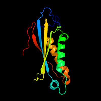
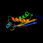
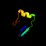
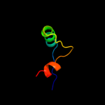
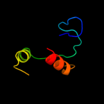
| 1 | c2eeyA_
|
|
 |
100.0 |
50 |
PDB header:biosynthetic protein
Chain: A: PDB Molecule:molybdopterin biosynthesis;
PDBTitle: structure of gk0241 protein from geobacillus kaustophilus
|
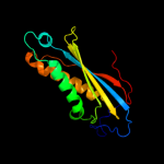
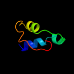
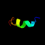
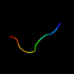
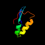
| 2 | c2ideE_
|
|
 |
100.0 |
54 |
PDB header:biosynthetic protein
Chain: E: PDB Molecule:molybdenum cofactor biosynthesis protein c;
PDBTitle: crystal structure of the molybdenum cofactor biosynthesis protein c2 (ttha1789) from thermus theromophilus hb8
|
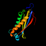
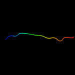
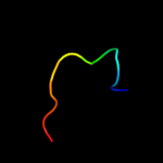
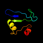
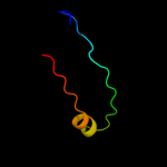
| 3 | d1ekra_
|
|
 |
100.0 |
100 |
Fold:Ferredoxin-like
Superfamily:Molybdenum cofactor biosynthesis protein C, MoaC
Family:Molybdenum cofactor biosynthesis protein C, MoaC |
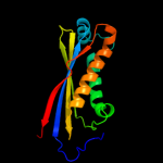
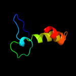
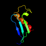
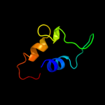
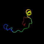
| 4 | c2eknC_
|
|
 |
100.0 |
41 |
PDB header:biosynthetic protein
Chain: C: PDB Molecule:probable molybdenum cofactor biosynthesis protein c;
PDBTitle: structure of ph1811 protein from pyrococcus horikoshii
|
| 5 | c2ohdB_
|
|
 |
100.0 |
45 |
PDB header:biosynthetic protein
Chain: B: PDB Molecule:probable molybdenum cofactor biosynthesis protein c;
PDBTitle: crystal structure of hypothetical molybdenum cofactor biosynthesis2 protein c from sulfolobus tokodaii
|
| 6 | c1sfeA_
|
|
 |
37.0 |
15 |
PDB header:dna-binding protein
Chain: A: PDB Molecule:ada o6-methylguanine-dna methyltransferase;
PDBTitle: ada o6-methylguanine-dna methyltransferase from escherichia coli
|
| 7 | d1oh4a_
|
|
 |
33.7 |
43 |
Fold:Galactose-binding domain-like
Superfamily:Galactose-binding domain-like
Family:Family 27 carbohydrate binding module, CBM27 |
| 8 | c3ct5A_
|
|
 |
31.7 |
30 |
PDB header:hydrolase
Chain: A: PDB Molecule:morphogenesis protein 1;
PDBTitle: crystal and cryoem structural studies of a cell wall degrading enzyme2 in the bacteriophage phi29 tail
|
| 9 | d2cqaa1
|
|
 |
22.5 |
15 |
Fold:OB-fold
Superfamily:Nucleic acid-binding proteins
Family:TIP49 domain |
| 10 | d1bu2a2
|
|
 |
14.8 |
47 |
Fold:Cyclin-like
Superfamily:Cyclin-like
Family:Cyclin |
| 11 | c2b5lC_
|
|
 |
13.0 |
31 |
PDB header:protein binding/viral protein
Chain: C: PDB Molecule:nonstructural protein v;
PDBTitle: crystal structure of ddb1 in complex with simian virus 5 v2 protein
|
| 12 | d1vqon1
|
|
 |
11.1 |
15 |
Fold:Ribonuclease H-like motif
Superfamily:Translational machinery components
Family:Ribosomal protein L18 and S11 |
| 13 | c3pn1A_
|
|
 |
10.3 |
19 |
PDB header:ligase/ligase inhibitor
Chain: A: PDB Molecule:dna ligase;
PDBTitle: novel bacterial nad+-dependent dna ligase inhibitors with broad2 spectrum potency and antibacterial efficacy in vivo
|
| 14 | c2p0xA_
|
|
 |
9.2 |
75 |
PDB header:de novo protein
Chain: A: PDB Molecule:abiotic atp-binding, folding optimized protein;
PDBTitle: solution structure of a non-biological atp-binding protein
|
| 15 | c3bdqB_
|
|
 |
9.0 |
24 |
PDB header:lipid transport
Chain: B: PDB Molecule:sterol carrier protein 2-like 2;
PDBTitle: room tempreture crystal structure of sterol carrier protein-2 2 like-2
|
| 16 | c2xuvB_
|
|
 |
8.8 |
22 |
PDB header:unknown function
Chain: B: PDB Molecule:hdeb;
PDBTitle: the structure of hdeb
|
| 17 | d1ikta_
|
|
 |
8.1 |
14 |
Fold:SCP-like
Superfamily:SCP-like
Family:Sterol carrier protein, SCP |
| 18 | d2f1fa2
|
|
 |
7.9 |
20 |
Fold:Ferredoxin-like
Superfamily:ACT-like
Family:IlvH-like |
| 19 | c2xskA_
|
|
 |
7.7 |
28 |
PDB header:chaperone
Chain: A: PDB Molecule:csgc;
PDBTitle: e. coli curli protein csgc - secys
|
| 20 | d2gp4a1
|
|
 |
7.6 |
25 |
Fold:The "swivelling" beta/beta/alpha domain
Superfamily:LeuD/IlvD-like
Family:IlvD/EDD C-terminal domain-like |
| 21 | d2bbya_ |
|
not modelled |
7.1 |
4 |
Fold:DNA/RNA-binding 3-helical bundle
Superfamily:"Winged helix" DNA-binding domain
Family:DNA-binding domain from rap30 |
| 22 | c2ketA_ |
|
not modelled |
7.1 |
45 |
PDB header:antibiotic
Chain: A: PDB Molecule:cathelicidin-6;
PDBTitle: solution structure of bmap-27
|
| 23 | c2kc5A_ |
|
not modelled |
7.0 |
13 |
PDB header:chaperone
Chain: A: PDB Molecule:hydrogenase-2 operon protein hybe;
PDBTitle: solution structure of hybe from escherichia coli
|
| 24 | d1pz4a_ |
|
not modelled |
6.9 |
23 |
Fold:SCP-like
Superfamily:SCP-like
Family:Sterol carrier protein, SCP |
| 25 | c1z4hA_ |
|
not modelled |
6.9 |
32 |
PDB header:protein binding, dna binding protein
Chain: A: PDB Molecule:tor inhibition protein;
PDBTitle: the response regulator tori belongs to a new family of2 atypical excisionase
|
| 26 | d1vlfn1 |
|
not modelled |
6.8 |
29 |
Fold:Prealbumin-like
Superfamily:Cna protein B-type domain
Family:Cna protein B-type domain |
| 27 | c2p09A_ |
|
not modelled |
6.7 |
75 |
PDB header:de novo protein
Chain: A: PDB Molecule:a non-biological atp binding protein with two mutations
PDBTitle: structural insights into the evolution of a non-biological protein
|
| 28 | d1wzua1 |
|
not modelled |
6.4 |
38 |
Fold:NadA-like
Superfamily:NadA-like
Family:NadA-like |
| 29 | d1uufa2 |
|
not modelled |
6.1 |
20 |
Fold:NAD(P)-binding Rossmann-fold domains
Superfamily:NAD(P)-binding Rossmann-fold domains
Family:Alcohol dehydrogenase-like, C-terminal domain |
| 30 | d4bcla_ |
|
not modelled |
5.9 |
23 |
Fold:Bacteriochlorophyll A protein
Superfamily:Bacteriochlorophyll A protein
Family:Bacteriochlorophyll A protein |
| 31 | c2gp4A_ |
|
not modelled |
5.8 |
25 |
PDB header:lyase
Chain: A: PDB Molecule:6-phosphogluconate dehydratase;
PDBTitle: structure of [fes]cluster-free apo form of 6-phosphogluconate2 dehydratase from shewanella oneidensis
|
| 32 | d1juva_ |
|
not modelled |
5.7 |
9 |
Fold:Dihydrofolate reductase-like
Superfamily:Dihydrofolate reductase-like
Family:Dihydrofolate reductases |
| 33 | d1cq3a_ |
|
not modelled |
5.2 |
83 |
Fold:Soluble secreted chemokine inhibitor, VCCI
Superfamily:Soluble secreted chemokine inhibitor, VCCI
Family:Soluble secreted chemokine inhibitor, VCCI |